Differentiation of Mouse Embryonic Stem Cells into
Endoderm and Mesoderm
Yang Liu
1, Qidong Liu
2, Wenwen Jia
2, Jie Chen
1, Jianmin Wang
1, Dan Ye
2, Xudong Guo
2, Wen Chen
2,
Guoping Li
2, Guiying Wang
2, Anmei Deng
1*, Jiuhong Kang
2*1 Department of Laboratory Medicine and Department of Hematology, Changhai Hospital, Second Military Medical University, Shanghai, People’s Republic of China, 2 Clinical and Translational Research Center of Shanghai First Maternity and Infant Health Hospital, Shanghai Key Laboratory of Signaling and Disease Research, School of Life Science and Technology, Tongji University, Shanghai, People’s Republic of China
Abstract
The mechanisms by which microRNAs (miRNAs) affect cell fate decisions remain poorly understood. Herein, we report that miR-200a can suppress the differentiation of mouse embryonic stem (ES) cells into endoderm and mesoderm. Interestingly, miR-200a directly targets growth factor receptor-bound protein 2 (Grb2), which is a key adaptor in the Erk signaling pathway. Furthermore, high levels of miR-200a dramatically decrease Grb2 levels and suppress the appearance of mesoderm and endoderm lineages in embryoid body formation, as well as suppressing the activation of Erk. Finally, Grb2 supplementation significantly rescues the miR-200a-induced layer-formation bias and the Erk suppression. Collectively, our results demonstrate that miR-200a plays critical roles in ES cell lineage commitment by directly regulating Grb2 expression and Erk signaling.
Citation: Liu Y, Liu Q, Jia W, Chen J, Wang J, et al. (2013) MicroRNA-200a Regulates Grb2 and Suppresses Differentiation of Mouse Embryonic Stem Cells into Endoderm and Mesoderm. PLoS ONE 8(7): e68990. doi:10.1371/journal.pone.0068990
Editor: Maurizio Pesce, Centro Cardiologico Monzino, Italy
Received March 23, 2013; Accepted June 3, 2013; Published July 18, 2013
Copyright: © 2013 Liu et al. This is an open-access article distributed under the terms of the Creative Commons Attribution License, which permits unrestricted use, distribution, and reproduction in any medium, provided the original author and source are credited.
Funding: This work was supported by grants from the Ministry of Science and Technology (2011CB965100, 2011DFA30480, 2010CB944900,
2010CB945000, 2012CB966603, 2011CBA01100, 2013CB967401 and 2013CB531606), the National Natural Science Foundation of China (31210103905, 91219305, 31201107, 31101061, 81170499, 31071306, 31000378, 31171432, and 81273282), the Science and Technology Commission of Shanghai Municipality (12ZR1450900, 11ZR1438500, 11XD1405300 and 11JC1410902), and IRT1168 and 20110072110039 from Ministry of Education. The work was also supported by Fundamental Research Funds for the Central Universities (2000219066, 2000219067 and 2000219077). The funders had no role in study design, data collection and analysis, decision to publish, or preparation of the manuscript.
Competing interests: The authors have declared that no competing interests exist. * E-mail: [email protected] (JK); [email protected] (AD)
Introduction
Embryonic stem (ES) cells are derived from the inner cell mass (ICM) of blastocysts, one of the early stages of embryonic development. These cells retain the features of pluripotency and self-renewal while serving as the progenitors of all cell types [1–3]. The regulatory mechanism for the differentiation of ES cells into functional cells remains unclear. Therefore, an in-depth understanding of the molecular mechanisms of cell lineage differentiation will facilitate clinical applications of stem cell therapy [4]. MicroRNAs (miRNAs), which are 21-23 nucleotide non-coding RNAs [5,6], have been identified as a class of gene regulators that act during the individual development and differentiation of specific cell types [7,8]. In the canonical pathway of miRNA biogenesis, the primary miRNAs processed into Drosha-DiGeorge syndrome critical region gene 8 (DGCR8) complexes to produce pre-miRNAs [9–13]. The pre-pre-miRNAs are then transported into the cytoplasm by Exportin-5 [14–16] and are further processed into
mature miRNAs by Dicer [17–20]. miRNAs are incorporated into the RNA-induced silencing complex (RISC), which then localizes to the 3’ untranslated region (UTR) of the target mRNA [21,22], leading to gene silencing [23–26] or degradation [27] at a post transcriptional level. miRNAs are a determinant of ES cell characteristics in early developmental processes [28].
cells [33–35]. miR-200 family members directly target Zeb1/ Zeb2 and enhance E-cadherin expression, resulting in the suppression of murine mammary tumor cell migration [33,36]. In contrast, Zeb1 suppresses the expression of miR-200 family members, forming a regulatory feedback loop [37]. A recent study demonstrated that miR-200a overexpression prevents the transformation of normal mammary cells and decreases cell migration by targeting the class III histone deacetylase silent information regulator 1 (Sirt1) [38]. miR-200a also targets p38alpha and regulates the oxidative stress response, affecting tumorigenesis and chemosensitivity [39]. miR-200a overexpression decreases Smad-3 activity and the matrix protein, including Collagen I, Collagen IV and Fibronectin, blocks the TGF-beta dependent epithelial-mesenchymal transition (EMT) process, and rescues early and advanced kidney disease in mouse models [40]. However, the function of miR-200a in the initiation of vertebrate embryo development has not been reported (A list of miR-200 family members targets in stem cells and development is shown in Table 1).
Growth factor receptor-bound protein 2 (Grb2) is a key adapter protein in intracellular signal transduction pathways, in which it links activated cell surface receptors to downstream targets by binding to specific phosphotyrosine-containing and proline-rich sequence motifs [41,42]. Deletion of the Grb2 gene leads to preimplantation embryonic lethality in vivo [43,44]. Signaling via Grb2 is essential to the segregation of epiblast and primitive endoderm progenitors [43]. Those findings suggest that Grb2 might support differentiation. However, the roles of Grb2 and miRNA-induced intracellular signaling cascades in lineage commitment are not well understood.
In this paper, we report that miR-200a was highly expressed in ES cells and gradually decreased in expression during embryoid body (EB) formation and that miR-200a suppressed endoderm and mesoderm lineage commitment. We further identified Grb2 as a direct regulatory target of miR-200a. In EB, knockdown of Grb2 with a specific shRNA had an identical effect to treatment with miR-200a. Finally, a rescue assay
Table 1. miR-200 family menbers in stem cells and development.
Target Member Cell type References
Zeb1/ Zeb2 miR-200a, miR-200b, miR-200c, miR-429, miR-141
Induced pluripotent stem cells, breast cancer stem cells, neuroectodermal precursors
Wang et al. 2013 [70], Radisky 2011 [71], Du et al. 2013 [72]
Bmi1 miR-200c embryonal carcinoma cells Shimono et al. 2009 [73]
Sox2 miR-200c, miR-141
neural stem/progenitor
cells Peng et al. 2012 [29]
E2F3 miR-200c, miR-141
neural stem/progenitor
cells Peng et al. 2012 [29] Suz12 miR-200b Breast cancer stem cells Iliopoulos et al. 2010 [74] Flk1 miR-200c embryonic stem cells Gill et al. 2012 [75] Ets1 miR-200c embryonic stem cells Gill et al. 2012 [75]
Srf miR-200b oligodendrocyte progenitor
cells Buller et al. 2012 [76]
showed that exogenous Grb2 could reverse the miR-200a-induced endoderm and mesoderm suppression. Similarly, the extracellular signal-regulated kinase (Erk) signaling, when activated with assistance from Grb2, also rescued miR-200a-induced effects. Taken together, these results suggest that miR-200a might control cell fate decisions affecting the early endoderm and mesoderm layers in a manner that is partly dependent on Erk signaling, by regulating Grb2 expression levels.
Materials and Methods
Cell culture
The mouse ES cell line ES-E14TG2a, purchased from the American Type Culture Collection (ATCC CRL-1821), was maintained on gelatin-coated plates in high glucose-Dulbecco’s Modified Eagle Medium (DMEM; Gibco) that was supplemented with 15% (vol/vol) ES qualified-fetal bovine serum (FBS; Gibco), 2 mM L-glutamine (Hyclone), 1x nonessential amino acids (Hyclone), 50 mM beta-mercaptoethanol (Gibco) and 500-1000 U/ml of leukocyte inhibitory factor (LIF; generated in house). ES cells were trypsinized and split every 2 days, and the culture medium was changed daily. For the formation of EB, ES cells were plated in 3.5 cm dishes at a density of 5 × 10⁴ cells in 2 ml medium
without LIF. HEK293T cells were cultured in DMEM (Gibco) that was supplemented with 100 U/ml of penicillin/streptomycin (Invitrogen) and 10% FBS (Gibco).
Transfection of miRNA mimics and inhibitors
miR-200a mimics and inhibitors (including the Negative control) were purchased from Ribo. ES cells were seeded into 6-well plates at a density of 5 × 10⁴ cells per well. For miRNA
overexpression or knockdown experiments, miRNA mimics or inhibitors and the scrambled negative control were gently mixed with Opti-MEM (Gibco) and X-tremeGENE 9 Transfection Reagents (Roche) according to the manufacturer’s instructions. At 12-72 hours post-transfection, the cells were harvested for Western blotting or quantitative real-time PCR analysis.
Plasmids
The miR-200a sequence was obtained from the mirbase website and cloned into the AgeI and EcoRI sites of the pLKO. 1 vector. To construct a miR-200a sponge vector, the following oligoribonucleotides for the miR-200a sponge were designed and synthesized: miR-200a sponges forward oligo, CCGGTACATCGTTACTCTCAGTGTTACCGACATCGTTACT CTCAGTGTTAGCGACATCGTTACTCTCAGTGTTAG; reverse oligo,
putative target sites of miR-200a, was amplified and cloned into the SacI and XbaI sites downstream of the luciferase reporter gene in pGL3. A pGL3-Grb2-3’UTR-Mutant vector, which carried a mutation in the complementary site for the seed region of miR-200a, was generated from the pGL3-Grb2-3’UTR-WT vector by mutation PCR. The Grb2 expression vector was created by cloning the Grb2 coding sequence into the BamHI and AgeI sites of pFUW.
Protein analysis
Cells were lysed in RIPA buffer that contained a protease inhibitor cocktail. Estimation of lysate protein concentrations was performed with a BCA Protein Assay Kit (Pierce). Approximately 100 µg of lysate was resolved on a 12% SDS/ PAGE gel and transferred to NC membranes (Bio-Rad). The membranes were blocked with blocking solution (3% BSA in TBST) and incubated with primary antibodies. The following primary antibodies were used: mouse anti-Oct4 (1:1000; Santa Cruz Biotechnology), rabbit anti-Sox2 (1:1000; Millipore), rabbit anti-Nanog (1:1000; Abcam), rabbit anti-Grb2 (1:1000; Bioworld), rabbit anti-Erk (1:1000; Bioworld), rabbit anti-p-Erk (1:1000; Cell signaling) and rabbit anti-GAPDH (1:1000; Miaotong). The blots were subsequently incubated with either HRP-conjugated anti-rabbit IgG or HRP-conjugated anti-mouse IgG (1:3000; Cell signaling). Labeled proteins were detected with the eECL Western Blot Kit (Cwbio).
Quantitative real-time PCR
Total RNA was extracted from ES cells and EBs with TRIzol reagent (Takara) according to the manufacturer’s instructions. cDNA synthesis was performed from 250 ng of total RNA in a single step (37 °C for 15 min) with the Takara reverse transcription kit. Quantitative real-time PCR was performed with iTaq reagent (Bio-Rad) and a STRATAGENE Mx3005p Real-Time PCR Cycler. The following PCR cycle conditions were used: initial denaturation for 30 sec at 95 °C, followed by 40 cycles of 5 sec at 95 °C and 30 sec at 60 °C. Mature miRNA primers, including stem-loop RT and special miRNA primers, were purchased from Ribo. Real-time PCR primers are shown in Table S1.
Dual luciferase reporter assay
At 12 hours prior to the transfections, HEK293T cells were plated at a density of 5 × 10⁴ cells per well in 24-well plates.
Cells were transfected via Lipofectamine 2000 Transfection Reagent (Invitrogen) and Opti-MEM (Gibco) with 50 nM miRNA mimics, 400 ng of the luciferase vector (pGL3 constructs) and 20 ng of the Renilla vector. At 24 hours post-transfection, the cells were harvested, and the luciferase activity was measured with a dual-luciferase reporter assay (Promega) according to the manufacturer’s instructions.
Alkaline phosphatase staining
ES cells were cultured at a clonal density (3000 cells/cm3).
Three days later, ES cells were fixed in 4% paraformaldehyde for 3 min at room temperature and then rinsed in PBS solution for 5 min. ES cells were stained with a staining solution of with
0.4% N,N-dimethylformamide (Sigma) and 0.06% Red Violet LB salt (Sigma). The result was obtained after 30 min of incubation at room temperature.
Immunofluorescence staining
Cells were fixed with 4% paraformaldehyde for 20 min, washed three times with PBS, permeabilized with 0.1% Triton X-100 (Amresco) for 8 min, and blocked by incubation with 10% FBS (Gibco) in PBS for one hour to prevent nonspecific binding. The cells were incubated overnight at 4 °C with the
primary antibodies alpha-fetoprotein (Afp; Sigma), anti-alpha-smooth muscle actin (alpha-Sma; Sigma), anti-Nestin (Millipore) and anti-Tuj1 (Millipore), which were diluted 1:1000 in PBS with 10% FBS (Gibco). Subsequently, cells were incubated for one hour at room temperature with the secondary Alexa Fluor 546-conjugated antibody (Invitrogen). The cells were counterstained with Hoechst 33342 to visualize the cell nuclei. Images were captured with a Nikon Eclipse Ti–S fluorescence microscope.
Results
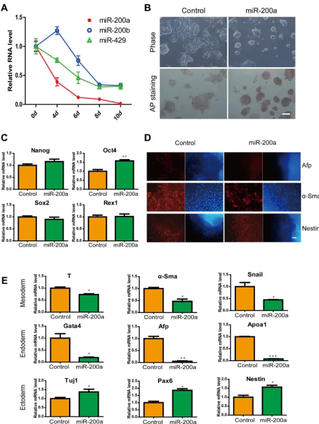
Overexpression of miR-200a in ES cells suppressed differentiation into endoderm and mesoderm
maintained ES cell pluripotency and suppressed differentiation capacity of endoderm and mesoderm.
Grb2 as a novel and important target gene for miR-200a
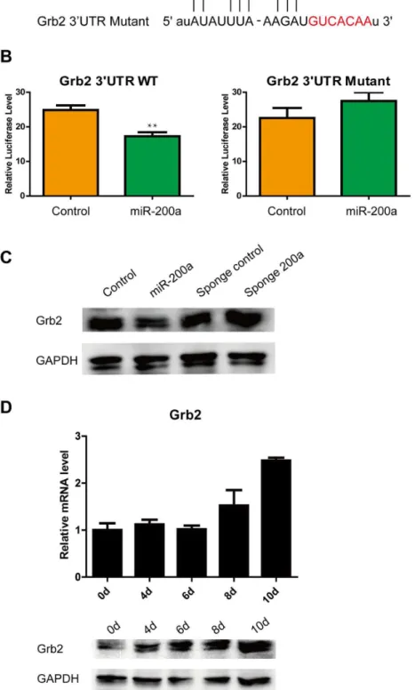
It was not clear whether miR-200a exerted its effects by negatively regulating multiple genes that are involved in ES cell identity. Grb2 had been suggested to be a putative target for miR-200a according to the miRDB, miRanda, TargetScan, PicTar and miRWalk algorithms, as well as a recent study [47]. The predicted interaction between the Grb2 3’ UTR and miR-200a is illustrated in Figure 2A. To confirm the post-translational repression of miR-200a, Grb2 3’ UTR reporter luciferase assays were performed. The delivery of miR-200a mimics significantly suppressed Grb2 3’ UTR reporter luciferase activity more than 25% over the empty vector, and a mutation in the miR-200a binding site blocked this suppression (Figure 2B). Additionally, we performed transient transfections of the miR-200a vector and sponge 200a vector into ES cells at a ratio of 3000 ng vectors per 5 × 10⁴ cells to investigate the
regulation of Grb2. At 48 h post-transfection, Western blotting analysis showed that miR-200a reduced the level of endogenous Grb2 protein, whereas the Grb2 protein level was rescued from endogenous miR-200a by the sponge 200a treatment of ES cells (Figure 2C Figure S1A). Furthermore, Grb2 expression was upregulated during EB formation, unlike miR-200a expression (Figure 2D). These findings indicated that miR-200a specifically bound the predicted translation repressor site on Grb2 and repressed the expression of Grb2 in ES cells.
Knockdown of Grb2 overlaps phenotypically with the enforced expression of miR-200a
To determine whether the knockdown of Grb2 affected layer formation, we established two candidate Grb2 shRNA vectors, shGrb2-1 and shGrb2-2. shGrb2-1 did not significantly affect Grb2 expression. However, the results demonstrated that shGrb2-2 effectively reduced Grb2 protein expression (Figure 3A Figure S1B), and thus shGrb2-2 was used in subsequent studies.
To evaluate the effects of Grb2 during differentiation, we collected RNA at day 10 and performed quantitative real-time PCR for lineage-specific marker analysis. Gene expression analysis showed that when Grb2 expression was knocked down, changes in the expression of genes associated with differentiation were observed. Expression levels of genes related to differentiated states were significantly changed. The expression levels of genes associated with endoderm formation, such as Gata4, Afp and Apoa1, were downregulated. The induction of genes involved in mesoderm specification, such as T, alpha-Sma and Snail, was decreased in Grb2-deficient EBs, compared to those with scrambled controls. However, expression levels of the neuronal cell markers, Tuj1 and Pax6 were upregulated in response to Grb2 knockdown (Figure 3B).
As expected, the immunostaining of proteins associated with differentiation genes showed the same results. Grb2 shRNA and control EBs were plated at spontaneous differentiation day 10. We performed immunofluorescent staining and observed that the Afp and alpha-Sma protein levels were significantly
decreased in the Grb2 shRNA EBs (Figure 3C). Grb2 is known to be involved in the Erk signaling pathway, and thus we measured the state of this signaling pathway to survey the role of Grb2 in ES cell differentiation. Similar to previous findings in tumors [48] and Grb2-null embryos [49], Grb2 knockdown led to reduced levels of phosphorylated Erk in spontaneous differentiated EBs (Figure 3D Figure S1C). Taken together, these data indicate that the loss of Grb2 suppressed differentiation toward the endoderm and mesoderm lineages under spontaneous differentiation conditions.
Neutralization of Grb2 rescues aberrant miR-200a-induced endoderm and mesoderm repression
To investigate whether the Grb2-miR-200a interaction is needed for the spontaneous differentiation of ES cells in vitro, ES cells were infected with a lentiviral vector that overexpressed Grb2 without the 3’ UTR. The cells were subsequently transfected with a miR-200a vector, followed by the immediate induction of spontaneous differentiation. We plated the ES cells at a density of 103 cells/cm2 in the absence
of LIF for 3 days. Under these conditions, in which miR-200a expression was rapidly induced, the Grb2 protein expression levels significantly decreased. An analysis of colony formation showed that ES cells that overexpressed miR-200a appeared to retain the classic compact morphology and well-defined borders of undifferentiated ES cell colonies. However, the colonies that overexpressed both Grb2 and miR-200a were flat and displayed abundant cytoplasmic prolongations when compared to the empty-vector control ES cells (Figure 4A). Next, we performed alkaline phosphatase staining to confirm this result. Only the miR-200a-overexpressing ES cell colonies showed strong staining; the majority of the control and Grb2/ miR-200a-overexpressing ES cell colonies showed faint or no staining under the same conditions.
Interestingly, during EB formation, lower levels of the primitive endoderm markers Afp, Gata4, Apoa1 and the mesoderm markers T, alpha-Sma and Snail were observed in response to miR-200a overexpression, as well as opposite effects on the ectoderm markers Tuj1, Pax6 and Nestin. By contrast, the overexpression of Grb2 significantly rescued the expression of those genes (Figure 4B).
Furthermore, we performed immunofluorescent staining to examine the effects of the Grb2 expression constructs on ES differentiation. Consistently, miR-200a-overexpressing ES cells displayed much lower Afp, Gata4 and alpha-Sma expression levels and higher Tuj1 and Nestin expression levels. In contrast, Grb2-overexpressing ES cells had similar expression levels of the above-mentioned genes to the control cells (Figure 4C). This finding suggested that the overexpression of Grb2 partially reversed the changes in morphology and losses of endoderm and mesoderm formation that were induced by miR-200a.
Figure 1. Effects of miR-200a in ES cells and ES cell differentiation. (A) The expression of miR-200a, miR-200b and miR-429 in ES cell diferentiation. (B) Brightfield images and alkaline phosphatase staining of ES cells without LIF at 72 hours post-transfection with miRNA-200a. The scale bar represents 100 µm. (C) Relative levels of Oct4, Nanog, Sox2 and Rex1 mRNA in control or miR-200a-transfected ES cells. (D) Representative immunofluorescence images of control and miR-200a overexpression after 10 days of EB formation. Red, layer markers; blue, nuclei. The scale bar represents 100 µm. (E) Expression levels of genes associated with the differentiated state in EBs in response to miR-200a expression. All data are shown as the means ± SD. Statistical significance was assessed by the two-tailed Student’s t test. ***, p < 0.001; **, p < 0.01; *, p < 0.05.
Figure 2. miR-200a targets Grb2 at the protein level. (A) Outline of the interaction of miR-200a with the Grb2 3’ UTR. (B) Repression of luciferase activity validates the interaction between miR-200a and the specific predicted sites in the Grb2 3’ UTR. (C) Overexpression of miR-200a downregulates Grb2 expression, and sponge 200a rescues Grb2 expression in ES cells. (D) The expression of Grb2 in ES cell diferentiation. All data are shown as the means ± SD. Statistical significance was assessed by the two-tailed Student’s t test. **, p < 0.01.
Figure 3. Knockdown of Grb2 during EB formation. (A) Western blotting analysis demonstrates that shGrb2 knocks down Grb2 protein levels in ES cells. (B) Relative levels of layer markers are varied in shGrb2-treated EBs. (C) The in situ expression of layer markers in day 10-differentiated EBs. Red, layer markers; blue, nuclear DNA staining. The scale bar represents 100 µm. (D) Erk activity decreases in response to Grb2 knockdown. All data are expressed as the means ± SD. Statistical significance was assessed by the two-tailed Student’s t test. ***, p < 0.001; **, p < 0.01; *, p < 0.05.
Figure 4. Grb2 can rescue cells from the effects of miR-200a. (A) Brightfield images and AP staining of wild-type ES cells, miR-200a-expressing ES cells and exogenous Grb2-expressing ES cells plus expressing miR-200a. The scale bar represents 100 µm. (B) Expression levels of genes associated with the differentiated state in EBs. (C) Immunofluorescence shows that Grb2 reversed the miR-200a-induced failures in endoderm and mesoderm differentiation. The scale bar represents 100 µm. (D) Erk activity can be rescued by Grb2 in miR-200a-expressing ES cells. All data are expressed as the means ± SD. Statistical significance was assessed by one-way ANOVA, followed by Tukey’s post-test. ***, p < 0.001; **, p < 0.01; *, p < 0.05.
via the repression of Grb2 and that the main defect in miR-200a-induced endoderm and mesoderm formation was due to decreased Erk activation in response to reduced Grb2 levels.
Discussion
During early embryogenesis, specific miRNAs have been shown to be essential for the maintenance of bias-in-fate decisions [50]. miR-206 promotes mesendoderm formation by targeting Prickle1a, which subsequently regulates Jnk2 phosphorylation, thereby indicating the potential function of miR-206 in embryonic axis formation [51]. miR-1 and miR-133 are essential to cardiac and skeletal muscle development [28]. During the differentiation of ectoderm, Let-7, miR-9 and miR-124a are specifically required for neuron production [52,53]. miR-124 targets Ptb and switches the balance between the expression of Ptb and nPtb to promote neuronal differentiation [54]. In our study, we found that miR-200a acted as an inhibitor of endoderm and mesoderm formation by repressing the expression of genes involved in mesoderm and endoderm formation. In contrast, ectoderm genes were enhanced in response to miR-200a. To further address the mechanisms underlying the effects of miR-200a, we predicted that Grb2 was a target of miR-200a repression and confirmed this by using a partial-length Grb2 3’ UTR reporter. The addition of miR-200a or Sponge 200a to ES cells further validated the potent and specific miR-200a-Grb2 connection at the protein level. To extend these findings, we investigated the effects of Grb2 knockdown and miR-200a overexpression, and we found that these induced similar characteristics during EB differentiation. Our data suggested that the effects of miR-200a might depend on the repression of Grb2.
Grb2 is an adapter protein that participates in the fibroblast growth factor (FGF) receptor signaling pathway and thus is involved in multiple aspects of cellular function. FGF, which controls the responsiveness of differentiation regulators, is of particular importance to mesoderm and endoderm formation [55]. Gene knockout experiments show that FGFR1 [56], FGFR2 [57] and FGF4 [58] mutant embryos fail to develop a primitive endoderm layer and die in blastocyst outgrowths or in vivo. Similarly, expression of negative FGF receptors does not contribute to the endoderm in chimeras due to a failure of the primitive streak [59]. On the other hand, FGF signaling in early embryogenesis initiates the activation of transcription factors that function in the differentiation of the endoderm and mesoderm layers. Our findings showed that, in response to Grb2 knockdown or miR-200a overexpression, Gata4 and Afp expression was decreased. Gata4 expression is upregulated by exogenous FGF1 in response to cardiac genes in differentiating embryonic carcinoma cell cultures [43], and Afp expression is dependent on FGFs that are produced by the cardiac mesoderm in embryonic endoderm cells [49]. Expression of the mesoderm marker T was also reduced; T is positively regulated by FGFR1 and controls mesodermal morphogenesis [60]. Similarly, decreases in Alpha-Sma expression were observed; in Xenopus, this gene is specifically induced in the ventrolateral mesoderm in response to bFGF
[61]. Our data showed that Grb2 knockdown or miR-200a overexpression likely mediated FGF signaling that stimulated layer formation. A further suggestion was that Grb2, as a main intermediary, is indispensable to FGF signaling-mediated endoderm and mesoderm formation. miR-200a is involved in endoderm and mesoderm formation in a Grb2-dependent manner.
Previous studies have demonstrated that Grb2 transmitted FGF signaling to Erk and thus controlled the basal balance between the ES cell state and inductive differentiation [61]. Autoactivation of the Erk pathway provokes ES cell differentiation [62,63] and eliminates self-renewal [64–66]. The SH2 (Src Homology 2) domain of Grb2 fused with Son of sevenless (Sos) to induce downstream Ras activation, which specifically induces the phosphorylation and activation of Erk and rescues the endoderm differentiation phenotype in Grb2 mutant ES cells [44]. Activated Ras mutant constructs induce ES cell differentiation to the primitive endoderm layer [67], and similar results are observed in response to the expression of an activated form of Erk [68]. Erk2-null embryos results in embryonic lethality at the gastrulation and ES cell deficient in Erk1 and Erk2 is defective in mesoderm differentiation [69]. In Erk2-/- ES cells, T expression is downregulated and
pluripotency markers Oct4 and Nanog are maintained and they fail to differentiate into lateral mesoderm cells under mesoderm differentiation condition [62]. In neural induction in vitro, FGF-induced Erk signalling is required in a short period in the absence of BMP [62,63]. Our results showed that the extraneous addition of miR-200a significantly repressed Erk activation, due to the loss of Grb2 expression. Subsequently, endoderm and mesoderm formation was repressed, and ectoderm formation was induced. Furthermore, Grb2 supplementation rescued miR-200a-induced Erk inactivation and losses in endoderm and mesoderm differentiation after 10 days of EB formation. These findings revealed that miR-200a directly targeted Grb2, thereby mediating Erk signaling. The miR-200a-Grb2-Erk axis is therefore indispensable to layer formation in embryogenesis.
Conclusion
Taking these findings together, we postulate that Erk signaling and miR-200a maintain a balance in specific cell fate decisions such that Erk signaling regulates differentiation into the mesoderm and endoderm lineages and miR-200a suppresses differentiation into these lineages. The link between these factors is Grb2, which delivers activation signals to Erk. Our findings suggest that miR-200a mediates Grb2 expression, thereby blocking Erk activation, which leads to the arrest of endoderm and mesoderm lineage differentiation and promotes ectoderm lineage commitment.
Supporting Information
Figure S1. Quantifications of protein levels. (A)
of Grb2 and p-Erk in shGrb2 treated ES cells in Figure 3D. (D) Quantifications of Grb2 and p-Erk in Grb2 rescue assay in Figure 4D. All data are expressed as the means ± SD. Statistical significance was assessed by the two-tailed Student’s t test. **, p < 0.01; *, p < 0.05.
(TIF)
Table S1. Real-Time PCR primers used in this study.
(DOC)
Author Contributions
Conceived and designed the experiments: YL QL WJ AD JK. Performed the experiments: YL QL JC JW WC. Analyzed the data: YL QL WJ DY XG WC GL. Contributed reagents/ materials/analysis tools: GW AD JK. Wrote the manuscript: YL WJ AD JK.
References
1. Gardner RL (1983) Origin and differentiation of extraembryonic tissues in the mouse. Int Rev Exp Pathol 24: 63-133. PubMed: 6302028. 2. Nichols J, Smith A (2011) The origin and identity of embryonic stem
cells. Development 138: 3-8. doi:10.1242/dev.050831. PubMed: 21138972.
3. Chambers I, Smith A (2004) Self-renewal of teratocarcinoma and embryonic stem cells. Oncogene 23: 7150-7160. doi:10.1038/sj.onc. 1207930. PubMed: 15378075.
4. Klimanskaya I, Rosenthal N, Lanza R (2008) Derive and conquer: sourcing and differentiating stem cells for therapeutic applications. Nat Rev Drug Discov 7: 131-142. doi:10.1038/nrd2403. PubMed: 18079756.
5. Bushati N, Cohen SM (2007) microRNA functions. Annu Rev Cell Dev Biol 23: 175-205. doi:10.1146/annurev.cellbio.23.090506.123406. PubMed: 17506695.
6. Rana TM (2007) Illuminating the silence: understanding the structure and function of small RNAs. Nat Rev Mol Cell Biol 8: 23-36. doi: 10.1038/nrg1947. PubMed: 17183358.
7. Wienholds E, Plasterk RH (2005) MicroRNA function in animal development. FEBS Lett 579: 5911-5922. doi:10.1016/j.febslet. 2005.07.070. PubMed: 16111679.
8. Alvarez-Garcia I, Miska EA (2005) MicroRNA functions in animal development and human disease. Development 132: 4653-4662. doi: 10.1242/dev.02073. PubMed: 16224045.
9. Denli AM, Tops BB, Plasterk RH, Ketting RF, Hannon GJ (2004) Processing of primary microRNAs by the Microprocessor complex. Nature 432: 231-235. doi:10.1038/nature03049. PubMed: 15531879. 10. Gregory RI, Yan KP, Amuthan G, Chendrimada T, Doratotaj B et al.
(2004) The Microprocessor complex mediates the genesis of microRNAs. Nature 432: 235-240. doi:10.1038/nature03120. PubMed: 15531877.
11. Han J, Lee Y, Yeom KH, Kim YK, Jin H et al. (2004) The Drosha-DGCR8 complex in primary microRNA processing. Genes Dev 18: 3016-3027. doi:10.1101/gad.1262504. PubMed: 15574589.
12. Landthaler M, Yalcin A, Tuschl T (2004) The human DiGeorge syndrome critical region gene 8 and Its D. melanogaster homolog are required for miRNA biogenesis. Curr Biol 14: 2162-2167. doi:10.1016/ j.cub.2004.11.001. PubMed: 15589161.
13. Lee Y, Ahn C, Han J, Choi H, Kim J et al. (2003) The nuclear RNase III Drosha initiates microRNA processing. Nature 425: 415-419. doi: 10.1038/nature01957. PubMed: 14508493.
14. Yi R, Qin Y, Macara IG, Cullen BR (2003) Exportin-5 mediates the nuclear export of pre-microRNAs and short hairpin RNAs. Genes Dev 17: 3011-3016. doi:10.1101/gad.1158803. PubMed: 14681208. 15. Bohnsack MT, Czaplinski K, Gorlich D (2004) Exportin 5 is a
RanGTP-dependent dsRNA-binding protein that mediates nuclear export of pre-miRNAs. RNA 10: 185-191. doi:10.1261/rna.5167604. PubMed: 14730017.
16. Lund E, Güttinger S, Calado A, Dahlberg JE, Kutay U (2004) Nuclear export of microRNA precursors. Science 303: 95-98. doi:10.1126/ science.1090599. PubMed: 14631048.
17. Jiang F, Ye X, Liu X, Fincher L, McKearin D et al. (2005) Dicer-1 and R3D1-L catalyze microRNA maturation in Drosophila. Genes Dev 19: 1674-1679. doi:10.1101/gad.1334005. PubMed: 15985611.
18. Hutvágner G, McLachlan J, Pasquinelli AE, Bálint E, Tuschl T et al. (2001) A cellular function for the RNA-interference enzyme Dicer in the maturation of the let-7 small temporal RNA. Science 293: 834-838. doi: 10.1126/science.1062961. PubMed: 11452083.
19. Ketting RF, Fischer SE, Bernstein E, Sijen T, Hannon GJ et al. (2001) Dicer functions in RNA interference and in synthesis of small RNA involved in developmental timing in C. elegans. Genes Dev 15: 2654-2659. doi:10.1101/gad.927801. PubMed: 11641272.
20. Saito K, Ishizuka A, Siomi H, Siomi MC (2005) Processing of pre-microRNAs by the Dicer-1-Loquacious complex in Drosophila cells. PLOS Biol 3: e235. doi:10.1371/journal.pbio.0030235. PubMed: 15918769.
21. Hutvágner G, Zamore PD (2002) A microRNA in a multiple-turnover RNAi enzyme complex. Science 297: 2056-2060. doi:10.1126/science. 1073827. PubMed: 12154197.
22. Martinez J, Tuschl T (2004) RISC is a 5' phosphomonoester-producing RNA endonuclease. Genes Dev 18: 975-980. doi:10.1101/gad. 1187904. PubMed: 15105377.
23. Gregory RI, Chendrimada TP, Cooch N, Shiekhattar R (2005) Human RISC couples microRNA biogenesis and posttranscriptional gene silencing. Cell 123: 631-640. doi:10.1016/j.cell.2005.10.022. PubMed: 16271387.
24. Lee Y, Hur I, Park SY, Kim YK, Suh MR et al. (2006) The role of PACT in the RNA silencing pathway. EMBO J 25: 522-532. doi:10.1038/ sj.emboj.7600942. PubMed: 16424907.
25. Kim DH, Saetrom P, Snøve O Jr., Rossi JJ (2008) MicroRNA-directed transcriptional gene silencing in mammalian cells. Proc Natl Acad Sci U S A 105: 16230-16235. doi:10.1073/pnas.0808830105. PubMed: 18852463.
26. Lingel A, Sattler M (2005) Novel modes of protein-RNA recognition in the RNAi pathway. Curr Opin Struct Biol 15: 107-115. doi:10.1016/j.sbi. 2005.01.010. PubMed: 15718141.
27. Guo H, Ingolia NT, Weissman JS, Bartel DP (2010) Mammalian microRNAs predominantly act to decrease target mRNA levels. Nature 466: 835-840. doi:10.1038/nature09267. PubMed: 20703300. 28. Ivey KN, Muth A, Arnold J, King FW, Yeh RF et al. (2008) MicroRNA
regulation of cell lineages in mouse and human embryonic stem cells. Cell Stem Cell 2: 219-229. doi:10.1016/j.stem.2008.01.016. PubMed: 18371447.
29. Peng C, Li N, Ng YK, Zhang J, Meier F et al. (2012) A unilateral negative feedback loop between miR-200 microRNAs and Sox2/E2F3 controls neural progenitor cell-cycle exit and differentiation. J Neurosci 32: 13292-13308. doi:10.1523/JNEUROSCI.2124-12.2012. PubMed: 22993445.
30. Dykxhoorn DM (2010) MicroRNAs and metastasis: little RNAs go a long way. Cancer Res 70: 6401-6406. doi: 10.1158/0008-5472.CAN-10-1346. PubMed: 20663901.
31. Leskelä S, Leandro-García LJ, Mendiola M, Barriuso J, Inglada-Pérez L et al. (2011) The miR-200 family controls beta-tubulin III expression and is associated with paclitaxel-based treatment response and progression-free survival in ovarian cancer patients. Endocr Relat Cancer 18: 85-95. PubMed: 21051560.
32. Korpal M, Kang Y (2008) The emerging role of miR-200 family of microRNAs in epithelial-mesenchymal transition and cancer metastasis. RNA Biol 5: 115-119. doi:10.4161/rna.5.3.6558. PubMed: 19182522.
33. Korpal M, Lee ES, Hu G, Kang Y (2008) The miR-200 family inhibits epithelial-mesenchymal transition and cancer cell migration by direct targeting of E-cadherin transcriptional repressors ZEB1 and ZEB2. J Biol Chem 283: 14910-14914. doi:10.1074/jbc.C800074200. PubMed: 18411277.
34. Ahmad A, Aboukameel A, Kong D, Wang Z, Sethi S et al. (2011) Phosphoglucose Isomerase/Autocrine Motility Factor mediates epithelial-mesenchymal transition regulated by miR-200 in breast cancer cells. Cancer Res 71: 3400-3409. doi: 10.1158/1538-7445.AM2011-3400. PubMed: 21389093.
36. Park SM, Gaur AB, Lengyel E, Peter ME (2008) The miR-200 family determines the epithelial phenotype of cancer cells by targeting the E-cadherin repressors ZEB1 and ZEB2. Genes Dev 22: 894-907. doi: 10.1101/gad.1640608. PubMed: 18381893.
37. Burk U, Schubert J, Wellner U, Schmalhofer O, Vincan E et al. (2008) A reciprocal repression between ZEB1 and members of the miR-200 family promotes EMT and invasion in cancer cells. EMBO Rep 9: 582-589. doi:10.1038/embor.2008.74. PubMed: 18483486.
38. Eades G, Yao Y, Yang M, Zhang Y, Chumsri S et al. (2011) miR-200a regulates SIRT1 expression and epithelial to mesenchymal transition (EMT)-like transformation in mammary epithelial cells. J Biol Chem 286: 25992-26002. doi:10.1074/jbc.M111.229401. PubMed: 21596753. 39. Mateescu B, Batista L, Cardon M, Gruosso T, de Feraudy Y et al.
(2011) miR-141 and miR-200a act on ovarian tumorigenesis by controlling oxidative stress response. Nat Med 17: 1627-1635. doi: 10.1038/nm.2512. PubMed: 22101765.
40. Wang B, Koh P, Winbanks C, Coughlan MT, McClelland A et al. (2011) miR-200a Prevents renal fibrogenesis through repression of TGF-beta2 expression. Diabetes 60: 280-287. doi:10.2337/db10-0892. PubMed: 20952520.
41. Lowenstein EJ, Daly RJ, Batzer AG, Li W, Margolis B et al. (1992) The SH2 and SH3 domain-containing protein GRB2 links receptor tyrosine kinases to ras signaling. Cell 70: 431-442. doi: 10.1016/0092-8674(92)90167-B. PubMed: 1322798.
42. Rozakis-Adcock M, Fernley R, Wade J, Pawson T, Bowtell D (1993) The SH2 and SH3 domains of mammalian Grb2 couple the EGF receptor to the Ras activator mSos1. Nature 363: 83-85. doi: 10.1038/363083a0. PubMed: 8479540.
43. Chazaud C, Yamanaka Y, Pawson T, Rossant J (2006) Early lineage segregation between epiblast and primitive endoderm in mouse blastocysts through the Grb2-MAPK pathway. Dev Cell 10: 615-624. doi:10.1016/j.devcel.2006.02.020. PubMed: 16678776.
44. Cheng AM, Saxton TM, Sakai R, Kulkarni S, Mbamalu G et al. (1998) Mammalian Grb2 regulates multiple steps in embryonic development and malignant transformation. Cell 95: 793-803. doi:10.1016/ S0092-8674(00)81702-X. PubMed: 9865697.
45. Gill JG, Langer EM, Lindsley RC, Cai M, Murphy TL et al. (2011) Snail and the microRNA-200 family act in opposition to regulate epithelial-to-mesenchymal transition and germ layer fate restriction in differentiating ESCs. Stem Cells 29: 764-776. doi:10.1002/stem.628. PubMed: 21394833.
46. Palmieri SL, Peter W, Hess H, Schöler HR (1994) Oct-4 transcription factor is differentially expressed in the mouse embryo during establishment of the first two extraembryonic cell lineages involved in implantation. Dev Biol 166: 259-267. doi:10.1006/dbio.1994.1312. PubMed: 7958450.
47. Tsang JS, Ebert MS, van Oudenaarden A (2010) Genome-wide dissection of microRNA functions and cotargeting networks using gene set signatures. Mol Cell 38: 140-153. doi:10.1016/j.molcel.2010.03.007. PubMed: 20385095.
48. Giubellino A, Burke TR Jr., Bottaro DP (2008) Grb2 signaling in cell motility and cancer. Expert Opin Ther Targets 12: 1021-1033. doi: 10.1517/14728222.12.8.1021. PubMed: 18620523.
49. Wang Y, Smedberg JL, Cai KQ, Capo-Chichi DC, Xu XX (2011) Ectopic expression of GATA6 bypasses requirement for Grb2 in primitive endoderm formation. Dev Dyn 240: 566-576. doi:10.1002/ dvdy.22447. PubMed: 20925113.
50. Ivey KN, Srivastava D (2010) MicroRNAs as regulators of differentiation and cell fate decisions. Cell Stem Cell 7: 36-41. doi:10.1016/j.stem. 2010.06.012. PubMed: 20621048.
51. Liu X, Ning G, Meng A, Wang Q (2012) MicroRNA-206 regulates cell movements during zebrafish gastrulation by targeting prickle1a and regulating c-Jun N-terminal kinase 2 phosphorylation. Mol Cell Biol 32: 2934-2942. doi:10.1128/MCB.00134-12. PubMed: 22615492. 52. Krichevsky AM, Sonntag KC, Isacson O, Kosik KS (2006) Specific
microRNAs modulate embryonic stem cell-derived neurogenesis. Stem Cells 24: 857-864. doi:10.1634/stemcells.2005-0441. PubMed: 16357340.
53. Zhao C, Sun G, Li S, Lang MF, Yang S et al. (2010) MicroRNA let-7b regulates neural stem cell proliferation and differentiation by targeting nuclear receptor TLX signaling. Proc Natl Acad Sci U S A 107: 1876-1881. doi:10.1073/pnas.0908750107. PubMed: 20133835. 54. Makeyev EV, Zhang J, Carrasco MA, Maniatis T (2007) The MicroRNA
miR-124 promotes neuronal differentiation by triggering brain-specific alternative pre-mRNA splicing. Mol Cell 27: 435-448. doi:10.1016/ j.molcel.2007.07.015. PubMed: 17679093.
55. Morrison GM, Oikonomopoulou I, Migueles RP, Soneji S, Livigni A et al. (2008) Anterior definitive endoderm from ESCs reveals a role for
FGF signaling. Cell Stem Cell 3: 402-415. doi:10.1016/j.stem. 2008.07.021. PubMed: 18940732.
56. Ciruna BG, Schwartz L, Harpal K, Yamaguchi TP, Rossant J (1997) Chimeric analysis of fibroblast growth factor receptor-1 (Fgfr1) function: a role for FGFR1 in morphogenetic movement through the primitive streak. Development 124: 2829-2841. PubMed: 9226454.
57. Arman E, Haffner-Krausz R, Chen Y, Heath JK, Lonai P (1998) Targeted disruption of fibroblast growth factor (FGF) receptor 2 suggests a role for FGF signaling in pregastrulation mammalian development. Proc Natl Acad Sci U S A 95: 5082-5087. doi:10.1073/ pnas.95.9.5082. PubMed: 9560232.
58. Li L, Arman E, Ekblom P, Edgar D, Murray P et al. (2004) Distinct GATA6- and laminin-dependent mechanisms regulate endodermal and ectodermal embryonic stem cell fates. Development 131: 5277-5286. doi:10.1242/dev.01415. PubMed: 15456727.
59. Chen Y, Li X, Eswarakumar VP, Seger R, Lonai P (2000) Fibroblast growth factor (FGF) signaling through PI 3-kinase and Akt/PKB is required for embryoid body differentiation. Oncogene 19: 3750-3756. doi:10.1038/sj.onc.1203726. PubMed: 10949929.
60. Ciruna B, Rossant J (2001) FGF signaling regulates mesoderm cell fate specification and morphogenetic movement at the primitive streak. Dev Cell 1: 37-49. doi:10.1016/S1534-5807(01)00017-X. PubMed: 11703922.
61. Nichols J, Silva J, Roode M, Smith A (2009) Suppression of Erk signalling promotes ground state pluripotency in the mouse embryo. Development 136: 3215-3222. doi:10.1242/dev.038893. PubMed: 19710168.
62. Kunath T, Saba-El-Leil MK, Almousailleakh M, Wray J, Meloche S et al. (2007) FGF stimulation of the Erk1/2 signalling cascade triggers transition of pluripotent embryonic stem cells from self-renewal to lineage commitment. Development 134: 2895-2902. doi:10.1242/dev. 02880. PubMed: 17660198.
63. Stavridis MP, Lunn JS, Collins BJ, Storey KG (2007) A discrete period of FGF-induced Erk1/2 signalling is required for vertebrate neural specification. Development 134: 2889-2894. doi:10.1242/dev.02858. PubMed: 17660197.
64. Ying QL, Wray J, Nichols J, Batlle-Morera L, Doble B et al. (2008) The ground state of embryonic stem cell self-renewal. Nature 453: 519-523. doi:10.1038/nature06968. PubMed: 18497825.
65. Burdon T, Stracey C, Chambers I, Nichols J, Smith A (1999) Suppression of SHP-2 and ERK signalling promotes self-renewal of mouse embryonic stem cells. Dev Biol 210: 30-43. doi:10.1006/dbio. 1999.9265. PubMed: 10364425.
66. Chen S, Do JT, Zhang Q, Yao S, Yan F et al. (2006) Self-renewal of embryonic stem cells by a small molecule. Proc Natl Acad Sci U S A 103: 17266-17271. doi:10.1073/pnas.0608156103. PubMed: 17088537.
67. Yoshida-Koide U, Matsuda T, Saikawa K, Nakanuma Y, Yokota T et al. (2004) Involvement of Ras in extraembryonic endoderm differentiation of embryonic stem cells. Biochem Biophys Res Commun 313: 475-481. doi:10.1016/j.bbrc.2003.11.138. PubMed: 14697213.
68. Hamazaki T, Kehoe SM, Nakano T, Terada N (2006) The Grb2/Mek pathway represses Nanog in murine embryonic stem cells. Mol Cell Biol 26: 7539-7549. doi:10.1128/MCB.00508-06. PubMed: 16908534. 69. Yao Y, Li W, Wu J, Germann UA, Su MS et al. (2003) Extracellular
signal-regulated kinase 2 is necessary for mesoderm differentiation. Proc Natl Acad Sci U S A 100: 12759-12764. doi:10.1073/pnas. 2134254100. PubMed: 14566055.
70. Wang G, Guo X, Hong W, Liu Q, Wei T et al. (2013) Critical regulation of miR-200/ZEB2 pathway in Oct4/Sox2-induced mesenchymal-to-epithelial transition and induced pluripotent stem cell generation. Proc Natl Acad Sci U S A 110: 2858-2863. doi:10.1073/pnas.1212769110. PubMed: 23386720.
71. Radisky DC (2011) miR-200c at the nexus of epithelial-mesenchymal transition, resistance to apoptosis, and the breast cancer stem cell phenotype. Breast Cancer Res 13: 110. doi:10.1186/bcr2885. PubMed: 21682933.
72. Du ZW, Ma LX, Phillips C, Zhang SC (2013) miR-200 and miR-96 families repress neural induction from human embryonic stem cells. Development, 140: 2611–8. PubMed: 23637338.
73. Shimono Y, Zabala M, Cho RW, Lobo N, Dalerba P et al. (2009) Downregulation of miRNA-200c links breast cancer stem cells with normal stem cells. Cell 138: 592-603. doi:10.1016/j.cell.2009.07.011. PubMed: 19665978.
75. Gill JG, Langer EM, Lindsley RC, Cai M, Murphy TL et al. (2012) Snail promotes the cell-autonomous generation of Flk1(+) endothelial cells through the repression of the microRNA-200 family. Stem Cells Dev 21: 167-176. doi:10.1089/scd.2011.0194. PubMed: 21861700.