Received on 6/23/2009. Approved on 8/12/2009. Study undertaken at the Rheumatology Department of the Medical School of Universidade de São Paulo with a grant from the Research Support Foundation of São Paulo (FAPESP, from the Portuguese).
1. Associate Rheumatology Professor of the Medical School of Universidade de São Paulo
2. Professor of Veterinary Medicine and Animal Health at the Faculdade de Medicina Veterinária e Zootecnia da Universidade de São Paulo 3. Retired Infectious Diseases Professor of Universidade Federal de São Paulo.
4. Assisting Physician of the Hospital das Clínicas of the Medical School of Universidade de São Paulo 5. Rheumatology PhD student at the Medical School of Universidade de São Paulo
6. Biologist of the Rheumatologic Investigation Laboratory of the Hospital das Clínicas of the Medical School of Universidade de São Paulo
Correspondence to: Prof. Natalino Hajime Yoshinari. Av. Dr. Arnaldo 455, sala 3184. Cerqueira Cesar, SP – CEP: 01246-903. E-mail: yoshinari@lim17.fm.usp.br
Report on the unusual presence of latent
microorganisms in animals: a risk to
research and health of employees?
Natalino Hajime Yoshinari1, Silvio Arruda Vasconcelos2, Arary da Cruz Tiriba3, GiancarlaGauditano4, Elenice Mantovani5, Virgínia Lúcia Nazário Bonoldi6
ABSTRACT
We report the unusual inding of mobile spirochetal microorganisms with different morphologies and sizes, on dark-ield microscopy of the blood of animals from the Vivarium of the Medical School of USP. The bacteria did not grow
in common culture media, shows faint staining to Giemsa and silver-derived stains, and serologies and molecular tests were negative for Borrelia and Leptospira.
Electron microscopy revealed the presence of microorganisms with Mycoplasma-like morphology and, due to its mobility, it was suggested that they represented Mollicutes of the genus Spiroplasma. Microorganisms with the same morphology were also observed in 15 out of 26 employees (57.6%) of the Vivarium of FMUSP; however, clinical and laboratorial exams indicated that those individuals were healthy. Additional studies undertaken at the Rheumatology
Department of FMUSP demonstrated the presence of the same structures identiied at the Vivarium in approximately
94% of the patients with Baggio-Yoshinary syndrome (BYS) and 20% of healthy individuals. Electron microscopy of the blood of BYS patients showed bacteria that shared similarities with Mycoplasma, Chlamydia, and Bacteroides. Since serologies and molecular tests were negative for those contaminants, and based on publications in the medical
literature, it was suggested that those latent infectious agents were L-form bacteria, deined as cell wall deicient bacteria,
assuming, therefore, Mycoplasma morphology and they are, for the most part, harmless to the host. We concluded that spirochetal microorganisms visualized in animals and employees of the Vivarium were non-pathogenic L-form bacteria from contaminants in the environment, regular infections, or endogenous microorganism from the normal saprophytic
lora. On the other hand, spirochetal organisms identiied in BYS, by preserving the capacity to invade cells in vitro, are potentially pathogenic and related to the etiology of BYS. We consider BYS as a novel Brazilian zoonosis caused by spirochetes adapted to their latent form, possibly due to bacterial mutations in response to ecologic and geographic conditions unique to Brazil.
Keywords: spirochete, spirochete-like, L-form bacteria, Mycoplasma, Lyme-like disease, Baggio-Yoshinari syndrome,
Borrelia, latent microorganism, Vivarium, laboratory animals, Brazil.
INTRODUCTION
On an administrative ruling published on 04/06/2002,
the dean of USP, Professor Adolfo José Meli, appointed
Professors Natalino Hajime Yoshinari (Medical School of
The problem in the Vivarium of FMUSP began in the beginning of 2001 when Dr. Ismar Cestari detected in vitro
the presence of spirochete-like microorganisms in culture of spleen cells of an animal from the Vivarium of the Medical School of USP (FMUSP).
To understand the extension of the problem, the Commission
examined the blood of the animals on dark-ield microscopy
and, indeed, confirmed the presence of spirochete-like microorganisms in 15 out of 15 mice blood samples (BALB/c, A/SNELL1, SWISS, and C57BI/6), in two out of 5 rat blood
samples (WISTAR), ive out of ive rabbit blood samples, and
in none of the guinea pig blood samples.
Blood samples were examined again on the second semester of 2002 and 28 out of 40 (70%) blood samples of different species of mice and rats were positive. The same procedure was used in animals from the Veterinary and Zootomy School of USP (FMVZUP, from the Portuguese)
and dark-ield microscopy showed “spirochetal structures” in
only two out of eight mice blood samples and in none of the hamsters, indicating a problem of Vivariums, although this
inding was more common at FMUSP.
Clinically, animals from the USP Vivarium were apparently healthy. The veterinary Sueli Blanes Dani et al.1 undertook preliminary biochemical analysis of serum pools of four female
mice and ive of WISTAR rats, showing increased levels of
BUN, alkaline phosphatase, aspartate aminotransferase (AST), and alanine aminotransferase (ALT), suggesting the presence of some infectious or toxic factor responsible for the development of liver function-related biochemical disruption. In the same study, electron microscopy and immunohystochemical tests of the lungs, liver, spleen, heart, and kidneys of the animals, and
they identiied Mycoplasma pulmonis in almost 100% of the animals in conventional vivariums, and in 18% of those that maintained adequate sanitary barriers. They stated that studies in the literature reported liver diseases caused by Mycoplasma
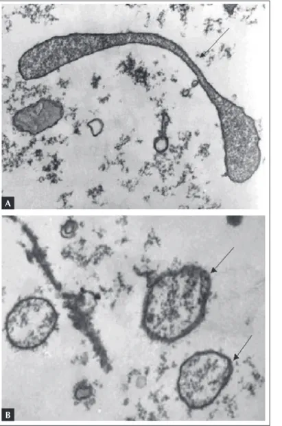
in sheep, doves, and goats, but they did not ind reports on rats. To conirm those indings, we repeated the electron
microscopy of the blood samples of rodents with spirochetal organisms (Figure 1).
Since optical microscopy revealed the presence of mobile microorganisms of different sizes, ranging from miniscule dots to elongated structures reaching up to 15-20 µm, in the blood sample of the animals, the Commission initially thought they could be spirochetes. Serology for Leptospira
and Borrelia, hemocultures in aerobic and anaerobic media and BSK, as well as molecular testing (PCR) for Leptospira
and Borrelia, performed at FMUSP, FMVZUSP, and Biological Institute were persistently negative. The uncommon size of
the structures identiied in the peripheral blood of the animals
called the attention of the members of the Commission because they were large and apparently incompatible with
Mycoplasma or Chlamydia. It was also interesting that it was
dificult to cultivate those structures in common culture media and they were dificult to identify using stains like Giemsa,
silver products, and vital stains, such as acridine orange.
Those indings, associated with the absence of information in
the medical literature, led many researchers to interpret those spirochetal structures as simple artifacts.
The major concerns were related to the anthropozoonotic aspects of this uncommon finding. Possible risks of this latent infectious process for employees who handled the animals, characterizing a work-related disease, as well as the interference of those contaminants on experimental studies, were hypothesized. It was interesting that animals underwent frequent bacteriological testing, but pathogenic microorganisms were never isolated. It is important to emphasize that the animals in the USP Vivarium were not isolated, nor were they “germ-free”, besides being raised and kept in the Vivarium for approximately 20 years, except the dogs that are brought from outside the FMUSP complex.
The present study reports the problems and actions adopted by the Commission to answer the questions formulated by the
Dean of USP regarding the risks to the employees and scientiic
investigations. The other objective was to discover the etiology and source of infection of the animals.
Due to the relevance of the subject, a complementary discussion resulting from the investigation of the Brazilian Lyme-like disease (Baggio-Yoshinari syndrome – BYS), which brought new understanding on the incidence of spirochetal organisms in laboratory animals and humans, was added. It was demonstrated that those structures are seen almost always in mammals, including humans (Figure 2A), and that they grow
briely in SP4 medium (Figure 2B).
When analyzed under electron microscopy, those structures are similar to Mycoplasma, which, in reality, represent
microorganisms that lost the cellular wall (cell wall deicient
bacteria or L-form) and are, for the most part, “harmless” to living beings. However, for unknown reasons, maybe due to ecologic and climatic factors inherent to the country, strong evidence indicate that the etiological factor of BYS is an atypical latent spirochete with characteristics suggestive of
The objective of this study was to demonstrate that the description of “harmless” L-form microorganisms in living beings is common. But, in some situations, as in the
Baggio-Yoshinari syndrome, cell wall deicient spirochete, possibly
bacteria of the genus Borrelia, would preserve their pathogenic properties, causing an extremely morbid disease distinct from Lyme disease seen in the northern hemisphere.
PATIENTS AND METHODS
All employees of the FMUSP Vivarium, evaluated according to the study protocol determined by the Commission instituted
by the Dean of USP, were included in this study to evaluate the health of the employees who handled the animals or worked at the Vivarium.
This protocol included a clinical investigation with complete history and physical exam. As for complementary exams, the following were evaluated: 1; Blood: CBC (automated test, microscopy, and Panotic stain); 2. Biochemical: BUN (kinetic assay), creatinine (colorimetric kinetic assay), glucose (colorimetric enzymatic assay), total cholesterol (colorimetric enzymatic assay), triglycerides (colorimetric enzymatic assay), uric acid (colorimetric enzymatic assay), sodium selective electrode assay), potassium
(ion-A
B
Figure 1.A) Electron photomicrography of the serum of Wistar rat from the Vivarium without treatment, showing microorganisms with
Mycoplasma morphology. Collected on 11/06/2002. B) Presence of several structures with irregular shapes and sizes surrounded by a membrane, and with granular contents, compatible with Mycoplasma, in a rodent form the Vivarium.
A
B
selective electrode assay), calcium (colorimetric assay), serum iron (colorimetric assay), ALT (kinetic assay), AST (kinetic assay), gamma-glutamyl transferase (GGT – kinetic test), and creatine phosphokinase (CPK – kinetic test); 3. Urine analysis: urine type I; 4. Bacteriologic: hemoculture, coproculture (stool culture), and stool parasitologic (Leishman
stain); 5. Immunologic and inlammatory: C-reactive protein
(nephelometric assay), erythrocyte sedimentation rate (Westergren assay), C3 and C4 complement (nephelometric assay), protein electrophoresis, protein immunoelectrophoresis, rheumatoid factor (agglutination assay), antinuclear factor in
Hep-2 cells (immunoluorescence assay), and anti-streptolysin
O (nephelometric assay); 6. Serologies for infectious diseases:
Lyme disease, at the Medical Investigation Laboratory (LIM-17, from the Portuguese) of HCFMUSP (immunoenzymatic assay – ELISA), leptospirosis, at the FMVZUSP (microscopic seroagglutination assay), and the remaining assays for syphilis (immunoenzymatic assay – ELISA), toxoplasmosis (microparticle immunoenzymatic assay – MEIA), hepatitis A and B (microparticle immunoenzymatic assay – MEIA), hepatitis C (chemiluminescence assay), and cytomegalovirus (immunoenzymatic assay – ELISA) at the Central Laboratory of HCFMUSP; 6. Imaging exams: chest X-ray and abdominal ultrasound (US) scan; and 7. Specialized medical evaluation when necessary.
A B
C D
Figure 3.A) Electron microscopy of the blood of a patient with BYS on SP4 culture medium showing structures suggestive of spirochetes and
This study was approved by the Ethics Commission for the Analysis of Experimental Studies – CAAPPesq (from the Portuguese) of HCFMUSP (342/09).
RESULTS
Thirty-seven employees of the FMUSP Vivarium, ive of
which were administrative employees, were evaluated at the Internal Medicine Outpatient Clinic of the Hospital das Clínicas of FMUSP. Since this was a conventional Vivarium, without isolation areas, the employees were not discriminated
according to their position. Twenty-ive were males and 12
females; their age ranged from 21 to 54 years (36.6 ± 9.87).
HISTORY
Twelve out of 27 (32.4%) employees complained of some allergic manifestation, such as asthma, rhinitis, or skin eruption. The frequency of “social drinking” was 62.5%, i.e., 15 out of 24 individuals who answered this question.
Out of 37 employees interviewed, ten complained of frequent fatigue and asthenia (27%), while three (8.1%) referred recurrent episodes of fever or chills.
As for the locomotor system, nine (24.5%) complained of constant back pain, eight (21.6%) had arthralgias, two had talalgia, and one had arthritis and myalgia. Sixteen employees (43.2%) did not have osteoarticular symptoms.
Regarding neurological manifestations, 12 (32.4%) employees had headaches regularly, nine (24.3%) reported
being forgetful, seven (18.9%) had sleep problems, five (13.5%) were nervous or irritable, three (8.1%) had lack of concentration, three (8.1%) experienced episodes of dizziness, and one had facial paralysis. Nine employees (24.3%) denied having any neurological symptoms.
As for cardiovascular symptoms, ive employees (13.5%)
experienced frequent episodes of palpitations and three (8.1%) had atypical chest pain.
Review of the other systems showed that 12 (32.4%) complained of increased in the daily number of evacuations (more than two), six (16.2%) had sore throat frequently, and four reported constant episodes of coughing or upper airways infection. Five (13.5%) employees did not have any clinical complaints.
PHYSICAL EXAM
The physical exam did not show signiicant changes; cutaneous
manifestations were observed in five patients (pustules, erythema macular, hyperhidrosis, pityriasis versicolor, and psoriasis). One employee had hepatomegaly and clubbing of the
ingers, lung auscultation was compatible with bronchospasm
in one employee, another had arthritis compatible with gout, and one had Heberden and Bouchard nodes (arthrosis of the hands).
LABORATORIAL TESTS
Complete blood count was normal, except in two cases in which it showed mild leukocytosis, three had leucopenia,
and ive had mild anemia. Changes in platelet count were not
observed.
Table 1 shows the main results of blood biochemistry, and the number of employees with increased levels of liver and muscle enzymes is striking.
INFLAMMATORY ACTIVITY AND IMMUNOLOGIC TESTS
Table 1 shows the changes in inflammatory activity and immunologic tests of the employees of the FMUSP Vivarium. Note that 26% of the employees presented positive C-reactive protein and 36% had increased IgE.
SEROLOGIES FOR INFECTIOUS DISEASES
Table 2 shows the results of the serologies for the Brazilian Lyme-like disease, leptospirosis, syphilis, toxoplasmosis, hepatitis A, B, and C, and cytomegalovirus. The frequency Figure 4. Electron microphotography of a spirochetal structure inside
of sera positive for Borrelia burgdorferi was similar to that observed in the normal control population. One patient tested positive for syphilis and two for toxoplasmosis, being referred to the Infectious Disease Department.
BACTERIOLOGY AND STOOL PARASITOLOGY
Table 2 describes the bacteriological and stool parasitologic
tests, visualization of spirochetal structures on dark-ield
microscopy, and PCR for leptospirosis of the employees of FMUSP Vivarium. Blood cultures and coprocultures were negative; spirochetal structures were observed in 57.6% of the cases (Figure 2A), and the presence of Blastocystis hominis
was demonstrated in 12% of the employees analyzed. Stool leukocytes were present in 36% of the cases.
IMAGING
Chest X-ray was normal in all employees who underwent this exam. Total abdominal US showed some changes in 10 out of 18 cases (55.5%), and the abnormalities observed included:
hepatic steatosis in ive cases, liver enlargement in four cases, peripancreatic lymph nodes in one case, hepatic calciications
in one case, dilated common hepatic duct in one, and hepatic nodules in one employee.
LIVER EVALUATION
Employees with abnormal abdominal US were referred to
the gastroenterologist, but signiicant changes, deserving
complementary investigation, were not observed.
Table 1
Biochemical parameters, urine analysis, inlammatory activity tests, and
immunologic tests of employees of the Vivarium of FMUSP
Biochemical parameters
and urine analysis Incidence of changes Normal levels Inflammatory activity tests and immunologic parameters Incidence of changes Normal levels
Elevated BUN 1/25 (4%) 10-45 mg/dL Elevated α2 globulin 6/24 (25%) 0.4-0.7 g/dL
Elevated creatinine 0/25 (0%) 0.6-1.4 mg/dL Elevated γ globulin 4/24 (16.6%) 0.7-1.6 g/dL
Elevated glucose 2/25 (8%) 70-110 mg/dL Elevated C-reactive protein 6/23 (26%) < 5 mcg/mL
Elevated cholesterol 8/28 (32%) até 200 mg/dL Elevated ESR 3/24 (12.5%) até 15 mm
Elevated triglycerides 5/25 (20%) até 200 mg/dL Elevated α1 glycoprotein 2/24 (8.3%) até 125 mg/dL
Elevated uric acid 4/24 (16.6%) H:3.4-7.0 mg/dL M:2.4-5.7 Elevated glycoprotein/ESR or CRPα2 globulin/ α1 12/24 (50%) –
Abnormal sodium 0/25 (0%) 135-145 mEq/L ≥ 2 of above parameters elevated 6/24 (25%) –
Abnormal potassium 0/25 (0%) 3.5-5.0 mEq/L Decreased C3 and C4 1/24 (4.1%) C3 < 90 mg/dL e C4 < 10 mg/dL
Abnormal calcium 4/23 (17.5%) 8.8-10.5 mg/dL Positive rheumatoid factor 2/23 (8.6%) –
Abnormal iron 0/24 (0%) acima 50 ug/dL Positive ANF, speckled pattern 2/22 (9.0%) IF acima de 1/40
Elevated AST 1/25(4%) 10-34 U/L Elevated IgA 4/25 (16%) 70-400 mg/dL
Elevated ALT 4/25 (16%) 10-44 U/L Elevated IgG 6/25 (24%) 700-1600 mg/dL
Elevated gamma GT 8/25 (32%) 11-50 U/L Elevated IgM 4/25 (20%) 40-230 mg/dL
Elevated LDH 0/25 (0%) 240-480 U/L Elevated IgE 9/25 (36%) até 156 KU/L
Elevated CPK 9/25 (48%) 24-204 U/L Elevated ASLO 2/24 (8.3%) até 200 U/mL
Elevation of at least 1 enzyme* 16/25 (64%) –
Elevation of two or more enzymes* 5/25 (20%) –
Urine leukocytes 1/24 (4.1%) até 5 por campo
Urine erythrocytes 1/24 (4.1%) até 5 por campo
Proteinuria 0/24 (0%) até 0.05 mg/L
DISCUSSION
A large number of professionals of different Research Institutions was mobilized to study the clinical condition of the
employees of the USP Vivarium due to the uncommon inding
of latent microorganisms in the animals of that institution, which might indicate an emerging zoonosis and a new work-related disease. Since references on microorganisms, with the characteristics described here, in the blood of animals and humans, were not found in the medical literature, it was very
dificult to explain where they came from and what would be
the pathogenic role of those microorganism in the host. An explanation was urgently needed, not only to reassure the employees regarding the risks to their health, but also to inform the scientists of the institution who used animals from the Vivarium of any interferences of this infection on animal research.
The poor sanitary conditions of the Vivarium suggested that
environmental factors could be inluencing the development
of those spirochetal structures, since the frequency of contaminants varied among the institutions investigated. It was surprising that the same structures present in animals
were identiied in 15 out of 26 employees (57.6%), indicating
possible work-related transmission of those microorganisms. The Commission also observed that the employees of the Vivarium had poor hygiene, since they ate and slept in the work place, sometimes manipulated animals without gloves, they did not wear boots when they were in direct contact with animal waste, and promiscuity among the different animal species was also observed. Therefore, educating the employees on proper hygiene and establishing strict rules regarding animal
care were the irst steps taken.
The medical exam of the employees demonstrated that they were in good clinical condition; however, the high frequency of allergic phenomena and diarrhea was striking. As for the laboratorial work up, some individuals had abnormal inflammatory activity assays, elevated liver and muscle enzymes, high levels of IgA and IgE, and leukocytes in the
Table 2
Column A: incidence of seropositivity for the Brazilian Lyme-like disease, leptospirosis, syphilis, toxoplasmosis,
viral hepatitis, and cytomegalovirus. Column B: hemocultures, coprocultures, dark- and light-ield
microscopy, stool parasitology, and PCR for Leptospira spp. on employees of the Vivarium of FMUSP
Parameters (A) Incidence Parameters (B) Incidence
Borrelia burgdorferi (IgG) 4/27 (14.8%) Blood culture (aerobic) (n=25) S. epidermidis (1 case)
Borrelia burgdorferi (IgM) 0/27 (0%) Blood culture (anaerobic) (n=25) Negative
Borrelia burgdorferi (IgG or IgM) 4/27 (14.8%) Coproculture (*) (n=25) Negative
Leptospirosis 0/27 (0%) Coproculture for sp. (n= 5)(**) Helicobacter Negative
Syphilis (serology) 1/25 (4%) PCR for leptospirosis (n=5)(**) Negative
Toxoplasmosis (IgM) 2/25 (8%) Spirochetal structures on dark-field microscopy 15/26 (57.6%)
Toxoplasmosis (IgG) 8/25 (32%) Light-field bacterioscopy (n= 5)(**) Negative
Hepatitis A (IgM) 0/25 (0%) Dark-field bacterioscopy (n= 5)(**) Negative
Hepatitis A (IgG) 20/25 (80%) Stool parasitology (Blastocystis hominis) 3/25 (12%)
HBs Ag 0/25 (0%) Stool parasitology (E. nana) 1/25 (4%)
Anti-HBs Ag 1/25 (4%) (vaccinated) Stool leukocytes 9/25 (36%)
Hepatitis C 0 (0%)
Cytomegalovirus (IgM) 0/25 (0%)
Cytomegalovirus (IgG) 21/25 (84%)
stools. Initially, we thought we were seeing a higher incidence of liver, intestinal, and allergic complications. However, this
impression was not conirmed due to the lack of a control
group, with the same demographic characteristics and habits, but not working at the Vivarium.
Some aspects stood out, such as the high incidence of social drinking (62.5%), continuous exposure of the employees to animal waste, rations, and different biological products, besides the poor hygiene of those individuals. We thought those factors partly explained the clinical complaints and laboratorial changes seen, and that preventive measures would contribute to reduce the incidence of animal and human infection, in addition to the “eventual normalization” of the abnormal tests. We were reassured when employees whose abdominal US and laboratorial tests indicated hepatic changes were evaluated by the Gastroenterology Department of HCFMUSP and were considered normal, without the need of further procedures.
The next step of the study was to try to elucidate the etiology
of the latent infection demonstrated by the inding of spirochetal
structures in animals and employees of the Vivarium (Figure 2A). They were of different sizes and morphologies, mobile, nonculturable, did not stain by the Giemsa method and, although they resembled spirochetes, laboratorial tests were persistently negative for Leptospira and Borrelia. Serologies for the Brazilian Lyme-like disease, leptospirosis, and syphilis were negative, as well as molecular biology tests for Leptospira spp. and Borrelia spp. (data not presented).
As mentioned before, Damy SB et al.1 identified
Mycoplasma pulmonis in 100% of laboratory animals raised conventionally, and they reported that those microorganisms could cause hepatic damage, justifying the enzymatic changes seen in the animals of the FMUSP Vivarium.
Mycoplasmata are considered the smaller self-replicating organisms, require cholesterol for their survival, and do not have cellular wall. Similar to Chlamydiae, they are intracellular organisms that infect several cells, such as endothelial and epithelial cells, and macrophage, besides representing important co-factors of the increased virulence of infections caused by other microorganisms2. Higuchi et al.3,4 reported that
those microorganisms inluence the development of unstable
atheroma plaques. They were able to visualize, on electron microscopy, elliptical and cylindrical forms of Mycoplasmata
of different sizes distributed in the extracellular matrix of affected human tissues.
However, we did not ind any references in the medical
literature to the possible role of Mycoplasma spp. and
Chlamydia spp. as zoonotic agents. It was also intriguing that those microorganisms were minute and non-mobile,
contradicting the indings of dark-ield microscopy, which
revealed mobile structures measuring up to 15 µm in length.
Thus, the hypothesis that the microorganisms identiied in
the blood of rodents from the FMUSP Vivarium (Figure 1) were Mycoplasmata was not conirmed. According to the
personnel of Professor J. Timenetsky of ICBUSP, a specialist in Mycoplasmata, the majority of rodents is infected by these
bacteria and, therefore, the indings on electron microscopy could be incidental, not related to our indings.
But we investigated the possibility of finding mobile bacteria with greater dimensions and Mycoplasma morphology. It is known that the Mollicutes group of bacteria is composed by Mycoplasma, Spiroplasma, and Acholeplasma, cell wall
deicient microorganisms surrounded by a cholesterol-rich
cellular membrane. According to Shlomo T & Rami G5, microorganisms of the genus Spiroplasma show circular and elliptical movement due to the presence of a cytoskeleton that works as a propeller, and they have chemotactic properties. They can reach up to 10 µm in length, are sensitive to erythromycin and tetracyclines, and some species are pathogenic for rats, mice, hamsters, and rabbits. They can also
be identiied in plants, bees, ticks, wasps, and mosquitoes6. It is curious that several Spiroplasma cells contain a virus (SpV)7, whose pathogenic meaning is unknown.
Since the Mollicutes hypothesis might not be completely satisfactory, the Commission considered other agents with similar morphology to that of spirochetes on dark-field microscopy. Among other possibilities, we thought of mobile spiral microorganisms, such as Helicobacter spp8., Serpuline spirochetes9, and Anaerobiospirillum10, whose common trait
includes dificulty growing in usual media and their role on the pathogenesis of human and animal inirmities. Spirochetes
of the genus Serpulina live in the digestive tract of animals, causing diarrhea11,12, and immunosuppressed patients may be
equally infected by those dificult to diagnose spirochetes12. Additionally, it is known that several spirochetes are among
the oral saprophytic lora of normal individuals13; however, we did not know whether those microorganisms would be able to invade the blood stream and express as spirochetal structures.
Despite different etiologic possibilities, none was satisfactory, except for the possibility that they might be
Spiroplasmas, mobile microorganisms similar to Mycoplasma. The other microorganisms mentioned have extremely different morphology on electron microscopy, since they have cellular
wall, some of them have lagella, and they are extremely small
(except for the genus Serpulina/Brachyspira).
yet, caused by bacteria morphologically similar to Mollicutes,
i.e., cell wall deicient, was present in animals and employees
of the Vivarium. Due to the electron microscopic morphology of the microorganisms, the possibility of infection by the
Spiroplasma genus was suggested. The medical evaluation of the employees allowed the prediction that those latent bacteria have a low pathogenic potential, but extended follow up of those individuals is warranted. As for animal studies, the Commission suggested the continuation of the studies since animals from other vivariums were also contaminated and, apparently, they were all in good physical condition.
The commission also recommended the urgent improvement of hygiene conditions of the employees, modernization and reformulation of the Vivarium, and stricter sanitary conditions as useful preventive measures to reduce the severity and frequency of contaminations. Although animal studies were not formally contraindicated, the members of the Commission
reminded investigators that speciic studies that depend on the total lack of microorganisms could be inluenced by this
latent infection. However, due to the characteristics of this infection, i.e., silent, occult, dificult to control, besides being
disseminated among different vivariums, the Commission raised the possibility that this infection could be present in axenic or germ-free animals.
COMPLEMENTARY DISCUSSION ABOUT NEW KNOWLEDGE ON THE BAGGIO-YOSHINARI SYNDROME
The manuscript above, with some modiications, represents the
Report sent to the Dean of USP and presented to the researchers of FMUSP, who use regularly animals from the Vivarium, and its employees.
Discovering the etiology of the Brazilian Lyme-like disease (BLLD), or Baggio-Yoshinari syndrome14, has been a great challenge. There are no doubts that symptoms compatible with Lyme disease, including typical erythema migrans and the development of multiple systemic complications, are seen in Brazil15,16. Unlike LD, the etiological agent of BYS has never
been identiied by microbiological (cultures) and molecular
(PCR) methods17.
Thus, in our opinion, this marked difference in the etiology of both tick-transmitted zoonoses would justify the large number of clinical and laboratorial particularities between the diseases seen in Brazil and the northern hemisphere. Clinically, the Brazilian zoonosis has a high incidence of relapses, which is rare in LD. As for laboratory exams, patients with BYS have low immunological reactivity to Borrelia burgdorferisensu lato
antigens and high frequency of autoimmune disorders, such as the development of autoantibodies against neuronal elements18.
We believe that, despite the differences, BYS is a zoonosis caused by spirochetes. We postulate that, due to the geographical, climatic, and ecological conditions seen in Brazil, such as the absence of the Ixodes ricinus tick, the main vector of LD in the northern hemisphere19, conditions for the development of exotic spirochetes, maybe mutants, capable of surviving in vertebrate and invertebrate hosts in the country, do exist. Currently, we know that Borrelia organisms are capable of modifying their genome and proteome during their life cycle, which involves infection of ticks and animals20,21,22,23,24,25.
To identify the etiological agent of BYS in the peripheral blood of affected patients on dark-field microscopy, we identified similar spirochetal structures in animals and employees of the Vivarium of FMUSP. Similarly, spirochetal organisms from patients with BYS did not grow in BSK medium or in any of several other culture media tested.
Initially, based on the conclusions of the report given to the Dean of USP, we believed those structures to be Mollicutes of the genus Spiroplasma. And reinforcing this hypothesis, we discovered that those latent bacteria were capable of growing and surviving for approximately 10 days in adequate medium for the development of Spiroplasma, known as SP4 (Figure 2B). On the other hand, seeding spirochete of the Borrelia burgdorferi sensu lato complex in SP4 medium caused their cellular degeneration to the point that they lost their typical helicoidal movement and became similar to those structures seen in BYS and in the animals of the Vivarium.
The presence of those latent microorganisms in the blood of normal individuals who were not employees of the Vivarium and who did not have a history of recent tick bite
was a surprising inding. Upon investigating those structures
in 52 patients with BYS and in 50 healthy individuals, we demonstrated the presence of those spirochetal structures in 49 of 52 (94.2%) samples of patients with BYS and in only 20% of healthy individuals (non-published data).
Analyzing the spirochetal structures seen in BYS on electron microscopy, Mantovani et al.26 visualized microorganisms whose morphology was suggestive of
When we performed serologies and molecular biology testing (PCR) for Mycoplasma spp. and Chlamydia spp. in patients with the BLLD and healthy subjects, we noticed that the behavior in both groups was similar, indicating that those spirochetal structures were not the microorganisms imagined previously (unpublished data). At that moment, the hypothesis that those latent bacteria belonged to genera Mycoplasma
and Chlamydia lost strength. By analogy, the hypotheses that animals and employees of the Vivarium were contaminated by
Mollicutes of the genus Spiroplasma was also under suspicion. Searching for answers, we discovered, after a deep review of the medical literature, that all bacteria can assume
Mycoplasma morphology when they lose components of the cellular wall, which might happen in adverse conditions27,28,29. Those morphologically altered bacteria, structurally similar to
Mycoplasma, are known as L-form, spheroplasts or cell wall
deicient bacteria. This phenotypic change is also observed in
spirochetes of the genera Treponema and Borrelia30,31,32. When spirochetes are cultivated under adverse conditions of pH, temperature, or in the presence of antibiotics, they undergo important morphological changes, giving rise to atypical structures of different sizes and shapes, ranging from miniscule dots and spores (known as blebs) to formations resembling elongated bacteria (spirochetal), dense corpuscles with a double membrane (similar to Chlamydia), and single-membrane cysts (suggestive of Mycoplasma) on electron microscopy33. Additionally, the presence of spirochetes with atypical morphology, such as those mentioned above, in the brain parenchyma of patients with neurological manifestations of syphilis and Lyme borreliosis, has been described34,335. Under favorable culture conditions, L-form spirochetes reassume the normal helicoidal morphology36.
The medical literature considers most L-form bacteria non-pathogenic, with rare exceptions27. The aggregated knowledge of LIM-17 HCFMUSP led us to postulate that the presence of L-form bacteria in animals and humans would be relatively common. We considered that regular and transitory infections of the respiratory, digestive, and urinary tract would be the usual source of contamination. Places with improper sanitary conditions, such as those found in the FMUSP vivarium, would certainly present a high environmental bacterial proliferation, as well as contamination of humans and animals, leading to a high incidence of spirochetal structures (L-form bacteria) in
the peripheral blood. The normal saprophytic lora would be
another suggested source of spirochetal structures.
In most cases, L-form microorganisms are not pathogenic, as we mentioned on our report to the Dean of USP. However, in our opinion, the behavior of spirochetal structures found in
BYS is different than normal since, by preserving the ability to invade endothelial cells, they reveal a high pathogenic potential. Since spirochetes in their helicoidal form were never cultivated and isolated in Brazil, we assumed that the etiology of BYS was linked to L-form spirochetes. We believe that the etiological agent of BYS adapted permanently to its atypical morphology due to the irreversible loss of genetic and cell wall lipoprotein contents (Osp) in order to survive in adverse conditions, such as the absence of Iodes ricinus ticks in Brazil. Recent publications demonstrated that the genetic diversity of different species of Borrelia burgdorferisensu lato
complex spirochetes is subjected to regional and continental
inluences37,38,39.
Finally, by accepting that BYS is caused by atypical spirochetes, we are able to justify all clinical and laboratorial particularities of this Brazilian zoonosis. This theory explains the clinical relapses and evolution of BYS into the so called
idiopathic chronic disorders; treatment dificulties, especially
in chronic diseases; the interference of microorganisms on the immune system, leading to the development of
immune-allergic reactions; why those bacteria are dificult to grow in
different media and stain poorly by common staining methods; why those microorganisms cause low immunologic reactivity to Borrelia burgdorferi; and why molecular tests, such as PCR, are persistently negative, possibly due to the partial loss of plasmids.
The theory that, in Brazil, BYS is caused by a mutant
spirochete, genetically modiied, and devoid of most of the cellular wall (Osp) and periplasmic lagella, is supported by the
medical literature, since a mutant form of Borrelia burgdorferi
and deicient on Osp, A, B, C, and D, has been described40. Those surface proteins are important to distinguish the different species of spirochetes of the Borrelia burgdorferi sensu lato complex and they participate on the pathogenicity and triggering of immunologic host reaction to the microorganism. Additionally, the mobility and helicoidal form of Borrelia
are dependent on the 7-11 periplasmic lagella41,42, and when mutation of the lab gene (main lagellin gene) is present, the
spirochete assumes a bacteroid morphology43, resembling the electron microscopic shape visualized in Brazil. Thus,
spirochetes that have lost their lagella and wall lipoproteins would assume a spirochetal aspect, on dark-ield microscopy,
and an aspect of Mycoplasma and Chlamydia, on electron microscopy.
Today, after 20 years of investigations, we dare to deine
produces clinical manifestations similar to those observed in LD, except for the high incidence of relapses, and a tendency for chronicity and immune-allergic reactions.
ACKNOWLEDGMENTS
Professor Flair Carrilho, MD - Professor of the Gastroenterology Department of FMUSP
Professor Milton Arruda Martins, MD – Professor of Internal Medicine of FMUSP
Dr. José Antonio Atta – Collaborating Professor of FMUSP
Professor Maria de Lourdes Higuchi, MD – Associate Professor of INCOR FMUSP
Eliana Scarcelli Pinheiro – Scientiic
Researcher of the Biological Institute
Margareth Elide Genovez – Scientiic
Researcher of the Biological Institute
Professor Paulo Yasuda, MD – Retired Professor of ICB USP
Professor Silvia Barreto C. Ortiz – Responsible for the Vivarium of FMUSP
Robson José da Cruz – Biologist of the Vivarium of FMUSP
Sueli Blanes Damy – Veterinarian of the Vivarium of FMUSP
Dr. Cristiano Correa de Azevedo Marques – General Director of the Adolfo Lutz Institute
Dr. Vera Simonsen – Head Bacteriologist of the Adolfo Lutz Institute
REFERÊNCIAS
REFERENCES
1. Damy SB, de Lourdes Higuchi M, Timenetsky J, Sambiase NV, Reis MM, Ortiz SC. Co-infection of Laboratory Rats with Mycoplasma pulmonis and Chlamydia pneumoniae. Contemporary Topics. 2003; 42:52-6.
2. Razin S, Yogev D & Naot Y. Molecular biology and pathogenicity of mycoplasms. Microbiology and Molecular Biology Reviews. 1998; 62:1094-156.
3. Higuchi M, Castelli JB, Aiello VD, et al. Great amount of C. pneumoniae in ruptured plaque vessel segments at autopsy. A comparative study of stable plaques. Arq bras Cardiol. 2000; 74: 149-11.
4. Higuchi ML, Sambiase N, Palomino S, et al. Detection of
Mycoplasma pneumoniae and Chlamydia pneumoniae in ruptured atherosclerotic plaques. Braz J med biol Res. 2000; 33: 1023-26.
5. Shlomo T & Rami G. A bacterial linear motor: cellular and molecular organization of contractile cytoskeleton of the helical bacterium
Spiroplasma melliferum BC3. 2001; 41:827-48.
6. Regassa LB, Gasparich GE. Spiroplasmas: evolutionary relationships and biodiversity. Front Biosci.2006; 11:2983-3002.
7. Renaudin J, Bové JM. SpV1 and SpV4, Spiroplasma viruses with circular, single-strained DNA genomes, and their contributions to the molecular biology of Spiroplasmas. Adv Virus Res. 1994; 44:429-63. 8. Fox JG. The non H pylory helicobacters: their expanding role in
gastrointestinal and systemic diseases. Gut. 2002; 273-83. 9. Scarcelli E, Bersano JG, Genovez ME, Mandorino I. Presença de
Campylobacterhyointestinalis e Serpulina hyodysenteriae em um suíno oriundo de um surto de gastroenterite. Arq Inst Biol. 1995; 62:97-100.
10. Pienaar C, Kruger AJ, Venter FC, Pitout JD. Anaerobiospirillum succiniciproducens bacteremia. J Clin Pathol. 2003; 316-8. 11. Mikosza AS, Hampson DJ. Human intestinal spirochetosis:
Brachyspira aalborgi and/or Brachyspira pilosicoli ? Anim Health Res Rev. 2001; 2:101-10.
12. Smith JL. Colonic spirochetosis in animal and humans. J Food Prot. 2005; 68:1525-34.
13. Schuster GS. Oral lora and pathogenic organisms. Infect Dis North America. 1999; 13:757-74.
14. Gauditano G, Bonoldi VLN, Costa IP et al. Síndrome de Lyme-símile ou complexo infecto-reacional do carrapato ou Síndrome Baggio-Yoshinari. Rev Paulista Reumatol. 2005; 4:16-7.
15. Yoshinari NH, Barros PJL, Bonoldi VLN. Peril da borreliose de Lyme no Brasil. Rev Hosp Clin Fac Med S Paulo 1997; 52:111-7. 16. Costa IP, Bonoldi VLN, Yoshinari NH. Peril clínico e laboratorial
da Doença de Lyme-símile no Estado de Mato Grosso do Sul: análise de 16 pacientes. Rev Bras Reumatol. 2001; 41:142-50.
17. Costa IP, Bonoldi VLN, Yoshinari NH. Search for Borrelia sp in ticks from potential reservoir in an urban forest in the State of Mato Grosso do Sul, Brazil: a short report. Mem Inst Oswaldo Cruz. 2002; 97:631-5.
18. Gauditano G, Bonoldi VLN, Hiratsuka RC, Kiss MH, Yoshinari NH. Aspectos imunológicos comuns entre a Doença de Lyme e a Febre Reumática. Rev Bras Reumatol. 2000; 40:1-7.
19. Lane RS, Piesman J, Burgdorfer W. Lyme borreliosis: relation of its causative agent to its vectors and hosts in North America and Europe. Annu Rev Entomol. 1991; 36:587-609.
20. de Silva AM, Fikrig E. Arthropod and host-speciic gene expression by Borrelia burgdorferi. J Clin Invest. 1997; 99:377-9.
21. Schwan TG, Piesman J. Temporal changes in outer surface proteins A and C of the Lyme disease-associated spirochete, Borrelia burgdorferi, during the chain of infection in ticks and mice. J Clin Microbiol. 2000; 38:382-8.
22. Hovius JW, van Dam AP, Fikrig E. Tick-host-pathogen interactions in Lyme borreliosis. Trends Parasitol. 2007; 23:434-8.
23. Hyde JA, Trzeciakowski JP, Share JT. Borrelia burgdorferi alters its gene expression and antigen proile in response to CO2 levels. J Bacteriol. 2007; 180:437-5.
25. Singh SK, Girschick HJ. Molecular survival strategies of the Lyme spirochete Borrelia burgdorferi. Lancet Infect Dis. 2004; 4:575-83. 26. Mantovani E, Costa IP, Gauditano G, Bonoldi VLN, Higuchi ML, Yoshinari NH. Description of Lyme disease-like syndrome in Brazil. Is it a new tick borne disease or Lyme disease variation? Braz J Med Biol Res 2007; 40:443-56.
27. Dominique GJ, Wood HB. Bacterial persistence and expression of disease. Clin Microbiol Ver 1997; 10:320-44.
28. Alla EJ, Hoischen C, Gumper J. Bacterial L form. Adv Appl Microbiol 2009; 68:1-39.
29. Austrauskiene D, Bernotiene E. New insights into bacterial persistence in reactive arthritis. Clin Exp Rheumatol 2007; 25:470-9. 30. Mursic VP, Wanner G, Reinhardt S, Wilske B, Busch U, Marget W. Formation and cultivation of Borrelia burgdorferi spheroplast-L-form variants. Infection. 1996; 218-6.
31. Murgia R, Cinco M. Induction of cystic forms by different stress conditions in Borrelia burgdorferi. APMIS. 2004; 112:57-62. 32. Ovcinnikov NM, Delektorskij VV. Current concepts on the
morphology and biology of Treponema pallidum based on electron microscopy. Br J Vener Dis. 1971; 47:315-28.
33. Kersten A, Poitschek C, Rauch S, Aberer E. Effects of penicillin, ceftriaxone, and doxycycline on morphology of Borrelia burgdorferi. Antimicrobial Agents & Chemotherapy. 1995; 39: 1127-33. 34. Miklossy J, Kasas S, Zurn AD, McCall S, Yu S, McGeer PL.
Persisting atypical and cystic forms of Borrelia burgdorferi and local inlammation in Lyme neuroborreliosis. J Neuroinlammation. 2008; 5:40; published online doi: 10.1186/1742-2094-5-40
35. Duray PH, Yin SR, Ito Y et al. Invasion of human tissue ex vivo by
Borrelia burgdorferi. J Infect Dis 2005; 191:747-54.
36. Brorson O, Brorson S. A rapid method for generating cystic forms of Borrelia burgdorferi, and their reversal to mobile spirochetes. APMIS 1998; 106:1131-41.
37. Derdáková M, Lencáková D. Association of genetic variability within the Borrelia burgdorferisensu lato with the ecology, epidemiology of Lyme borreliosis in Europe. Ann Agric Environ Med. 2005; 12:165-72.
38. Kurtenbach K, Hanincová K, Tsao JI, Margos G, Fish D, Ogden NH. Fundamental processes in the evolutionary ecology of Lyme borreliosis. Nature Reviews/Microbiology. 2006; 4:660-9. 39. Malawista SE, Montgomery RR, Wang XM, Fu LL, Giles SS.
Geographic clustering of an outer surface protein A mutant of
Borrelia burgdorferi. Possible implications of multiple variants for Lyme disease persistence. Rheumatology. 2000; 39:537-41. 40. Sadziene A, Denée T, Barbour AG. Borrelia burgdorferi mutant
lacking Osp: Biological and Immunological characterization. Infection and Immunity. 1995; 63:1573-80.
41. Sal MS, Li C, Motalab MA, Shibata S, Aizawa S, Charon NW.
Borrelia burgdorferi uniquely regulates its motility genes and has an intricate lagellar hook-basal body structure. J Bacteriol. 2008; 190:1912-21.
42. Motaleb MA, Corum L, Bono JL et al. Borrelia burgdorferi periplasmic lagella have both skeletal and motility functions. Proc Natl Acad Sci USA. 2000; 97:10899-904.