Episodic Future Thinking in Semantic Dementia: A
Cognitive and fMRI Study
Armelle Viard1,2,3,4*, Pascale Piolino5,6, Serge Belliard1,2,3,4,7, Vincent de La Sayette1,2,3,4, Be´atrice Desgranges1,2,3,4, Francis Eustache1,2,3,4
1Inserm, U1077, Caen, France,2Universite´ de Caen Basse-Normandie, UMR-S1077, Caen, France,3Ecole Pratique des Hautes Etudes, UMR-S1077, Caen, France,4Centre Hospitalier Universitaire, U1077, Caen, France,5Universite´ Paris Descartes, Institut de Psychologie, Memory and Cognition Lab, Paris, France,6Inserm UMR S894, Centre de Psychiatrie et Neurosciences, Paris, France,7CHU Pontchaillou, Rennes, France
Abstract
Semantic dementia (SD) is characterized by gradual loss of semantic memory. While episodic autobiographical memory seems relatively preserved, behavioral studies suggest that episodic future thinking is impaired. We used fMRI to measure brain activity in four SD patients (JPL, EP, LL, EG) while they envisioned future events and remembered personal past events. Twelve healthy elders served as controls. Episodic quality, emotion, mental imagery and level of consciousness (via remember/know judgements) were checked at debriefing. We analyzed the future compared to the past for each patient. All patients presented lateral temporal atrophy, but varied in terms of frontal and anterior hippocampal atrophy. Patient JPL presented atrophy in bilateral superior medial frontal gyri and left anterior hippocampus and was unable to engage in episodic future thinking, despite hyperactivations in frontal and occipital regions. Patient EP presented no atrophy in the anterior hippocampus, but atrophy in bilateral superior medial frontal gyrus and had difficulties to engage in episodic future thinking. Patient LL presented atrophy in left anterior hippocampus, but hyperactivated its right counterpart for future compared to past thinking, permitting her to project efficiently in the future in an episodic way. Patient EG presented no atrophy in the superior medial frontal gyri or anterior hippocampi and was able to engage in episodic future thinking. Altogether, patients’ future projections differed depending on the severity and localization of their atrophy. The functional integrity of bilateral superior medial frontal gyri and anterior hippocampus appear crucial for episodic future thinking: atrophy of both structures strongly impairs future projection, while integrity of these structures or hyperactivation of residual tissue normalizes episodic future projection.
Citation:Viard A, Piolino P, Belliard S, de La Sayette V, Desgranges B, et al. (2014) Episodic Future Thinking in Semantic Dementia: A Cognitive and fMRI Study. PLoS ONE 9(10): e111046. doi:10.1371/journal.pone.0111046
Editor:Jean-Claude Baron, INSERM U894, Centre de Psychiatrie et Neurosciences, Hopital Sainte-Anne and Universite´ Paris 5, France ReceivedMay 21, 2014;AcceptedSeptember 13, 2014;PublishedOctober 21, 2014
Copyright:ß2014 Viard et al. This is an open-access article distributed under the terms of the Creative Commons Attribution License, which permits unrestricted use, distribution, and reproduction in any medium, provided the original author and source are credited.
Data Availability:The authors confirm that all data underlying the findings are fully available without restriction. Ethical restrictions prevent public deposition of data. A de-identified data set will be made available upon request to Dr. Armelle Viard, [email protected].
Funding:This work was supported by the University Hospital Center of Caen, Ecole Pratique de Hautes Etudes (EPHE), Fondation pour la Recherche sur le Cerveau (FRC), Inserm, UCBN (research unit U1077). The funders had no role in study design, data collection and analysis, decision to publish, or preparation of the manuscript.
Competing Interests:The authors have declared that no competing interests exist. * Email: [email protected]
Introduction
Semantic dementia (SD) is a variant of fronto-temporal dementia characterized by a gradual loss of semantic memory [1], with progressive anomia and deterioration of vocabulary [2]). An asymmetrical atrophy of the lateral temporal lobe is generally observed [3] with an antero-posterior gradient, the highest changes located in its anterior portion [4]. A relative preservation of episodic memory is observed [5,6], with intact recent and day-to-day memory. Episodic memory is characterized by a particular self-reflective mental state, termed autonoetic consciousness, which implies that a person recollects his/her personal events with a sense of re-experiencing, by mentally ‘‘travelling in time’’ in the past [7,8]. Converging lines of evidence from different fields of research indicate that remembering the past or envisioning the future share common cognitive [9,10,11] and neural underpin-nings [12,13,14,15]. Indeed, past and future thinking build on similar information stored in episodic memory and rely on similar cognitive processes (i.e., self-referential processing, imagery and
flexible recombination of stored details). Yet, in semantic dementia, behavioral studies have consistently shown that future thinking is impaired [16,17,18].
Duval et al. [18] examined self-representations and states of consciousness (measured via remember/know judgements) in SD patients for past, present and future periods. The future period elicited poorer performances than the past and present periods in terms of episodic self-representations and autonoetic conscious-ness. SD patients had difficulties in projecting themselves into the future and had an impaired level of consciousness associated to the future. These findings suggest that personal semantic memory is necessary to imagine one’s future self, as confirmed by Irish et al. [16]. Indeed, Irish et al. [16] also indicated that SD patients had difficulties in episodic future simulation, despite a relative preservation of past episodic retrieval. They showed that SD patients’ future thinking deficit was driven by their difficulty in providing episodic (specific) details, contrasting with elevated semantic (factual) details. SD patients’ phenomenological past experiencing however (measured by subjective ratings of vividness,
valence, emotional intensity, personal significance) did not differ from controls. This reveals a disconnect between objective task performances and subjective phenomenological pre-experiencing when generating future events in this population. Irish et al. [17] showed relatively intact episodic retrieval but significant impair-ments for episodic future thinking in their group of SD patients. Atrophy in areas of the lateral temporal cortex (left inferior temporal gyrus and bilateral temporal pole) was found to correlate with deficits in episodic future thinking. This region is known for its role in semantic processing and thus confirms previous findings that semantic knowledge is critical for the construction of novel future events [19].
Atrophy in the lateral temporal cortex is characteristic of semantic dementia and as indicated above may play a role in SD’s episodic future thinking deficit, although other regions are also important (e.g., hippocampus, medial prefrontal cortex). Evidence of hippocampal atrophy is now well documented in SD, even in the early stages of the disease [20,21,22,23,24,25,26,4,27,28,29,6], although some patients were reported to have no hippocampal atrophy [30,6]. The hippocampus supports relational processing [31,32,33], including flexible recombination of details for past and future event construction [34]. Addis and Schacter [35] showed that future-associated activity in the anterior hippocampus was associated with higher demands on recombination of details. Hence, atrophy in this region may strongly affect episodic future thinking. In joint collaboration with the hippocampus, the medial prefrontal cortex is also critical in episodic future thinking. Its anterior part seems more involved in information integration and self-referential thinking, while its dorsal part has a role in generative construction [36].
Studies on future thinking in SD are scarce and no fMRI study is yet available. Neuroimaging studies on future thinking in healthy adults have shown a neural network which has striking similarities with the one recruited during episodic past remembering, including prefrontal and medial temporal cortices, medial parietal (posterior cingulate and retrosplenial cortices), posterior parietal (precuneus and temporo-parietal junction), occipital regions and the cerebellum [12,13,14,15,37,38]. We previously showed that a common network of brain regions was activated for past and future thinking in healthy older adults, reflecting the use of similar cognitive processes [19]. Our results also showed that the episodic nature of future events depended on the inferior frontal and lateral temporal gyri, involved in semantic memory retrieval.
Here, we examined episodic future thinking in four SD patients using fMRI and compared it to past remembering. A group of 12 healthy elders served as a comparison group. In the scanner, upon presentation of a cue-phrase prompting a specific past or future event (obtained by questioning a close family member), patients were asked to mentally recall specific events from the past 12 months and specific plans they intended to pursue in the next 12 months. We also collected several behavioral data to assess the degree of phenomenological (re/pre)experiencing, through ratings of episodic quality, emotional intensity, valence, mental visual imagery, level of consciousness and repetition. Beyond lateral temporal atrophy which was present in all patients and may impact episodic future thinking, each had atrophy in different regions of the episodic future thinking network. We predicted that anterior hippocampal and/or medial prefrontal atrophy would strongly impair episodic future thinking given the crucial role these regions play. We expected that, in the presence of atrophied regions within this network, different processes might arise: either activation in residual tissue, up-regulation of areas within the network or recruitment of additional brain areas.
Materials and Methods
Participants
Patient details. We studied four patients suffering from SD selected according to a codified procedure in French qualified centres by senior neurologists (VDLS & SB) whose major activity is dedicated to the diagnosis and follow-up of patients suffering from neurodegenerative disorders including semantic dementia, in addition to a neuropsychologist and a speech therapist. The four patients all presented the two core features of SD (i.e., impaired picture naming and single-word comprehension) and the three following additional criteria, as specified by Gorno-Tempini et al. [39]: impaired object knowledge, spared repetition, spared speech production (i.e., fluent) and/or surface dyslexia and severe anterior temporal atrophy (see VBM analyses). The clinical and neuropsy-chological follow-up of our patients, who have been re-examined between 5 to 8 years after their first consultation, has confirmed the initial diagnosis (i.e., predominant semantic memory deficits, and preserved spatial orientation and autonomy). Table 1 summarizes patients’ main neuropsychological scores and z-scores. Of note, all participants were scanned within the same week as neuropsychological testing.
JPL, a 62-year-old right-handed male retired cutter with 10 years of formal education, was seen at the University Hospital Center of Caen in November 2006. JPL reported progressive memory loss, particularly involving remembering peoples’ names or familiar places, difficulty in word-finding and impaired object knowledge (e.g., he would eat an orange without pealing it). His wife reported a tendency to irritability, lack of initiative and poor conversational interactions, but his day-to-day memory and spatial orientation were preserved. As a leisure activity, he played sudoku. On formal neuropsychological testing, JPL showed relatively well preserved episodic memory (good free recall, but impaired immediate recall) as measured by a procedure derived from the Grober and Buschke test [40] (see Table S9 in File S1 for brief explanation of this test and what it measures). Visuospatial abilities were good as measured by copying and delayed recall of the Rey complex figure [41]. He scored poorly on semantic memory tests, as measured by category (names of animals) and letter (words beginning by letter p) fluency tasks [42] (see Table S9 in File S1 for brief explanation of this test and what it measures). On the Semantic Knowledge Task [43] (see Table S9 in File S1 for brief explanation of this test and what it measures), he was impaired for picture naming and object knowledge, as shown by his difficulty in providing specific attributes of words. He was impaired at recognizing famous faces on the French Celebrities Questionnaire [44] (see Table S9 in File S1 for brief explanation of this test and what it measures). He performed poorly on the Mill Hill test [45], a multiple-choice synonym vocabulary questionnaire which assesses verbal knowledge. His language abilities were tested with the LEXIS battery [46] (see Table S9 in File S1 for brief explanation of this test and what it measures). He produced semantic paraphasias and presented difficulties in picture naming, word-finding, word definitions (e.g., watermelon, deer, hammock, city hall…) and single-word comprehension (e.g., when asked ‘‘what is the capital of France?’’, he would answer: ‘‘what does capital mean?’’ or when asked ‘‘in which country is Venice?’’, he would answer, ‘‘what is Venice?’’). His speech production was fluent. Repetition, reading of a text and its restitution were spared. He presented no deficit on the Trail Making Test [47].
Table 1.Scores on neuropsychological tests for patients JPL, EP, LL, and EG.
Neuropsychological tests HE (mean±s.d.) JPL z-score EP z-score LL z-score EG z-score
Episodic memory
Grober and Buschke
Immediate recall (/16) 15.4260.99 12 23.455 16 0.586 16 0.586 16 0.586
Free recall (/48) 31.7565.36 24 21.446 31 20.140 29 20.513 30 20.326
Total recall (/48) 46.6761.92 38 24.516 47 0.172 48 0.693 47 0.172
Delayed recall (/16) 1162.22 9 20.901 14 1.351 6 22.252 8 21.351
Rey complex figure
Copy (sec) 33.9062.44 36 0.875 36 0.875 36 0.875 33 20.375
Recall (sec) 16.9367.33 25 1.101 18 0.146 10.5 20.877 20 0.419
Semantic memory
Fluency tasks
Category fluency (/2 min) 32.64+/27.9 17 21.980 9 22.992 16 22.106 9 22.992
Letter fluency (/2 min) 23.78+/28.35 7 22.010 26 0.266 8 21.890 5 22.249
Semantic Knowledge Task
Picture naming (/36) 36+/20.0 30 pathological 32 pathological 35 pathological NA
-Superordinate category (/54) 54+/20.0 54 OK 54 OK 54 OK NA
-Specific attributes (/54) 54+/20.0 49 pathological 50 pathological 50 pathological NA -BECS-GRECO Semantic Battery
Words/40) 39.4+/20.9 NA - NA - NA - 22 219.33
Pictures (/40) 39.2+/20.9 NA - NA - NA - 37 22.44
French celebrities questionnaire (%) 100+/23.0 39.1 219.96 NA 34.8 221.4 2.5 231.17
Mill Hill (/33) 25.6465.84 16 21.651 29 OK 20 OK 29 OK
Language
Speech production NA Fluent Fluent Fluent Fluent
Repetition NA OK OK OK OK
Reading NA OK OK OK OK
LEXIS battery
Picture naming (/80) 72.863.28 55 25.427 60 23.902 NA - 47 27.866
Picture naming (/64) 58.563.21 NA - NA - 47 23.583 NA
-Executive functions
TMT B-A (sec) 76664 30 20.719 35 20.641 63 20.203 66 20.156
Other
MMSE (/30) 28.4261.38 27 21.029 29 0.420 25 22.478 28 20.304
Episodic
Future
Thinking
in
Semantic
Dementia
PLOS
ONE
|
www.ploson
e.org
3
October
2014
|
Volume
9
|
Issue
10
|
animal names and geographic locations which she once knew well. She had impaired object knowledge (e.g., for EP, zebras live in French prairies; salads and carrots grow in forests). She continued to have stimulating intellectual activities by reading and attending university senior courses, although she complained not being able to learn efficiently. She was autonomous in daily life and her day-to-day memory was preserved. On formal neuropsychological testing, EP showed relatively well preserved episodic memory on the Grober and Buschke test. Visuospatial abilities were good as measured by copying and delayed recall of the Rey complex figure. Given her high level of education, she scored poorly on semantic memory, as measured by category fluency tasks (names of animals). She produced errors on picture naming and on object knowledge, as shown by her difficulty in providing specific attributes of words on the Semantic Knowledge Task. She performed well on the Mill Hill vocabulary test. Her language abilities, tested with the LEXIS battery, confirmed her difficulties in picture naming. She produced paraphasias (semantic, visual, visuo-semantic), had difficulties on single-word comprehension and in providing word definitions (zebra, hippopotamus, elephant, kiwi…), with many specific attributes being lost. Her speech production was fluent. Repetition and reading were spared. She presented no deficit on the TMT.
LL, a 73-year-old right-handed female retired secretary with 9 years of formal education, was seen at the University Hospital Center of Caen in September 2007. LL reported difficulties in word-finding, object knowledge (e.g., she will cook cucumber and radish; she will put sugar on avocados) and recognizing people. She was fond of logical games (e.g., sudoku). She was autonomous in daily life and her day-to-day memory was preserved. On formal neuropsychological testing, LL had preserved episodic memory (on immediate and free recalls) on the Grober and Buschke test. Visuospatial abilities were good as measured by copying and delayed recall of the Rey complex figure. She scored poorly on semantic memory tests, both on category (names of animals) and letter (words beginning by letter p) fluency tasks. She produced errors on picture naming and on object knowledge (difficulty in providing specific attributes of words) on the Semantic Knowledge Task. She was impaired at recognizing famous faces on the French Celebrities Questionnaire. She performed well on the Mill Hill vocabulary test. Her language abilities, tested with the LEXIS battery confirmed her difficulties in picture naming. She also showed difficulties on single-word comprehension and made errors on word definitions (e.g., bull, kangourou, harp, volcano…). Her speech production was fluent. Repetition, reading of a text and its restitution were spared. She presented no deficit on the TMT.
EG, a 62-year-old right-handed female retired secretary with 9 years of formal education, was seen at the University Hospital Center of Rennes in October 2008. EG reported difficulties in word-finding, names of places and recognizing people. She also showed oral and written comprehension difficulties. Her spatial orientation was good and she mentioned that she frequently consulted atlases to resituate her trip itineraries. She was autonomous in daily life and her day-to-day memory was preserved. Her leisure activities consisted in playing with logical games (e.g., sudoku, video games). On formal neuropsychological testing, EG showed preserved episodic memory (on immediate, free and delayed recalls) on the Grober and Buschke test. Visuospatial abilities were good as measured by copying and delayed recall of the Rey complex figure. She scored poorly on semantic memory tests, both on category (names of animals) and letter (words beginning by letter p) fluency tasks. Being a patient from Rennes (not Caen, like the three other patients), clinicians there proposed the BECS GRECO Semantic Battery [48] to EG,
instead of the Semantic Knowledge Task. On the BECS GRECO Semantic Battery, she was impaired on single-word comprehen-sion (difficulty on semantic matching of words) and on object knowledge (difficulty on semantic matching of pictures). She was extremely impaired at recognizing famous faces on the French Celebrities Questionnaire. She presented surface dyslexia (e.g., baptism; note that in French, the [p] is not pronounced). She performed well on the Mill Hill vocabulary test. Her language abilities, tested with the LEXIS battery), confirmed her difficulties in picture naming. Her speech production was fluent. Repetition and reading were spared. She presented no deficit on the TMT.
For all patients, an indicator of disease severity was obtained with the Mini-Mental Status Exam [49] (MMSE; total score/30) which assesses both comprehension and expression abilities through eight different questions. The MMSE has already been used as an indicator of disease severity. Matuszewski et al. [50] subdivided their two groups of SD patients into a mild group (mean score on the MMSE of 2661.91) and a moderate group (mean score of 21.2961.07). Based on the MMSE (see Table 1), the four patients (JPL = 27; EP = 29; LL = 25; EG = 28) show high scores, indicative of an early stage of dementia (i.e., mild stage). Note that all patients were autonomous in daily life. Global cognitive functioning was assessed via the Mattis dementia rating scale [51] (total score/144; see Table S9 in File S1 for brief explanation of this test and what it measures). Signs of depression were estimated with the Geriatric Depression Scale [52] (GDS; total score/30). Table 1 summarizes patients’ neuropsychological scores and z-scores according to normative data provided for each test.
Healthy elders. Twelve right-handed healthy elders (females, mean age6s.d. = 67.265.2 years; ranging from 60 to 75 years old), mean years of education (11.4262.81), with no history of psychiatric or neurological disorder were recruited through a senior university, a retirement association or a newspaper advertisement. Participants had no abnormality on their T1 -weighted high-resolution magnetic resonance imaging (MRI). They underwent a battery of neuropsychological tests to assess their cognitive abilities and all performed in the normal range (see Viard et al. [53] for a full description). Each participant resided at home and all were active in cultural pursuits, continuing education or with responsibilities in diverse associations.
The study was approved by the Regional Ethics Committee of Lower Normandy and written informed consent was obtained from all participants prior to their participation in the study. Patients understood the experiment and had full capacity to consent (i.e., no surrogate consent procedure was needed). Data on healthy elders were obtained as part of a broader experiment exploring five past periods previously published [53,54] and a future period [19]. These healthy subjects were scanned with the intention to be compared with the patients. Results of two SD patients (JPL and EP) on a task exploring episodic autobiograph-ical memory retrieval over five past periods have been published previously [6]. Here, we present new results concerning four SD patients on the episodic future thinking task and compare them to the group of healthy elders. Of note, given the rarity of patients with semantic dementia, we were not able to perfectly match each patient to the control group. This group of healthy elders can nonetheless serve as a comparison group to our patients, given that disparities have been controlled for by adding covariates (age and years of education) in statistical analyses (see below).
Task and experimental design
Autobiographical memories from the last 12 months and future plans scheduled in the next 12 months were obtained through an
interview with a close family member a week before the scanning session. The family member, in most cases the spouse, was close enough to provide specific personal past memories of the participant and actual future plans that the participant intended to pursue (i.e., events were not made up by family members, but corresponded to actual past or future events in the participant’s life). Among events provided by the family member, only those which corresponded to specific (i.e., episodic) events, unique in time and space, were selected. Before scanning, participants were familiarized with the task in a training phase outside of the scanner, using different events than in the experimental task. Special care was taken to explain to the patients the tasks they had to perform. The instructions given to participants for the past period were as follows: ‘‘You will see on the screen short sentences which refer to specific past events which happened to you in the last 12 months, followed by a black screen. Press on the button as soon as you can access a specific personal past event, which lasted less than 24 hours, related to the cue and think about all the details that come to mind, until the cross appears. Try to remember the event as if you were reliving it.’’ The instructions given to participants for the future period were as follows: ‘‘You will see on the screen short sentences which refer to future plans which you intend to pursue in the next 12 months, followed by a black screen. Press on the button as soon as you can access a specific personal future event, which will last less than 24 hours, related to the cue and think about all the details that come to mind, until the cross appears. Try to envision the event as if you were living it in advance.’’ In the scanner, personal sentence-cues were presented visually in white on a black background, using Superlab software (3.0 version, Cedrus). During the whole experiment, participants were asked to keep their eyes open to be able to read the sentence cues. Upon presentation of the visual cue, participants were instructed to recall or envision a specific detailed event, unique in time and space, that had either occurred in the past 12 months (past period) or was scheduled in the next 12 months (future period). For both past and future events, they were asked to experience the event with as much details as possible. Past and future events were sufficiently different to refrain participants from simply recalling a similar past event when instructed to envision a future event. Two functional runs, one per period, each lasting 5 minutes, were composed of 5 experimental (i.e., 5 events) and 5 control blocks, randomly intermixed across subjects. In the experimental condition, one block consisted of a sentence-cue (5 s) followed by a blank screen (19 s) during which participants had to mentally experience the corresponding specific personal event (e.g., past: ‘‘my grandson’s last birthday party’’; future: ‘‘my fifty-third wedding anniversary’’). In total, they had to recall five episodic past events and envision five episodic future events. They were asked to press on a button as soon as they gained access to the prompted event. In the control condition, participants were asked to detect the presence of two consecutive letters (‘‘mb’’) in pseudo-words of six letters (for example, ‘‘speugr’’ or ‘‘mbieha’’) and were instructed to press on a button when ‘‘mb’’ was present in the pseudo-word. Five pseudowords were presented in each control block, each lasting 24 seconds (1 second for cue presen-tation, followed by 3.8 seconds for the response). All participants were scanned on the same scanner (1.5 tesla) at the University Hospital Center of Caen.
Following the scanning session, the debriefing took place (mean duration = 2,5 hours) in order to verify participants’ engagement in the tasks and to identify the nature of the evocations experienced during scanning. Participants were asked to retrieve the five past and five future events again, using the same sentence-cues as in the scanner, but this time aloud in order to check that Episodic Future Thinking in Semantic Dementia
the events corresponded to the expected cued events and to assess their episodic nature (i.e., specificity and detail). Their episodic nature (or ‘‘episodicity’’) was estimated 1) with ‘‘objective’’ measures, using a 5-point scale (generating overall autobiograph-ical and strictly episodic scores; see below) and 2) with ‘‘subjective’’ measures, by collecting participants’ ratings on several behavioral scales (evaluating emotion, mental imagery, level of consciousness and effect of repetition; see below).
First, the specificity of each evocation was measured by the investigators (objective measurement of episodicity) using a fine-grained 5-point scale (0-1-2-3-4) similar to previous episodic scales used with healthy subjects and patients with cerebral diseases [55,56,57,58,59,50,53,19,6]. This scale takes into account the specificity of the content (single or repeated event), the spatiotem-poral situation and the presence of details (perceptions, thoughts, feelings). A specific event, situated in time and space, with sensory details is given a score of 4. A specific event with few details, but situated in time and space, scores 3. A generic event (repeated or prolonged over time, situated in time and space) scores 2. A vague event (repeated or prolonged over time, not situated in time and space) scores 1. Absence of memory, or general information about a theme, scores 0 (see Table S10 in File S1). Two different total scores are obtained per period. First, the overall autobiographical score (AS: maximum score per period 465 = 20) includes all the events (specific and generic) and corresponds to the classic episodic memory score used in the well-known Autobiographical Memory Interview [56] (AMI). The AS score is expressed in terms of ratio (i.e., scores obtained for each of the five events per period were summed and divided by the maximum score per period which is 20). Second, the strictly episodic score (ES: maximum score per period 465 = 20) includes only the number of specific and detailed events scoring 4, using a more stringent criterion. ES is expressed in terms of ratio of strictly episodic events per period (i.e., number of strictly episodic events divided by the number of retrieved events). Two independent experts rated each event recalled at debriefing and any difference of opinion between them was discussed until a consensus was reached. A final score was calculated based on the number of episodic (past or future) events experienced by participants in the scanner (e.g., since EP experienced one episodic future event, her score was of one for the future; see Table 2).
Second, subjective measures of episodicity were used to specify the different aspects of the recollective experience. Participants rated their own evocations on several behavioral scales (10-cm lines; self-ratings) known to be crucial to control the degree of episodic (re/pre)experiencing. Emotion is an important phenom-enological characteristic of vivid and persistent personal events [60]. Thus, participants were asked to rate their evocations on scales measuring emotional intensity (0 = no emotion to 10 = very strong emotion) and valence (0 = very negative to 10 = very positive). Visual mental imagery is known to increase the recall of specific details [61,62,63]. Several scales were used to measure mental visual imagery: mental strategy used (0 = verbal to 10 = visual), number of images (0 = no images to 10 = over 10 images), image quality (0 = very blurry to 10 = very clear) and perspective of mental images [64] (‘‘field’’ or ‘‘observer’’ perspec-tives). Regarding perspective, three choices were proposed: observer (0), field and observer (1) or field (2). The autonoetic and noetic levels of consciousness, which characterize episodic and semantic memory respectively, can be distinguished by the remember or know (R/K) paradigm. Participants were asked to rate their evocations on a scale measuring the level of conscious-ness between the autonoetic and noetic states (scale range from 0 = knowing to 10 = remembering). Unlike the ‘‘knowing’’ state,
the ‘‘remembering’’ state is characterized by phenomenal elements associated with (re/pre)experiencing of specific events (visual images, sensations, feelings). For the future, a ‘‘Remember’’ response indicates that the participant was able to pre-experience future events in advance. Recent reactivation (i.e., was an event evoked recently or not) was evaluated using two scales: (1) the frequency of rehearsal scale (0 = never to 10 = very frequent), to determine how frequently an event was rehearsed, prior to scanning and (2) the last recall scale (0 = today to 10 = over 10 years ago), to determine when each event was last recalled. Unfortunately, for JPL, subjective rating scales for the future are not available due to lost material. However, the subjective rating scales for the past and objective measures for both periods are available.
MRI data acquisition
A blocked functional MRI design was used. Lying in the scanner, participants viewed the display via a mirror to an active matrix video projector. Stimulus onset was synchronized with the acquisition of the first slice. Anatomical and functional MRIs were acquired on a General Electrics Signa 1.5 tesla MRI scanner (GE, BUC, France). First, a high-resolution T1-weighted MRI scan (T1-MRI) was acquired with a three-dimensional inversion recovery spoiled gradient echo sequence (matrix size = 25662566128; slice thickness = 1.5 mm). Second, a proton density/T2-weighted MRI scan (PD-MRI, T2-MRI) was acquired with 32 axial slices covering the entire brain and the superior part of the cerebellum (slice thickness = 3.8 mm). Finally, functional images were acquired with echo planar imaging blood oxygen level dependent (BOLD) sequence (repetition time = 6 s, echo time = 60 ms, flip angle = 90u, matrix size = 64664632, 50 volumes, 3.8-mm-thick slices) covering the same field of view as the T2-MRI acquisition.
Construction of an old-adult template
Using voxel-based morphometry [65] (VBM5), each individual T1-MRIs of healthy elders were segmented according to the unified segmentation procedure [66] with spatial normalization included. Mean templates were calculated based on the individual segmented and normalized T1-MRIs, creating three separate old-adult templates according to tissue type (e.g. grey and white matters, cerebro-spinal fluid) which were then spatially smoothed using an 8-mm3full width at half maximum (FWHM) Gaussian kernel.
Functional image pre-processing
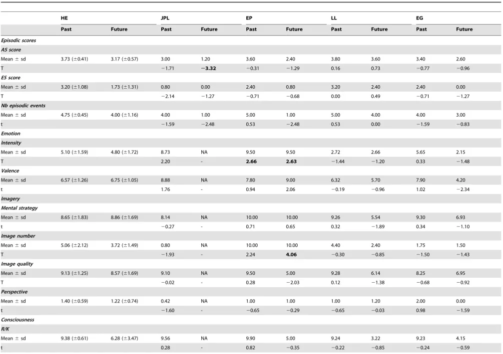
Table 2.Behavioral ratings for patients JPL, EP, LL, EG and healthy elders (HE) and t values comparing each patient to the control group for each period.
HE JPL EP LL EG
Past Future Past Future Past Future Past Future Past Future
Episodic scores
AS score
Mean6sd 3.73 (60.41) 3.17 (60.57) 3.00 1.20 3.60 2.40 3.80 3.60 3.40 2.60
T 21.71 23.32 20.31 21.29 0.16 0.73 20.77 20.96
ES score
Mean6sd 3.20 (61.08) 1.73 (61.31) 0.80 0.00 2.40 0.80 3.20 2.40 2.40 0.00
T 22.14 21.27 20.71 20.68 0.00 0.49 20.71 21.27
Nb episodic events
Mean6sd 4.75 (60.45) 4.00 (61.16) 4.00 1.00 5.00 1.00 5.00 4.00 4.00 3.00
t 21.59 22.48 0.53 22.48 0.53 0.00 21.59 20.83
Emotion
Intensity
Mean6sd 5.10 (61.59) 4.80 (61.72) 8.73 NA 9.50 9.50 2.72 2.66 5.65 2.15
T 2.20 - 2.66 2.63 21.44 21.20 0.33 21.48
Valence
Mean6sd 6.57 (61.26) 6.75 (61.05) 8.88 NA 7.80 9.00 6.32 5.70 7.90 4.20
t 1.76 - 0.94 2.06 20.19 20.96 1.02 22.34
Imagery
Mental strategy
Mean6sd 8.65 (61.83) 8.86 (61.69) 8.14 NA 10.00 10.00 9.26 5.54 9.30 6.93
t 20.27 - 0.71 0.65 0.32 21.89 0.34 21.10
Image number
Mean6sd 5.06 (62.12) 3.72 (61.49) 0.80 NA 10.00 10.00 4.40 2.40 1.75 1.50
T 21.93 - 2.24 4.06 20.30 20.85 21.50 21.43
Image quality
Mean6sd 9.13 (61.25) 8.57 (61.69) 9.10 NA 9.50 5.00 9.28 6.14 8.25 6.95
T 20.02 - 0.28 22.03 0.12 21.38 20.68 20.92
Perspective
Mean6sd 1.40 (60.59) 1.22 (60.74) 0.42 NA 1.00 1.00 1.00 1.20 2.00 0.00
t 21.60 - 20.65 20.29 20.65 20.03 0.98 21.59
Consciousness
R/K
Mean6sd 9.38 (60.61) 6.28 (63.47) 9.56 NA 9.90 5.00 9.24 3.22 9.23 4.15
t 0.28 - 0.82 20.35 20.22 20.85 20.24 20.59
Episodic
Future
Thinking
in
Semantic
Dementia
PLOS
ONE
|
www.ploson
e.org
7
October
2014
|
Volume
9
|
Issue
10
|
Data analyses
Behavioral data analysis. An adjusted t-test intended for single case analyses [67] was used for comparisons between each patient and healthy elders (for autobiographical scores and behavioral scales). Within-subject differences across past and future conditions were conducted using the modified t test [68] which assesses whether the difference between past and future periods in each patient differs significantly from the control group pattern. Results were considered significant at a conservative threshold of t.62.576, p,0.01 to guard against Type 1 errors for multiple comparisons.
VBM analyses. In order to formally assess the extent of atrophy in each SD patient across the whole brain, we compared their structural MRI scans with those of the group of 12 healthy elders using voxel-based morphometry [66] (VBM5). Structural MRI images were analyzed using the optimised VBM procedure implemented in SPM5. Briefly, this involves a number of fully automated preprocessing steps including extraction of brain, spatial normalization into stereotactic (MNI) space, segmentation into grey and white matters and CSF compartments, correction for volume changes induced by spatial normalization (modulation), and smoothing with a 8-mm3FWHM isotropic Gaussian kernel. The preprocessing procedures used in SPM5 have been shown to produce good results when matching brains with lesions to standardised templates [69]. We used the optional ‘‘modulation of non-linear effects only’’ which takes into account intracranial volume, hence controlling for differences in brain sizes. Analyses focussed on grey matter. Each patient’s structural scan was compared to healthy elders’ scans using a two sample t-test to investigate differences in grey matter volume. The significance level was set at p,0.05 FWE corrected for multiple comparisons, k.50 voxels (see Tables S2 to S5 in File S1).
fMRI data analyses. fMRI time series were modelled by a general linear model (GLM) including separate regressors for each of the experimental (past and future periods) and control conditions using SPM5. All regressors were convolved with the canonical hemodynamic response function (HRF). Data were high-pass filtered (cut-off period = 96 s). Coefficients for each regressor were estimated for each participant using maximum likelihood estimates to account for serial correlations in the data. At the first level, linear contrasts of the parameter estimates for each experimental regressor of interest were calculated for each participant, subtracting the corresponding control regressor (resulting in ‘‘period minus control task’’ contrasts). To examine the future thinking network in each patient at the individual level, t-statistic maps were generated for the following contrasts of interest: future minus control task and future minus past period (subtracting for each period the corresponding control regressor) for each participant. The second level random effects analysis was conducted over contrast images obtained at the first level. Each patient was compared to the group of healthy elders (i.e., patients were not grouped together) using the two sample t-test model of SPM5, as recommended by Henson’s guidelines (2006; http://www.mrc-cbu.cam.ac.uk//personal/rik.henson/personal/ Henson_Singlecase_06.pdf) and previously used to report single case fMRI data, notably in semantic dementia [70,6]. Inter-group subtraction analyses were computed to determine which regions were differentially activated by each patient and healthy elders when comparing past and future periods. The significance level was set at p,0.05 FWE corrected for multiple comparisons, k.10 voxels. Coordinates of brain regions are reported in the MNI space. To control for the age difference between patients and healthy elders, age was included as covariate. EP’s duration of education was higher compared to healthy elders. Since education
and general intelligence are generally closely inter-related [71], variations due to differences in general intelligence between EP and healthy elders were controlled for by adding years of education as covariate for analyses comparing EP to healthy elders. Finally, to reveal pathophysiological properties of the future thinking brain network in each patient using a different method, plots of activation magnitude were obtained for each patient and average of healthy elders in several regions of interest (ROIs) of the conventional future thinking network (medial frontal gyrus, hippocampus, precuneus). For each participant, mean activation values corresponding to the difference in BOLD activation between the experimental (future period) and control tasks (for Figure S1: control task; for Figure S2: past period; see File S1), were extracted within each ROI using the ‘‘anatomical VOI analysis’’ of the fMRIroi SPM toolbox [54].
Results
Behavioral results
All patients were able to perform the tasks, both recalling past memories and envisioning future events, differences lay in the episodic nature of the events experienced. On the objective measures of episodicity, JPL and EP were able to pre-experience only one episodic future event (rated 3 or 4 on the episodic scale) and four semantic future events (rated 1 or 2). On the contrary, LL was able to pre-experience four episodic future events and one semantic future event. EG was able to pre-experience three episodic future events and two semantic future events. Of note, despite using verbal cues, all patients were all able to read and understand the cues.
Comparisons between each patient and healthy elders showed, on the objective measures of episodicity, significantly lower overall autobiographical score (AS) for the future and a trend for the strictly episodic score (ES) for the past for JPL (see Table 2). Based on the number of episodic events experienced, a trend showed that JPL and EP produced less episodic future events compared to healthy elders. On the subjective rating scales, main results indicated that emotional intensity was greater for the past and the future for EP compared to healthy elders. Concerning mental imagery, EP evoked a greater number of mental images for the future compared to healthy elders, although a trend showed that image quality of future mental images was less clear for EP compared to healthy elders. Within-subject comparisons, using the modified t test procedure [68], did not reveal statistical differences between past and future conditions for each patient (data not shown). Note that adding education as covariate in behavioral analyses for EP, following Crawford et al.’s [72] guidelines, did not change results (see Table S1 in File S1).
VBM results
Results depicted on Figure 1 and Tables S2 to S5 in File S1 reveal areas of grey matter volume loss in JPL, EP, LL and EG compared to healthy elders. All patients present lateral temporal atrophy, predominantly in its anterior portion (see Tables S2 to S5 in File S1). Of the four patients, three have predominant left-lateralized temporal atrophy (JPL, LL, EG) and EP has predominant right-lateralized temporal atrophy. JPL also present-ed atrophy in the left anterior hippocampus, left amygdala and frontal regions, in particular bilateral superior medial gyri.
Apart from predominantly right-lateralized temporal atrophy, EP also presented atrophy in frontal, in particular bilateral superior medial gyri and parietal regions (bilateral supramarginal, left inferior gyri) (see Table S3). EP showed preservation of anterior hippocampi.
Apart from predominantly left-lateralized temporal atrophy, LL also presented atrophy in left anterior hippocampus, left amygdala and bilateral inferior frontal gyri (see Table S4).
Apart from predominantly left-lateralized temporal atrophy, EG also presented atrophy in frontal regions (bilateral superior, left medial, bilateral inferior gyri) (see Table S5).
Strikingly, VBM analyses reveal that the two patients (JPL and EP) who have difficulties in envisioning the future in an episodic way present atrophy in bilateral superior medial frontal gyri which appears preserved in patients (LL and EG) who can project in the future in an episodic manner. Furthermore, the (left) anterior hippocampus, which is atrophied in JPL (and LL) may additionally explain his difficulties in episodic future projection (LL appears to compensate for this atrophy by hyperactivating the right anterior hippocampus, see below).
fMRI results
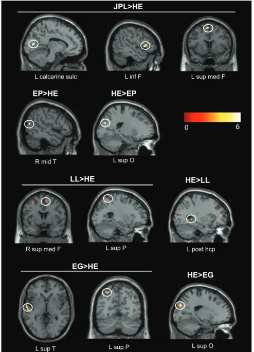
Patient JPL. At the individual level, results for the contrast future minus control task for JPL showed an essentially left-lateralized network of regions comprising the left (inferior, middle, superior) frontal gyri, middle temporal gyrus, precuneus, occipital areas and cerebellum (see Table S6). Results for the contrast future minus past period for JPL did not reveal any activation at the given threshold (see Table S7).
At the group level, the past period showed greater activations for JPL than healthy elders mainly in the left middle and inferior frontal gyri, bilateral cuneus and left insula compared to the control task (see Table 3). The future period showed greater activations for JPL than healthy elders mainly in the left calcarine sulcus, inferior and medial frontal gyri compared to the control task (see Table 4, Figure 2). Plots of activation magnitude confirm this medial frontal hyperactivation in JPL compared to healthy elders (see Figure S1). The past period showed greater activations for JPL, compared to healthy elders, in the right middle temporal gyrus compared to the future period (see Table 5). The future period showed no significant activations compared to the past period. No regions were less active in JPL compared to healthy elders.
Patient EP. At the individual level, results for the contrast future minus control task for EP showed activation in the right precuneus (see Table S6). No activation was detected for the contrast future minus past period (see Table S7).
At the group level, the past period showed greater activations for EP than healthy elders in the left middle occipital and precental gyri compared to the control task (see Table 3). No regions were less active in EP compared to healthy elders for this contrast.
The future period showed greater activations for EP than healthy elders in the right middle temporal gyrus compared to the control task (see Table 4, Figure 2). EP showed less activation than healthy elders in the left superior occipital gyrus and right insula for the future period compared to the control task (see Table 4, Figure 2).
The past period revealed greater activations for EP than healthy elders in left middle frontal gyrus compared to the future period (see Table 5). No regions were less active in EP compared to healthy elders for this contrast.
Conversely, the future period showed greater activations in EP than healthy elders in the left parietal gyrus compared to the past period (see Table 6, Figure 3). No regions were less active in EP compared to healthy elders for this contrast.
Patient LL. At the individual level, results for the contrast future minus control task for LL showed activation in the left supplementary motor area (see Table S6). Compared to the past Episodic Future Thinking in Semantic Dementia
period, the future period showed hyperactivation in the right middle occipital and supramarginal gyri (see Table S7).
At the group level, the past period showed greater activations for LL than healthy elders mainly in the bilateral middle temporal gyri, left middle and inferior frontal gyri, right cuneus and left caudate compared to the control task (see Table 3). LL showed less
activation than healthy elders in essentially the right anterior hippocampus, left parahippocampal gyrus, lateral temporal and occipital gyri for the past period compared to the control task (see Table 3).
The future period revealed greater activations for LL than healthy elders mainly in the right supramarginal and left superior Figure 1. Structural brain scans of the four SD patients.Left panels show coronal sections through the brains of JPL, EP, LL and EG. Right panels show results of the VBM analysis superimposed on MRI scans for each patient at a corrected FWE threshold of p,0.05. The pronounced atrophy in superior medial frontal regions is apparent for JPL and EP and atrophy in left anterior hippocampus apparent for JPL and LL. Color scale shows voxel z-scores. See Tables S2 to S5 in File S1 for detailed VBM results. Abbreviations: amg = amygdala; ant = anterior; F = frontal; hcp = hippocampus; inf = inferior; L = left; med = medial; mid = middle; parah = parahippocampal; R = right; sup = superior; T = temporal.
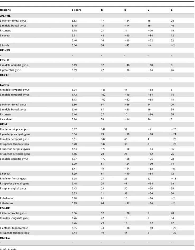
Table 3.Results for the comparisons past.control task for JPL, EP, LL, EG compared to healthy elders (HE) with age and years of education (for EP) as covariates at a corrected FWE statistical threshold of p,0.05, k.10.
Regions z-score k x y z
JPL.HE
L inferior frontal gyrus 5.83 17 234 16 28
L middle frontal gyrus 5.48 15 244 16 40
R cuneus 5.78 21 14 276 18
L cuneus 5.71 42 210 284 12
5.40 16 210 272 22
L insula 5.66 24 242 24 22
HE.JPL
- - -
-EP.HE
L middle occipital gyrus 6.19 32 246 280 8
L precentral gyrus 5.59 47 236 214 46
HE.EP
- - -
-LL.HE
R middle temporal gyrus 5.94 186 44 258 8
L middle temporal gyrus 5.42 102 240 254 14
5.13 102 252 258 18
L inferior frontal gyrus 5.86 67 236 14 20
L middle frontal gyrus 5.40 67 230 16 34
R cuneus 5.46 27 10 286 28
L caudate 5.90 74 216 26 2
HE.LL
R anterior hippocampus 6.87 142 32 24 220
L parahippocampal gyrus 5.64 73 230 210 224
R middle temporal gyrus 5.51 142 46 4 220
R superior temporal pole 5.28 142 38 8 220
L superior occipital gyrus 6.44 170 220 284 36
R superior occipital gyrus 5.66 20 26 282 26
L middle occipital gyrus 5.37 170 228 276 28
5.09 61 224 290 14
5.41 19 214 288 26
L cuneus 5.29 61 210 284 12
R inferior frontal gyrus 5.98 27 26 22 218
R superior parietal gyrus 5.48 24 48 238 58
R supramarginal gyrus 5.43 23 50 234 38
5.25 11 58 236 30
R thalamus 5.98 81 16 214 22
L thalamus 5.19 64 212 214 22
EG.HE
L inferior frontal gyrus 6.66 52 238 8 20
R middle cingulate gyrus 6.26 42 18 8 34
5.76 29 16 212 42
L anterior hippocampus 5.35 34 230 210 222
R superior temporal pole 5.44 19 44 8 222
HE.EG
- - -
-L: left, R: right.
doi:10.1371/journal.pone.0111046.t003
Episodic Future Thinking in Semantic Dementia
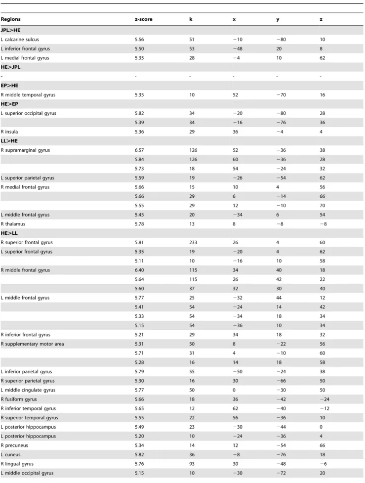
Table 4.Results for the comparisons future.control task for JPL, EP, LL, EG compared to healthy elders (HE) with age and years of education (for EP) as covariates at a corrected FWE statistical threshold of p,0.05, k.10.
Regions z-score k x y z
JPL.HE
L calcarine sulcus 5.56 51 210 280 10
L inferior frontal gyrus 5.50 53 248 20 8
L medial frontal gyrus 5.35 28 24 10 62
HE.JPL
- - -
-EP.HE
R middle temporal gyrus 5.35 10 52 270 16
HE.EP
L superior occipital gyrus 5.82 34 220 280 28
5.39 34 216 276 36
R insula 5.36 29 36 24 4
LL.HE
R supramarginal gyrus 6.57 126 52 236 38
5.84 126 60 236 28
5.73 18 54 224 32
L superior parietal gyrus 5.59 19 226 254 62
R medial frontal gyrus 5.66 15 10 4 56
5.66 29 6 214 66
5.55 29 12 210 70
L middle frontal gyrus 5.45 20 234 6 54
R thalamus 5.78 13 8 28 28
HE.LL
R superior frontal gyrus 5.81 233 26 4 60
L superior frontal gyrus 5.35 19 220 4 62
5.11 10 216 10 58
R middle frontal gyrus 6.40 115 34 40 18
5.64 115 26 42 22
5.60 37 32 30 40
L middle frontal gyrus 5.77 25 232 44 12
5.41 54 224 14 42
5.33 54 234 18 34
5.15 54 236 10 34
R inferior frontal gyrus 5.21 29 34 18 32
R supplementary motor area 5.31 50 8 222 56
5.71 31 4 210 60
5.28 16 14 18 58
L inferior parietal gyrus 5.79 55 250 224 38
R superior parietal gyrus 5.30 16 30 266 50
L middle cingulate gyrus 5.77 50 0 230 50
R fusiform gyrus 5.66 18 36 242 224
R inferior temporal gyrus 5.65 12 62 240 212
R superior temporal gyrus 5.55 22 56 236 10
L posterior hippocampus 5.49 23 230 244 0
L posterior hippocampus 5.20 10 224 236 4
R precuneus 5.34 14 12 254 66
L cuneus 5.82 36 28 276 18
R lingual gyrus 5.76 93 30 248 26
parietal gyri, right medial and left middle frontal gyri and right thalamus compared to the control task (see Table 4, Figure 2). LL showed less activation than healthy elders in essentially the left posterior hippocampus, frontal (bilateral superior, middle and right inferior) gyri, right precuneus, lateral parietal gyrus, right lateral temporal and occipital regions for the future period compared to the control task (see Table 4, Figure 2). Plots of activation magnitude confirm the significant lower activation in the left posterior hippocampus and right precuneus in LL compared to healthy elders (see Figure S1).
The past period showed greater activations for LL than healthy elders in the left posterior hippocampus, frontal (bilateral superior medial, middle, left superior and inferior frontal gyri), temporal (right middle, superior and left inferior temporal gyri), occipital (bilateral cuneus, middle and inferior occipital gyri) and parietal regions (angular, inferior parietal and supramarginal gyri, bilateral precuneus) compared to the future period (see Table 5). No regions were less active in LL compared to healthy elders for this contrast.
Conversely, the future period showed greater activations for LL than healthy elders in right anterior hippocampus, bilateral middle occipital gyri, right middle temporal pole and parietal regions (supramarginal and superior parietal gyri) compared to the past period (see Table 6, Figure 3). Plots of activation magnitude confirm this right anterior hippocampal hyperactivation in LL compared to healthy elders (see Figure S2). No regions were less active in LL compared to healthy elders for this contrast.
Patient EG. At the individual level, results for the contrast future minus control task for EG showed an essentially left-lateralized network of regions comprising the left middle frontal and temporal gyri (see Table S6). No activation was detected for the contrast future minus past period (see Table S7).
At the group level, the past period showed greater activations for EG than healthy elders in the left anterior hippocampus, left inferior frontal gyrus, right middle cingulate cortex and superior temporal pole compared to the control task (see Table 3). No regions were less active in EG compared to healthy elders for this contrast.
The future period showed greater activations for EG than healthy elders in the left superior temporal and superior parietal gyri compared to the control task (see Table 4, Figure 2). EG showed less activation than healthy elders in the left superior occipital gyrus for the future period compared to the control task (see Table 4, Figure 2).
The past period showed greater activations for EG than healthy elders in the left superior occipital gyrus compared to the future period (see Table 5). No regions were less active in EG compared to healthy elders for this contrast.
Conversely, the future period showed greater activations for EG than healthy elders in the right middle temporal gyrus and left calcarine sulcus compared to the past period (see Table 6, Figure 3). No regions were less active in EG compared to healthy elders for this contrast.
For sake of clarity, we also added results of the contrast future minus control task in healthy elders (see Table S8 in File S1). Results for the contrast future minus past period in healthy elders have been published previously [19].
Discussion
This work is, to our knowledge, the first fMRI study examining future projection in semantic dementia. While the sparse behavioral studies found that future projection was consistently impaired in SD [18,16,17], here we show that the capability of patients to project into their future largely depends on the structural integrity of certain brain regions, in particular the superior medial frontal cortex and anterior hippocampus. JPL presented atrophy in bilateral superior medial frontal gyri and left anterior hippocampus and had difficulties in experiencing episodic future events and to a certain extent past episodic events. Hyperactivations of neocortical (frontal and occipital) regions appeared inefficient in compensating for his deficit. EP presented atrophy in bilateral superior medial frontal gyri and, like JPL, could pre-experience only one episodic future event, but past episodic remembering was spared. However, behavioral ratings for the future were higher than those of healthy elders (in terms of Table 4.Cont.
Regions z-score k x y z
R middle occipital gyrus 5.11 10 34 276 26
R insula 5.97 54 34 218 18
L caudate 6.04 102 210 12 28
5.73 102 24 6 28
R caudate 5.17 102 8 10 210
5.91 58 14 22 10
R thalamus 5.55 19 2 214 4
5.45 27 12 234 4
R vermis 5.64 36 6 258 216
EG.HE
L superior temporal gyrus 5.64 45 258 210 0
5.60 45 256 218 0
L superior parietal gyrus 5.48 16 230 266 52
HE.EG
L superior occipital gyrus 6.96 118 218 282 54
L: left, R: right.
doi:10.1371/journal.pone.0111046.t004
Episodic Future Thinking in Semantic Dementia
emotion and mental imagery), suggesting that EP may have overestimated her capacities to project into the future. On the contrary, LL was able to pre-experience episodic future events. Although she had left anterior hippocampal atrophy, hyperactiva-tion of its right counterpart during future compared to past thinking compensated efficiently for this atrophy. Finally, EG who presented integrity of superior medial frontal gyri and anterior hippocampi was also able to pre-experience episodic future events. Figure 4 depicts summary representations of patients’ ability to engage in episodic future thinking (disturbed or spared) depicting sites of atrophy with associated hypothesized function and brain activations for each patient compared to healthy elders. Overall, VBM analyses showed that patients who had difficulties in envisioning the future in an episodic way (JPL and EP) presented specific atrophy in superior medial prefrontal cortex, while this
region was relatively preserved in LL and EG who could engage in episodic future projection. Furthermore, JPL and LL presented atrophy in (left) anterior hippocampus, a region known to be crucial for episodic past and future thinking, but LL was able to compensate efficiently by hyperactivation of its right counterpart, while JPL could not. We will mainly focus on these two key regions in the following discussion, distinguishing patients who cannot (JPL and EP) or can (LL and EG) engage in episodic future thinking.
Patients impaired in episodic future projection
JPL presented atrophy mainly in left anterior hippocampus, lateral (middle and inferior) temporal and frontal cortices, including in the bilateral superior medial frontal gyri. Behavioral ratings showed that he was impaired at episodic future projection Figure 2. Results of the comparisons between the future period and the control task for EP, LL and EG depicting hyperactivations (Patient.HE) and lower activations (HE.Patient) compared to healthy elders (HE) at a corrected FWE threshold of p,0.05, k.10 voxels.Color scale shows voxel z-scores. See Table 4 for full details. Abbreviations: F = frontal; hcp = hippocampus; sup = superior; inf = inferior; L = left; med = medial; mid = middle; O = occipital; P = parietal; post = posterior; R = right; sulc = sulcus; T = temporal.
Table 5.Results for contrast past.future for JPL, EP, LL, EG compared to healthy elders (HE) with age and years of education (for EP) as covariates at a corrected FWE statistical threshold of p,0.05, k.10.
Regions z-score k x y z
JPL.HE
R middle temporal gyrus 5.62 14 44 270 14
EP.HE
L middle frontal gyrus 5.83 52 234 26 44
LL.HE
R superior medial frontal gyrus 6.30 65 10 62 26
5.46 83 4 66 4
L superior medial frontal gyrus 5.53 142 24 64 28
5.15 142 26 62 20
5.43 83 28 62 0
L superior frontal gyrus 5.73 151 216 58 16
5.27 151 220 60 24
5.48 12 218 36 52
5.04 39 220 48 16
L medial frontal gyrus 5.16 33 212 50 26
R medial frontal gyrus 5.41 12 2 220 56
L middle frontal gyrus 6.19 161 230 16 36
6.11 151 236 58 14
6.08 52 230 44 10
5.45 29 246 30 30
5.40 39 220 48 26
R middle frontal gyrus 6.00 34 32 16 54
5.99 162 42 14 42
5.69 34 32 10 48
L inferior frontal gyrus 5.15 17 256 18 30
5.26 12 246 28 16
L posterior hippocampus 6.07 108 226 240 22
5.83 108 224 238 6
R parahippocampal gyrus 5.82 308 36 238 212
R middle temporal gyrus 5.66 51 54 254 8
5.23 51 44 256 10
5.39 42 18 274 58
R superior temporal gyrus 5.56 40 58 234 10
L inferior temporal gyrus 5.35 21 46 268 8
5.29 12 246 260 212
R fusiform gyrus 5.34 35 26 278 26
R cuneus 6.08 59 12 288 30
L cuneus 5.72 21 210 278 20
L middle occipital gyrus 5.98 48 236 274 24
5.35 55 224 258 32
5.14 80 238 278 36
R inferior occipital gyrus 5.43 35 34 286 24
5.15 12 38 276 24
R angular gyrus 6.55 180 40 262 40
6.01 180 46 272 36
L angular gyrus 5.57 55 234 254 36
L inferior parietal gyrus 5.67 60 252 224 38
5.51 80 232 278 42
R inferior parietal gyrus 5.39 39 56 252 46
Episodic Future Thinking in Semantic Dementia
and past remembering. The overall autobiographical score (AS) for the future and a trend for the strictly episodic autobiographical score (ES) for the past were significantly lower compared to healthy elders, indicating that JPL had difficulties in providing episodic details for future and past periods. A trend also showed that he produced less episodic future events compared to healthy elders. Irish et al. [16] showed that the future thinking deficit in SD was driven by a difficulty to provide ‘‘internal’’ details (i.e., episodic details, central to the event), while ‘‘external’’ details (i.e.,
semantic facts) were more numerous. With JPL, we confirm behavioral studies showing that SD patients are impaired at projecting into the future in an episodic way [18,16,17] and provide the neural correlates of these cognitive findings. JPL presented atrophy in bilateral superior medial frontal gyri (approximately corresponding to Brodmann areas 8 and 9). This area has been previously reported in episodic prospection studies [73,74,75,36] and has a role in processing coherent contexts [36] (BA9), manipulating processes in working memory [76] (BA8), Table 5.Cont.
Regions z-score k x y z
5.19 39 56 244 44
R supramarginal gyrus 5.55 19 52 222 22
R precuneus 5.74 35 12 250 44
5.48 13 8 256 56
L precuneus 5.62 66 212 256 40
L anterior cingulate gyrus 5.43 20 26 44 6
L middle cingulate gyrus 5.76 58 24 242 46
R thalamus 5.64 29 12 232 4
5.44 20 18 220 14
R cerebellum 6.44 205 12 262 216
5.75 205 18 264 222
5.53 13 34 242 226
EG.HE
L superior occipital gyrus 6.29 28 216 284 34
No regions were less active by patients compared to healthy elders for this contrast. L: left, R: right. doi:10.1371/journal.pone.0111046.t005
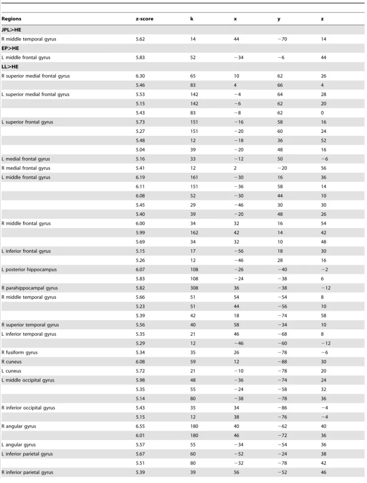
Table 6.Results for contrast future.past for JPL, EP, LL, EG compared to healthy elders (HE) with age and years of education (for EP) as covariates at a corrected FWE statistical threshold of p,0.05, k.10.
Regions z-score k x y z
JPL.HE
- - -
-EP.HE
L inferior parietal cortex 6.90 91 252 234 46
6.54 91 244 242 52
LL.HE
L middle occipital gyrus 6.77 130 222 282 36
R middle occipital gyrus 5.72 12 26 292 6
R supramarginal gyrus 6.68 262 52 236 38
6.28 262 60 236 30
6.11 262 54 222 32
L superior parietal gyrus 5.44 15 224 254 60
R posterior hippocampus 5.80 23 24 244 4
R anterior hippocampus 5.42 21 30 24 220
R middle temporal pole 5.28 15 46 4 218
EG.HE
R middle temporal gyrus 5.52 21 60 252 8
L calcarine sulcus 5.35 14 26 290 26
creative story generation [77] (BA8) and autonoetic consciousness [8,37], essential to pre-experience episodic future events. Bilateral atrophy in this region may also have been responsible for JPL’s inability to project into the future. JPL also presents atrophy in the left anterior hippocampus. We previously showed that anterior hippocampal integrity in SD is crucial for episodic past remembering [6,70]. Here, we expand these findings to episodic future thinking which also requires anterior hippocampal integrity. The anterior hippocampus supports relational processing [31,32,33], including flexible recombination of details for past and future event construction [34]. Addis and Schacter [35] showed that future-associated activity in the anterior hippocampus was associated with higher demands on recombination of details. Left atrophy in this region may be in part responsible for JPL’s inability to project into the future in an episodic way. Thus, we confirm that atrophy in crucial regions of the future thinking network (e.g., superior medial frontal gyrus and anterior hippo-campus), overlapping with the autobiographical memory network, impairs episodic future projection. Examination of activation patterns at the individual level indicated that JPL failed to activate the core episodic future thinking network observed in healthy elders (reported in Table S8 and Viard et al. [19]), although several regions may show similar activation patterns across patient and healthy elders (e.g., precuneus, middle cingulate gyrus).
Compared to healthy elders, JPL hyperactivated left (medial and inferior) frontal and occipital regions during future thinking compared to the control task. Plots of activation magnitude confirmed hyperactivation in the medial frontal gyrus in JPL compared to healthy elders. Yet, these hyperactivations did not efficiently compensate for atrophied regions, since he was unable to pre-experience episodic future events, as shown by behavioral results.
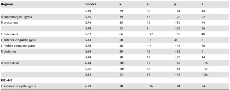
EP also presented atrophy in frontal cortices, including bilateral superior medial frontal gyri, and lateral (middle and superior) temporal cortices, but sparing of anterior hippocampi. EP was able to pre-experience only one episodic future event, most being semantic. Indeed, like JPL, a trend showed that EP produced less episodic future events compared to healthy elders. She was however not impaired at episodic past remembering. It appears that integrity of anterior hippocampi may have permitted EP to remember episodic past memories, but atrophy of bilateral superior medial frontal gyri may have been in part responsible for EP’s deficit in episodic future thinking. Examination of activation patterns at the individual level indicated that, except for the right precuneus, EP failed to activate the core future thinking network observed in healthy elders (Table S8; [19]). Direct comparisons with healthy elders showed that EP hyper-actived the inferior parietal and middle temporal cortices for the Figure 3. Results of the comparisons between the future and past periods for JPL, EP, LL and EG compared to healthy elders (HE) at a corrected FWE threshold of p,0.05, k.10 voxels.See Table 6 for full details. Abbreviations: ant = anterior; hcp = hippocampus; inf = inferior; L = left; mid = middle; O = occipital; P = parietal; post = posterior; R = right; sulc = sulcus; T = temporal.
doi:10.1371/journal.pone.0111046.g003
Episodic Future Thinking in Semantic Dementia
Figure 4. Summary representations of patients’ ability to engage in episodic future thinking (left: disturbed; right: spared) depicting (A) localization of preserved (+) or atrophied (2) brain regions and associated hypothesized function (superior medial prefrontal cortex (mPFC): coherent context processing; hippocampus: binding; amygdala: emotional processing; lateral temporal cortex: semantic processing) and (B) brain activations for each patient compared to healthy elders for the contrast Future.Past thinking (see results on Table 6).