Altered Activation in Cerebellum
Contralateral to Unilateral Thalamotomy
May Mediate Tremor Suppression in
Parkinson
’
s Disease: A Short-Term Regional
Homogeneity fMRI Study
Zhi Wen1, Jie Zhang2*, Jielan Li1, Jiankun Dai3,4, Fuchun Lin3, Guangyao Wu1*
1Department of Magnetic Resonance Imaging, Zhongnan Hospital of Wuhan University, Wuhan, Hubei, China,2Department of Neurosurgery, Zhongnan Hospital of Wuhan University, Wuhan, Hubei, China, 3National Center for Magnetic Resonance, Key State Laboratory of Atomic and Molecular Physics, Wuhan Institute of Physics and Mathematics, Chinese Academy of Sciences, Wuhan, Hubei, China,4University of Chinese Academy of Sciences, Beijing, China
*wuguangy2002@163.com(GW);zhangjie8790@163.com(JZ)
Abstract
Background
Ventral intermediate nucleus thalamotomy is an effective treatment for Parkinson’s disease tremor. However, its mechanism is still unclear.
Purpose
We used resting-state fMRI to investigate short-term ReHo changes after unilateral thala-motomy in tremor-dominant PD, and to speculate about its possible mechanism on tremor suppression.
Methods
26 patients and 31 healthy subjects (HS) were recruited. Patients were divided into two groups according to right- (rPD) and left-side (lPD) thalamotomy. Tremor was assessed using the 7-item scale from the Unified Parkinson’s disease rating scale motor score (mUPDRS). Patients were scanned using resting state fMRI after 12h withdrawal of medica-tion, both preoperatively (PDpre) and 7- day postoperatively (PDpost), whereas healthy
sub-jects were scanned once. The regions associated with tremor and altered ReHo due to thalamic ablation were examined.
Results
The impact of unilateral VIM thalamotomy was characterized in the frontal, parietal, tempo-ral regions, basal ganglia, thalamus, and cerebellum. Compared with PDpre, significantly
reduced ReHo was found in the left cerebellum in patients with rPDpost, and slightly a11111
OPEN ACCESS
Citation:Wen Z, Zhang J, Li J, Dai J, Lin F, Wu G (2016) Altered Activation in Cerebellum Contralateral to Unilateral Thalamotomy May Mediate Tremor Suppression in Parkinson’s Disease: A Short-Term Regional Homogeneity fMRI Study. PLoS ONE 11(6): e0157562. doi:10.1371/journal.pone.0157562
Editor:Xi-Nian Zuo, Institute of Psychology, Chinese Academy of Sciences, CHINA
Received:January 26, 2016
Accepted:June 1, 2016
Published:June 16, 2016
Copyright:© 2016 Wen et al. This is an open access article distributed under the terms of the
Creative Commons Attribution License, which permits unrestricted use, distribution, and reproduction in any medium, provided the original author and source are credited.
decreased ReHo in the cerebellum vermis in patients with lPDpost, which was significantly
higher than HS. We demonstrated a positive correlation between the ReHo values in the cerebellum (in rPD, peak coordinate [-12, -54, -21], R = 0.64,P= 0.0025, and peak coordi-nate [-9, -54, -18], R = 0.71,P= 0.0025; in lPD, peak coordinate [3, -45, -15], R = 0.71,P= 0.004) in the pre-surgical condition, changes of ReHo induced by thalamotomy (in rPD, R = 0.63,P= 0.021, R = 0.6,P= 0.009; in lPD, R = 0.58,P= 0.028) and tremor scores contralat-eral to the surgical side, respectively.
Conclusion
The specific area that may be associated with PD tremor and altered ReHo due to thalamic ablation is the cerebellum. The neural basis underlying thalamotomy is complex; cerebel-lum involvement is far beyond cerebello-thalamic tract breakage.
Introduction
Parkinson's disease (PD) is a progressive neurodegenerative disease in the elderly. Almost all patients experience tremor at rest in the disease process [1]. The depletion of dopamine in the substantia nigra is the predominant neurochemical hallmark of PD, and as the disease pro-gresses, it leads to dysfunction of striatal-thalamo-cortical circuits, involving non-dopaminer-gic brain areas, resulting in motor and non-motor symptoms [2]. Although this hypothesis serves as a critical reason for motor dysfunctions in PD, i.e. akinesia and rigidity, conversely, it fails to explain tremor [3]. Evidence suggests that loss of dopaminergic neurons in the basal ganglia correlates consistently with clinical ratings of akinesia and rigidity, rather than tremor [4]. Indeed, clinical studies provide evidence that the level of tremor severity is independent of the amount of dopamine deficiency [5] and is often refractory to dopamine replacement treat-ment [6]. Thus, tremor might be a unique symptom in PD.
Evidence that the thalamus is associated with PD tremor originates from the impact of ste-reotactic thalamotomy [7]. Neuromodulation in the form of steste-reotactic thalamotomy is an approved therapy to alleviate intractable tremor in developing countries [8]. Previous literature review has demonstrated that tremor suppression is observed immediately after creation of the lesion, and is still effective postoperatively [9]. Thalamic ablation, which targets the posterior ventral lateral (VLp) or ventral intermediate (VIM) nucleus, is able to suppress PD tremor [9]. The VIM nucleus is a subdivision of the motor thalamus, according to Hassler’s classification, containing complex overlaying distinct tracts, as cerebello-thalamic excitatory afferents termi-nate here and project to motor cortical areas [10]. This indicates that the cerebello-thalamo-cortical circuits may be involved in tremor generation. However, it remains elusive how dys-function of striatal-thalamo-cortical circuits in PD can drive the distinct cerebello-thalamo-cortical circuits into generating resting tremor; and the physiological mechanism of VIM thala-motomy that leads to the tremor suppression in PD.
In the last decade, the development of resting state functional magnetic resonance imaging (rs-fMRI) has provided researchers with a non-invasivein-vivotool to explore human brain
function by using a blood oxygen level dependent (BOLD) effect at rest. The regional homoge-neity (ReHo), which applies Kendall’s coefficient concordance (KCC) [11], measures the simi-larity of time series of a voxel and its given cluster of neighbor voxels [12]. It is a data-driven method of rsfMRI without a priori knowledge of the experimental design. The test–retest
Funding:This work was supported by the National Science Fund of China (http://www.nsfc.gov.cn/) Grant Nos. 81171315 to GW. The funders had no role in study design, data collection and analysis, decision to publish, or preparation of the manuscript.
reliability for ReHo to reveal the regional synchronization of spontaneous brain activity has also been validated [13,14]. This method has been successfully used to investigate the func-tional modulations in the resting state in patients with PD and motor subtypes [15]. Prior rs-fMRI study has revealed abnormal neural activity concerning PD tremor in several regions, namely the thalamus, basal ganglia, cortex, and cerebellum [3]. Indeed, many functional imag-ing studies have discussed the activation differences between tremor-dominant (TD) and non-tremor subtypes [3,4,16]. For example, Zhang et al. [4] illustrated that TD PD had increased ReHo in the left thalamus, primary motor cortex (M1), and cerebellum, whereas decreased ReHo in the left primary sensorimotor cortex (SM1) and putamen, when compared with aki-netic-rigid PD. It suggests that PD tremor is associated with dysfunction of striatal-thalamo-cortical and cerebello-thalamo-striatal-thalamo-cortical circuits [17]. However, there has been no published rs-fMRI study on thalamic ablation in TD PD, because the surgical changes relative to tremor suppression may be confounded by the non-specific effects derived from tissue lesions.
Based on previous studies, we hypothesized that clinical suppression of PD tremor due to VIM thalamotomy may be reflected by the ReHo alterations mainly in the cortex, basal ganglia, and cerebellum. During this study, we: (1) clarified the surgical impact by examining the ReHo differences in asymmetric TD PD preoperatively versus 7–day postoperatively (PDpostversus
PDpre); (2) examined the ReHo differences of TD PD from healthy subjects (HS) (PDpreversus
HS) and association with tremor scores; (3) speculated about the possible brain areas that mediate the surgical impact on tremor suppression.
Materials and Methods
Ethics statement
This study was approved by Zhongnan Hospital of Wuhan University Ethics Committee (Eth-ics No. [2014003]). All study procedures were in accordance with the Declaration of Helsinki. For the purpose of the study, all the patients were required to stop taking dopamine medication for at least 12 hours. All patients were well informed of the risks, such as orthostatic hypoten-sion, paroxysmal nocturnal dyspnea, and urinary disorders. All participants gave written con-sent before the study.
Subjects
31 patients with TD PD (12 females, 19 males, age 60.1 ± 8.17 years) who underwent unilateral VIM thalamotomy were recruited at the Department of Neurosurgery in Zhongnan Hospital of Wuhan University from March 2014 to August 2015. An additional 31 healthy subjects (16 females, 15 males, age 59.6 ± 7.65 years) were also measured. All patients were diagnosed based on UK Brain Bank Criteria for idiopathic PD [12] and demonstrated an asymmetric resting tremor during physical examination. The exclusion criteria were: (1) cognitive dysfunction measured as a Mini-Mental State Exam (MMSE) [13] score<26; (2) history of brain trauma or surgery; (3) history of neurological and neuropsychiatric diseases; and (4) clinically silent lesion evident on conventional MRI. Clinical descriptive information for healthy subjects and patients with Parkinson’s disease is shown inTable 1.
determined. Based on this method, predominantly left-side affected subjects had a ratio<1, whereas right-side affected subjects had a ratio>1. The surgical side was determined to sup-press the contralateral tremor-dominant symptom and patients were then further divided into two groups: patients with right-side VIM thalamotomy (rPD) and patients with left-side VIM thalamotomy (lPD). The surgical procedure is mentioned in theS1 Text. There were no com-plications such as hemorrhage, edema, or intracranial infection, identified by conventional MRI, either immediately or 7-day postoperatively. We sent a questionnaire to PD patients via mail to enquire about the ablation effect at 3 months after surgery. All patients had diminished tremor and the tremor score was equal to 0, which guaranteed the impact of tremor alleviation.
Functional MRI acquisition
During the whole study procedure, patients were accompanied by at least one experienced neu-rosurgeon. The rs-fMRI was performed immediately after clinical assessments with a 3.0 T MRI scanner (Magneto Trio, Siemens Erlangen, Germany), using a gradient-echo echo-planar sequence sensitive to BOLD contrast with the following parameters: TR/TE = 2000/30 ms, flip angle = 90°, matrix size = 64 × 64, FOV = 240 mm × 240 mm, slice thickness = 5 mm with no gap. Each fMRI scan lasted 7 minutes, and 30 axial slices were collected. A standard 8-channel head coil was used with foam padding to restrict head motion. During rs-fMRI, participants were instructed to keep their eyes closed, to keep still, and to clear their minds of thought.
Anatomic imaging was acquired using T1-3D MPRAGE with the following parameters: TR/ TE/TI = 1900/2.1/900 ms, flip angle = 9°, matrix size = 256 × 256, FOV = 240 mm × 240 mm, slice thickness = 1 mm with no gap. T2- weighted images were also obtained in every subject to detect clinically silent lesions.
Data analysis and statistical analysis
Image data were analyzed using DPARSF [19] and REST [20] based on Matlab 2010a (Math-works, Natick, Massachusetts, USA). The data for each fMRI scan contained 210 time points. The first 10 time points of fMRI data were discarded because of the instability of the transient signal and to allow subjects to get used to the scanning noise. The image pre-processing proce-dure included slice timing, head motion correction, co-registration to individuals’T1 image, Table 1. Clinical details of healthy subjects and patients with Parkinson’s disease.
HS (n= 31) rPD (n= 12) lPD (n= 14) P
Age (y) 59.6±7.65 60.8±7.02 61.4±7.77 0.87a
Gender (M/F) 15/16 8/4 6/8 0.44b
Handedness Right Right Right
MMSE >26 >26 >26
Disease/ Tremor duration (y) - 5 (1–17) 5.5 (1–14) 0.74c
mUPDRS - 28.9±10.9 26.4±15.6 0.47c
Tremor score contralateral to the surgical side - 2.92±0.79 3±0.68 0.76c
aANOVA;
bPearson Chi-square test; cMann-Whitney U test.
mUPDRS, Unified Parkinson’s Disease Rating Scale motor score; HS, healthy subjects; rPD, patients receiving right-side VIM thalamotomy; lPD, patients receiving left-side VIM thalamotomy; F, female; M, male.
and spatial normalization to the Montreal Neurological Institute (MNI) space. No subjects exceeded the head motion threshold of 3 mm of displacement or 3 degrees of rotation.
Linear drift was removed. Low-frequency drift and high-frequency physiological noise were removed by using a temporal filter (0.01 Hz<f<0.08 Hz). Kendall’s coefficient concordance (KCC) [11] was calculated to generate individual ReHo maps between the time series of a given voxel and those of its 26 nearest neighbors. For standardization purposes, each individual ReHo map was divided by its own mean ReHo within the mask that was extracted from the intracranial voxels. The head motion parameter [21], as well as white matter and CSF signals, were regressed out to remove confounding artifacts. Spatial smoothing was then applied to the ReHo maps (full width at half maximum, FWHM = 4 mm), to reduce noise and residual differ-ences in gyral anatomy.
Voxel-by-voxel based comparisons of ReHo differences were performed between PD groups in the pre- and post-surgical conditions using Student’s pairedt-test in REST software (PDpost
versus PDpre). Comparisons of ReHo differences were also performed between the healthy
sub-jects and PD patients in the pre-surgical condition (PDpreversus HS), and between the healthy
subjects and PD patients in the post-surgical condition (PDpostversus HS), using a two-sample
t-test with age and gender as covariates. The significance of group differences was set at P<0.005, cluster size23 voxels, corresponding to AlphaSim correctedP<0.05 (http://afni. nimh.nih.gov/pub/dist/doc/manual/AlphaSim.pdf). In addition, voxel-by-voxel correlation coefficients were also calculated between ReHo of PDpreand tremor score contralateral to the
surgery side (P<0.005, uncorrected, cluster size8 voxels).
The effect of surgery on PD tremor was explored. Firstly, the overlapping brain areas in PDpre, where ReHo values were altered relative to HS and correlated with tremor scores, were
identified. Secondly, the overlapping brain areas in PDpost, where there was aΔReHo relative to
PDpreand which correlated withΔtremor scores, were identified. Spherical ROI was drawn
using the peak coordinate as the origin and radius = 6 mm. The mean of the ReHo was extracted among HS, PDpreand PDpost. One-way ANOVA and post-hoc analysis were
per-formed using SPSS 19.0 (Chicago, IL, USA) (P<0.05, Bonferroni corrected). The change of ReHo between the pre- and post-surgical condition (ΔReHo) was calculated as the correlation coefficient with tremor score (P<0.05, Spearman corrected).
Results
Clinical assessments
5 patients were excluded due to their intolerance of the second MRI scanning. Their MRI data was not collected completely. Finally, 26 patients with PD and 31 HS were included in subse-quent data analysis. No difference was found in either age or gender among HS, rPD and lPD (P>0.05). There were no significant differences in the disease duration, total mUPDRS, and tremor score contralateral to the surgical side, between rPD and lPD (P>0.05). Moreover, compared with PDpre, total, the mUPDRS and tremor score of TD PD contralateral to the
sur-gical side reduced significantly after unilateral VIM thalamotomy (P<0.005) (Table 2).
Changes in ReHo of brain areas between the pre- and post-surgical
conditions in PD (PD
postversus PD
pre)
Compared with rPDpre, the rPDpostshowed increased ReHo in the right superior frontal gyrus
details of brain areas with significant ReHo differences are listed inS1 Table. In lPDpost, higher
ReHo was detected in the left MFG, left SFG, left inferior frontal gyrus (IFG), left inferior parie-tal lobule (IPL), bilateral postcentral gyrus, right MTG, bilateral angular gyrus, left middle occipital gyrus (MOG), right superior occipital gyrus (SOG), left ACC, and left cuneus; whereas lower ReHo was found in the left MFG, left thalamus, bilateral caudate, right ACC, and bilat-eral cerebellum. The details of brain areas with significant ReHo differences are listed inS1 Table. The PDpost−PDprecontrast images are shown in Figs1Band2B.
Changes in ReHo of brain areas for PD patients relative to healthy
subjects
Compared with HS, the rPDprehad increased ReHo in the right MTG and bilateral cerebellum,
as well as decreased ReHo in the right MFG and right STG. For the lPDpre, the authors
observed higher ReHo in the left SFG, right postcentral gyrus, left ACC, and right cerebellum, co-varying with lower ReHo in the right MFG, right IFG, right superior parietal lobule (SPL), right precentral gyrus, right MTG, left SOG and bilateral MOG. The details of brain areas with significant ReHo differences are listed inS2 Table. The PDpre−HS contrast images are shown
in Figs1Aand2A.
Compared with HS, the rPDpostrevealed significantly increased ReHo in the bilateral SFG,
left MFG, right STG, bilateral MTG, right IPL, right SOG and right IPL, and reduced ReHo in the right MFG, right paracentral lobule, left hippocampus, right thalamus, left calcarine and left cerebellum. The lPDpostgroup exhibited significantly increased ReHo in the right SFG, left
STG, left IPL, bilateral angular gyrus, bilateral fusiform gyrus, left insula, and left cerebellum, and decreased ReHo in the left MFG, left thalamus, and right cerebellum. The details of brain areas with significant ReHo differences are listed inS3 Table. The PDpost−HS contrast images
are shown in Figs1Cand2C.
Association between ReHo and tremor scores contralateral to the
surgical side
A significant negative correlation was shown between tremor scores contralateral to the surgi-cal side and ReHo in the right caudate in rPDpreand right precuneus in lPDpre, accompanied
by a positive correlation in the left IPL, left cerebellum anterior lobe in rPDpreand cerebellum
vermis in lPDpre. The details of brain areas correlated with tremor score are listed inS4 Table.
TheΔReHo in the left MFG, right postcentral gyrus, right IOG in rPD, as well as right SPL, right precuneus, right postcentral gyrus, and left MOG in lPD, were negatively correlated with Table 2. Severity of disease in patients with PD, pre- and postoperatively.
Severity of disease Pre Post P
mUPDRS
rPD 28.9±10.9 11.5±6 0.002a
lPD 26.4±15.6 10.6±8.3 0.001a
Tremor score contralateral to the surgical side
rPD 2.92±0.79 0 0.001a
lPD 3±0.68 0 0.002a
aWilcoxon Signed Ranks test.
mUPDRS, Unified Parkinson’s Disease Rating Scale motor score; rPD, patients receiving right-side VIM thalamotomy; lPD, patients receiving left-side VIM thalamotomy.
tremor score contralateral to the surgical side. Conversely,ΔReHo in the cerebellum culmen, left cerebellum posterior lobe in rPD, as well as right cerebellum posterior lobe in lPD, were positively correlated with tremor score.
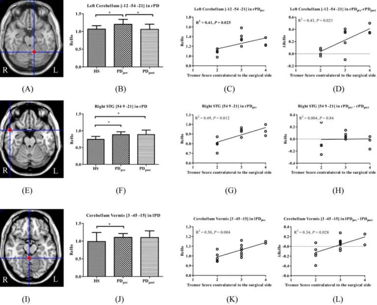
The effect of surgery on brain areas relative to PD tremor
There were overlapping regions of the altered ReHo values relative to HS that correlated with tremor scores in PD. In rPD, these were located in the left cerebellum anterior lobe (peak Fig 1. T- statistics maps of rPDpreversus HS (A), rPDpost−rPDpre(B), and rPDpost−HS (C).T-score bar: hot and cold colors
indicate ReHo increases and decreases, respectively. rPD: patients receiving right-side VIM thalamotomy.
coordinate [-12, -54, -21]) and right STG (peak coordinate [54, 9, -21]), but in the cerebellum vermis (peak coordinate [3, -45, -15]) in lPD. Compared with HS, significantly increased ReHo was found in the left cerebellum in rPDpre, which was significantly decreased in rPDpost. The
cerebellum vermis in lPD manifested a similar change pattern, but was not statistically signifi-cant. Moreover, the right STG showed significantly higher ReHo in rPDprerelative to HS,
which was even higher in rPDpost(Fig 3).
Fig 2. T- statistics maps of lPDpreversus HS (A), lPDpost−lPDpre(B), and lPDpost−HS (C).T-score bar: hot and cold colors
indicate ReHo increases and decreases, respectively. lPD: patients receiving left-side VIM thalamotomy.
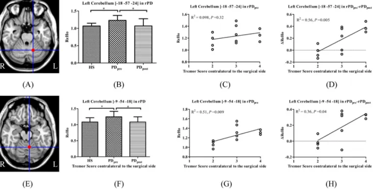
Moreover, the left cerebellum (peak coordinate [-18, -57, -24] and [-9, -54, -18]) in rPD are identified as the brain areas of significantΔReHo due to surgery, and correlated withΔtremor scores. Compared with HS, the left cerebellum showed significantly increased ReHo in rPDpre,
which was significantly decreased in rPDpost(Fig 4).
Discussion
In this study, the ReHo method was used to explore short-term (7 days) brain functional changes after unilateral VIM thalamotomy and to investigate its efficacy on PD tremor sup-pression. Our results reveal that the impact of unilateral VIM thalamotomy is characterized in the frontal, parietal, temporal, occipital regions, basal ganglia, thalamus, and cerebellum. The Fig 3. Brain areas which demonstrated altered ReHo relative to HS and correlated with tremor.(A) left cerebellum_4_5 in rPD; (E) right STG in rPD; (I) cerebellum vermis_3 in lPD; (B, F, J) ReHo among HS, PDpre, and PDpost; (C, G, K) ReHo in PDprecorrelated with tremor; (D, H, L)ΔReHo
correlated with tremor.
specific area that is associated with PD tremor and altered ReHo due to thalamic ablation may be the cerebellum. However, the change in pattern does not imply conformity in rPD and lPD.
Altered ReHo in the cerebellum
The tremor and cerebellar involvement are discussed in PD. It is suggested that a secondary dysfunction in the cerebello-thalamo-cortical (CTC) circuit is responsible for the occurrence of resting tremor, involving the VIM of thalamus, motor cortex, and cerebellum [3]. Systematic review suggested that the role of the cerebellum in PD tremor is a combination of pathological and compensatory effects [10].
Our study reports multiple clusters in the cerebellum in PDprewhere ReHo increased
signif-icantly relative to HS, such as bilateral cerebellum_6, vermis_3, and right cerebellum_crust1 in rPD, as well as right cerebellum_6 in lPD. Previous studies have documented that regional hypermetabolism in the basal ganglia, cerebellum, and primary motor cortex [22] and func-tional connectivity increase within the cerebello-thalamic circuit [3] in TD PD. Zhang et al. [23] suggest common“overheated”functional activities in TD PD, which is in accordance with our results. Our study provides evidence that the hyperactivation in the cerebellum is associ-ated with tremor.
The VIM nucleus of the thalamus is one of the targets for ablation and stimulation to sup-press PD tremor [9]. Cerebello-thalamic excitatory afferents terminate here and project to motor cortical areas [10]. Clinical studies which interfered in the cerebello-thalamic circuit [24] could effectively suppress resting tremor. A review describes it in terms of functional normaliza-tion [10]. Compared with PDpre, our study showed multiple clusters in the cerebellum where
ReHo reduced significantly in PDpost, such as left cerebellum_4_5, left cerebellum_crust1, left
cerebellum_6, and left cerebellum_10 in rPD and right cerebellum_6, bilateral cerebellum_8 in Fig 4. Brain areas which showed significantΔReHo in PDpostversus PDpreand correlated with tremor.(A) left cerebellum_6 in rPD; (E) left cerebellum_4_5 in rPD; (B, F) ReHo among HS, PDpre, and PDpost; (C, G) ReHo in PDprecorrelated with tremor; (D, H)ΔReHo correlated with tremor.
lPD. The decreased ReHo in the cerebellum after the VIM ablation supports a functional nor-malization. Of note, with the administration of thalamotomy, the ReHo in the cerebellum of the PD patients reduced, but was still higher than that of healthy subjects.
In order to explore the neural basis of ReHo alteration in the cerebellum due to thalamic ablation, we distinguished its effect on PD tremor from breakage of cerebello-thalamic fibers. Hoshi et al. [25] applied transneuronal transport of rabies virus in macaques, indicating that the outflow pathway was from deep cerebellar nuclei to thalamus via dentate neurons. In our study, we roughly observed that the ReHo in the same right cerebellum_8 decreased in lPDpost
relative to lPDpreand HS. There is a high probability that the decreased ReHo here after surgery
is due to the lesion of the cerebello-thalamic fibers. This finding highlights the important role of cerebello-thalamic circuits in understanding PD tremor, and indicates that tractography studies should be drawn in future.
We obtained intersections at different contrasts to discriminate the possible brain areas that may mediate tremor alleviation induced by surgery. We identified that in the left cerebel-lum_4_5 (peak coordinate [-12, -54, -21]) and vermis_3 (peak coordinate [3, -45, -15]), ReHo values relative to HS and were positively correlated with tremor scores in rPD and lPD, respec-tively. Afterwards, although there was slightly reduced amplitude of ReHo in vermis_3 in lPD after surgery, which was not statistically significant,ΔReHo was positively correlated withΔ tre-mor scores in rPD and lPD. The left cerebellum_6 (peak coordinate [-18, -57, -24]) and left cer-ebellum_4_5 (peak coordinate [-9, -54, -18]) revealed significantΔReHo due to surgery, and positively correlated withΔtremor scores in rPD instead of lPD. However, only the ReHo of the left cerebellum_4_5 (peak coordinate [-9, -54, -18]) in rPDprewas positively correlated with
tremor scores. Together, these findings favor the view of a critical role for the cerebellum in the generation of Parkinsonian tremor. The left cerebellum anterior lobe may mediate tremor alle-viation caused by right-sided thalamotomy. Compared with right-sided surgery, a complex mechanism may underlie ReHo changes in the right cerebellum after left-sided thalamotomy in PD.
Altered ReHo in the cortical areas and basal ganglia
According to the pathological theory of Braak et al. [26], PD involves the functional alter-ation of whole brain networks rather than localized neurodegeneralter-ation. Tessitore et al. [27] reported reduced resting state functional connectivity of right MTG and bilateral IPL within the default mode network (DMN) in PD, compared with HS. Besides DMN, specific changes have been found in the fronto-parietal, sensorimotor, visual and auditory networks [16]. In our study, altered ReHo was found in the right MFG, right MTG and right STG in rPD, as well as in the frontal (i.e. left SFG, right IFG, right MFG, right precentral gyrus) cortex, pari-etal (i.e. right postcentral gyrus, right SPL), right MTG, occipital (i.e. left SOG, bilateral MOG) cortex, and ACC in lPD, relative to HS, which is coherent with previous studies [28,29].
In addition, the cerebello-thalamo-cortical (i.e. cerebellum, thalamus, SM1, and M1) and striatal-thalamo-cortical (i.e. striatum, thalamus, SMA, SM1, M1) circuits are distinct, but with considerable overlapping in the motor cortical areas. Fukuda et al. [30] performed PET study combined with triaxial accelerometry to examine the effect of thalamic stimulation on PD tremor. The result showed that the activation in SM1 was associated with tremor, indicating that the interference with cerebello-thalamo-cortical circuits (VIM of the thalamus) may mod-ulate the striatal-thalamo-cortical circuits at the cortical level. Similarly, we detected the ReHo increases in the bilateral postcentral gyri and decreases in the bilateral caudate in lPDpost
Altered ReHo in PD
postversus HS
The PDpostversus HS contrast comparison is complex in this study. Ideally, there are no
differ-ences between PDpostand HS if patients with PD have fully recovered, as they are exactly the
same as HS. In fact, our study found ReHo changes in the frontal, parietal, limbic, and insular cortices. There are several confounding factors which need to be taken into account, such as the surgical brain tissue lesion, surrounding edema and inflammation, and more importantly, the surgical impact on PD tremor (positive aspect) and side effects (negative aspect). Unfortu-nately, we could not identify the specific neuro-mechanism underlying the PDpostrelative to
HS based on current data.
Limitations
There are two main issues that need to be considered in future. Firstly, PD is a common neuro-degenerative disease with a high degree of heterogeneity [31]. However, the current sample size is relatively small, which limits the power of the study. Secondly, although our study identified cerebellum involvement, it did not clarify the possible mechanism underlying tremor genera-tion and alleviagenera-tion by thalamic ablagenera-tion. Indeed, our conclusion is drawn based on the differ-ences of 7 days; a longitudinal study is needed to provide insights on the long-term impact of thalamic ablation on PD tremor.
Conclusion
This study provides evidence that the impact of thalamotomy is observed in the cortical, sub-cortical regions and cerebellum. Altered activation in the cerebellum may mediate generation and alleviation of PD tremor. However, the role of the cerebellum may be complex. Cerebellum involvement is far beyond cerebello-thalamic tract breakage. Further studies are needed with a large sample size, longitudinal observation, and multi-modality imaging analysis.
Supporting Information
S1 Text. Surgical procedure of VIM thalamotomy.
(DOCX)
S1 Table. ReHo differences of PD patients between the pre- and post-surgical conditions.
(DOCX)
S2 Table. ReHo differences between HS and PD patients in the pre-surgical condition.
(DOCX)
S3 Table. ReHo differences between HS and PD patients in the post-surgical condition.
(DOCX)
S4 Table. Brain areas showing significant correlations between ReHo in PDpreand tremor
score contralateral to surgical side.
(DOCX)
Acknowledgments
Author Contributions
Conceived and designed the experiments: JZ FL GW. Performed the experiments: ZW JL JD. Analyzed the data: ZW JD FL GW. Contributed reagents/materials/analysis tools: JZ. Wrote the paper: ZW JZ.
References
1. Jankovic J. Parkinson's disease: clinical features and diagnosis. J Neurol Neurosurg Psychiatry. 2008; 79(4):368–76. doi:10.1136/jnnp.2007.131045PMID:18344392.
2. Hacker CD, Perlmutter JS, Criswell SR, Ances BM, Snyder AZ. Resting state functional connectivity of the striatum in Parkinson's disease. Brain. 2012; 135(Pt 12):3699–711. doi:10.1093/brain/aws281
PMID:23195207; PubMed Central PMCID: PMC3525055.
3. Helmich RC, Janssen MJ, Oyen WJ, Bloem BR, Toni I. Pallidal dysfunction drives a cerebellothalamic circuit into Parkinson tremor. Annals of neurology. 2011; 69(2):269–81. doi:10.1002/ana.22361PMID:
21387372.
4. Zhang JQ, Wei LQ, Hu XF, Xie B, Zhang YL, Wu GR, et al. Akinetic-rigid and tremor-dominant Parkin-son's disease patients show different patterns of intrinsic brain activity. Parkinsonism Relat D. 2015; 21 (1):23–30. doi:10.1016/j.parkreldis.2014.10.017WOS:000348259600004.
5. Helmich RC, Hallett M, Deuschl G, Toni I, Bloem BR. Cerebral causes and consequences of parkinso-nian resting tremor: a tale of two circuits? Brain. 2012; 135(Pt 11):3206–26. doi:10.1093/brain/aws023
PMID:22382359; PubMed Central PMCID: PMC3501966.
6. Rodriguez-Oroz MC, Jahanshahi M, Krack P, Litvan I, Macias R, Bezard E, et al. Initial clinical manifes-tations of Parkinson's disease: features and pathophysiological mechanisms. Lancet Neurol. 2009; 8 (12):1128–39. doi:10.1016/S1474-4422(09)70293-5PMID:19909911.
7. Hassler R. Anatomy of the thalamus. In: Schaltenbrand G, editor. Introduction to Stereotaxis with an Altas of the Human Brain. Stuttgart: Thieme; 1959. p. 230–90.
8. Dwarakanath S, Zafar A, Yadav R, Arivazhagan A, Netravathi M, Sampath S, et al. Does lesioning sur-gery have a role in the management of multietiological tremor in the era of Deep Brain Stimulation? Clin Neurol Neurosur. 2014; 125:131–6. doi:10.1016/j.clineuro.2014.07.016WOS:000343346900025. 9. Duval C, Daneault JF, Hutchison WD, Sadikot AF. A brain network model explaining tremor in
Parkin-son's disease. Neurobiol Dis. 2016; 85:49–59. doi:10.1016/j.nbd.2015.10.009PMID:26459110. 10. Wu T, Hallett M. The cerebellum in Parkinson's disease. Brain. 2013; 136(Pt 3):696–709. doi:10.1093/
brain/aws360PMID:23404337.
11. Kendall M, Gibbons JD. Rank Correlation Methods. Oxford: Oxford Univ. Press; 1990.
12. Zang Y, Jiang T, Lu Y, He Y, Tian L. Regional homogeneity approach to fMRI data analysis. Neuro-Image. 2004; 22(1):394–400. doi:10.1016/j.neuroimage.2003.12.030PMID:15110032.
13. Zuo XN, Xu T, Jiang L, Yang Z, Cao XY, He Y, et al. Toward reliable characterization of functional homogeneity in the human brain: preprocessing, scan duration, imaging resolution and computational space. NeuroImage. 2013; 65:374–86. doi:10.1016/j.neuroimage.2012.10.017PMID:23085497; PubMed Central PMCID: PMC3609711.
14. Jiang L, Zuo XN. Regional Homogeneity: A Multimodal, Multiscale Neuroimaging Marker of the Human Connectome. The Neuroscientist: a review journal bringing neurobiology, neurology and psychiatry. 2015. doi:10.1177/1073858415595004PMID:26170004.
15. Zhang J, Wei L, Hu X, Xie B, Zhang Y, Wu GR, et al. Akinetic-rigid and tremor-dominant Parkinson's disease patients show different patterns of intrinsic brain activity. Parkinsonism Relat Disord. 2015; 21 (1):23–30. doi:10.1016/j.parkreldis.2014.10.017PMID:25465747.
16. Prodoehl J, Planetta PJ, Kurani AS, Comella CL, Corcos DM, Vaillancourt DE. Differences in brain acti-vation between tremor- and nontremor-dominant Parkinson disease. JAMA neurology. 2013; 70 (1):100–6. doi:10.1001/jamaneurol.2013.582PMID:23318516; PubMed Central PMCID: PMC3645004.
17. Helmich RC, Toni I, Deuschl G, Bloem BR. The pathophysiology of essential tremor and Parkinson's tremor. Curr Neurol Neurosci Rep. 2013; 13(9):378. doi:10.1007/s11910-013-0378-8PMID:
23893097.
19. Chao-Gan Y, Yu-Feng Z. DPARSF: A MATLAB Toolbox for "Pipeline" Data Analysis of Resting-State fMRI. Front Syst Neurosci. 2010; 4:13. doi:10.3389/fnsys.2010.00013PMID:20577591; PubMed Cen-tral PMCID: PMCPMC2889691.
20. Song XW, Dong ZY, Long XY, Li SF, Zuo XN, Zhu CZ, et al. REST: a toolkit for resting-state functional magnetic resonance imaging data processing. PLOS One. 2011; 6(9):e25031. doi:10.1371/journal. pone.0025031PMID:21949842; PubMed Central PMCID: PMC3176805.
21. Van Dijk KR, Sabuncu MR, Buckner RL. The influence of head motion on intrinsic functional connectiv-ity MRI. NeuroImage. 2012; 59(1):431–8. doi:10.1016/j.neuroimage.2011.07.044PMID:21810475; PubMed Central PMCID: PMC3683830.
22. Mure H, Hirano S, Tang CC, Isaias IU, Antonini A, Ma Y, et al. Parkinson's disease tremor-related meta-bolic network: characterization, progression, and treatment effects. NeuroImage. 2011; 54(2):1244–53. doi:10.1016/j.neuroimage.2010.09.028PMID:20851193; PubMed Central PMCID: PMC2997135. 23. Zhang D, Liu X, Chen J, Liu B, Wang J. Widespread increase of functional connectivity in Parkinson's
disease with tremor: a resting-state FMRI study. Frontiers in aging neuroscience. 2015; 7:6. doi:10. 3389/fnagi.2015.00006PMID:25691867; PubMed Central PMCID: PMC4315047.
24. Deiber MP, Pollak P, Passingham R, Landais P, Gervason C, Cinotti L, et al. Thalamic stimulation and suppression of parkinsonian tremor. Evidence of a cerebellar deactivation using positron emission tomography. Brain. 1993; 116 (Pt 1):267–79. PMID:8453462.
25. Hoshi E, Tremblay L, Feger J, Carras PL, Strick PL. The cerebellum communicates with the basal gan-glia. Nature neuroscience. 2005; 8(11):1491–3. doi:10.1038/nn1544PMID:16205719.
26. Braak H, Del Tredici K, Rub U, de Vos RA, Jansen Steur EN, Braak E. Staging of brain pathology related to sporadic Parkinson's disease. Neurobiol Aging. 2003; 24(2):197–211. PMID:12498954. 27. Tessitore A, Esposito F, Vitale C, Santangelo G, Amboni M, Russo A, et al. Default-mode network
con-nectivity in cognitively unimpaired patients with Parkinson disease. Neurology. 2012; 79(23):2226–32. doi:10.1212/WNL.0b013e31827689d6WOS:000312273800006. PMID:23100395
28. Herz DM, Eickhoff SB, Lokkegaard A, Siebner HR. Functional neuroimaging of motor control in Parkin-son's disease: a meta-analysis. Hum Brain Mapp. 2014; 35(7):3227–37. doi:10.1002/hbm.22397
PMID:24123553.
29. Tahmasian M, Bettray LM, van Eimeren T, Drzezga A, Timmermann L, Eickhoff CR, et al. A systematic review on the applications of resting-state fMRI in Parkinson's disease: Does dopamine replacement therapy play a role? Cortex. 2015; 73:80–105. doi:10.1016/j.cortex.2015.08.005
WOS:000367860300009. PMID:26386442
30. Fukuda M, Barnes A, Simon ES, Holmes A, Dhawan V, Giladi N, et al. Thalamic stimulation for parkin-sonian tremor: correlation between regional cerebral blood flow and physiological tremor characteris-tics. Neuroimage. 2004; 21(2):608–15. doi:10.1016/j.neuroimage.2003.09.068PMID:14980563. 31. Wu T, Long X, Zang Y, Wang L, Hallett M, Li K, et al. Regional homogeneity changes in patients with
Parkinson's disease. Hum Brain Mapp. 2009; 30(5):1502–10. doi:10.1002/hbm.20622PMID: