Arq Neuropsiquiatr 2004;62(1):167-169
Neurosurgery Department, Santa Casa de Belo Horizonte/Faculdade de Ciências Médicas de Minas Gerais (FCMMG), Belo Horizonte MG, Brazil: 1Neurosurgery Resident, Santa Casa de Belo Horizonte; 2M.D.,PhD., Professor at the Department of Neurosurgery,
FCM-MG; 3M.D., Professor at the Department of Neurosurgery, FCMMG.
Received 27 December 2002, received in final form 27 July 2003. Accepted 5 September 2003. Dr. Ricardo Souza Quadros - Rua Monte Sião 212/302 - 30240-050 Belo Horizonte MG - Brasil.
MULTIFOCAL OSTEOCLASTOMA OF THE SKULL
Case report
Ricardo Souza Quadros
1, Atos Alves de Sousa
2,
Gervásio Telles Cardoso Carvalho
3, Marcos Antônio Dellaretti Filho
1ABSTRACT - We describe the case of a 35 years old man with a nonspecific complaint of a slow growing solid mass in the frontal region. Radiological exams evidenced two more lesions : in the superior and lat-eral walls of the orbit. Treated with total excision of the lesions and a cranioplastic procedure at the same act, with favorable outcome. Microscopic findings suggested giant cell tumor in the three lesions that was confirmed by imunohistochemical examination.
KEY WORDS: osteoclastoma, giant cell tumor, multifocal, skull.
Osteoclastoma craniano multifocal: relato de caso
RESUMO - Descrevemos o caso de homem de 35 anos, com história de lesão endurecida de crescimento progressivo na região frontal direita. Exames radiológicos evidenciaram duas outras lesões: uma na parede superior e outra na parede lateral da órbita direita. Submetido a exérese total das lesões com margem de segurança e cranioplastia no mesmo tempo cirúrgico. O exame anatomo patológico sugeriu tumor de célu-las gigantes nas três lesões, diagnóstico confirmado pela imuno-histoquímica.
PALAVRAS-CHAVE: osteoclastoma, tumor de células gigantes, multifocal, crânio.
Osteoclastoma or giant cell tumor is a benign but often locally aggressive neoplasm of the bone. It occurs more frequently between the third and fifth decades of life, sometimes after patients are fifty years old and rarely after they are sixty years old. This is one of the few osseous neoplasms sho-wing higher occurrence in women1-3. Giant cell
tu-mor accounts for about 5 % of biopsed primary bo-ne tumors and about 20% of benign bobo-ne tumors. Most of the osteoclastomas are located near the articular end of tubular bones. The skull is rarely involved, only 1 to 2% of the cases described in the literature3-6. We describe a case of osteoclastoma.
CASE
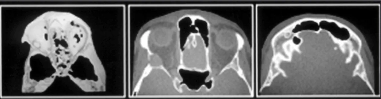
A 35 year-old white man presented with a 1 year his-tory of a slow growing solid mass at the right frontal region with about 2 cm of diameter. The lesion was sur-gically resected in another hospital, but probably par-tially because 4 months later it was back. The CT scan at our service showed two other lesions in the skull: in the superior and lateral walls of the right orbit (Figs 1
and 2). All the three lesions were completely resected and a cranioplastic procedure was performed at the sa-me act. Microscopic findings suggested giant cell tumor in the three lesions, and that was confirmed by imuno-histochemical exams (Fig 3).
DISCUSSION
Giant cell tumor or osteoclastoma is a rare tu-mor, representing only 3 - 7% of all bone tumors. About 70 - 90% are located at or close the extrem-ities of long bones, 10 - 30% are located in the sa-crum, region of the knee, small bones of the hands and vertebras, 2% affect the skull, more common-ly the mandible and maxilla1,2,7,8. A review of the
literature demonstrated 7 series of giant cell tumors totalizing 2404 cases1,2,6-10, and 24 (1%) occurred
in the skull, excluding the ones found in the man-dible. When located in the skull they are more frequently found at the sphenoid and temporal bones, and rarely at the ethmoidal, frontal or occi-pital bones5.
occur-168 Arq Neuropsiquiatr 2004;62(1)
ring in the hands and feet10. The multifocal
osteo-clastoma should not be confused with giant cell re-parative granuloma, which is often multifocal and presents a high incidence of recidivation. In an ex-tensive review of the literature no other case of multifocal osteoclastoma of the skull was found, and we believe this is the first published case. The most common radiological appearance of osteo-clastoma in the skull is an expanding and/or lytic neoplasm that may appear to extend into the
sur-rounding soft tissues, dura or sinuses. The rarity of these lesions explains the few numbers of MRI and CT descriptions of skull giant cell tumors7,11.
The presence of giant cells in the histochemical study does not confirm the diagnosis. The differen-tial diagnosis includes: giant cell reparative granulo-ma, benign fibrous histiocytoma and osteosarco-ma with prominent giant cells6. The most
com-mon macroscopic appearance of osteoclastoma is a grayish, soft lesion with small cysts and occasion-Fig 1. CT showing lesions in the superior and lateral walls of the orbit and right frontal bone.
Fig 2. Bone scyntilography showing the three lesions.
Arq Neuropsiquiatr 2004;62(1) 169
al necrotic areas. Our patient presented in the mi-croscopic exam: multinucleated giant cells, mono-nuclear cells and few histiocytes with hemossiderin deposition. The imunohistochemical study con-firmed CD68, a bone giant cell marker.
More than 60% of giant cell tumors will reci-divate if treated with partial resection. In addition to local intraosseous recurrence, surgery-related contamination may lead to implantation of cells of the tumor in the perilesional soft tissues in 10 - 15% of patients. The use of radiotherapy should probably be limited to lesions that cannot be sub-jected to total excision, and that is an issue for fur-ther literature discussion12-15.
REFERENCES
1. Campanacci M, Baldini N, Boriani S, Sudanese A. Giant-cell tumor of bone. J Bone Joint Surg Am 1987;69:106-114.
2. Unni KK. Giant cell tumor. In Unni KK (ed) Dahlin’s bone tumors. gen-eral aspects and data on 11,087 cases. Ed 5. Philadelphia: Lippincott-Raven: 1996;263-289.
3. Osaka S, Toriyama M, Taira K, Sano S, Saotome K. Analysis of giant cell tumor of bone with pulmonary metastases. Clin Orthop 1997;335:253-261. 4. Kuzeyli K, Baykal S, Duru S, Ceylan S, Aktürk F. Giant cell tumor of
the fronto-parietal bone. Neurosurg Rev 1995;18:265-268.
5. Vernet O, Ducrey N, Déruaz JP,Tribolet N. Giant cell tumor of the orbit. Neurosurgery 1993;5:848-852.
6. Goldenberg RR, Campbell CJ, Bonfiglio M. Giant-cell tumor of bone. J Bone Joint Surg Am 1970;52:619-664.
7. Mulder JD, Kroon HM, Schütte HE, Taconis WK. Giant cell tumor. In: Kroon HM (ed) Radiologic atlas of bone tumors. Amsterdam: Elsevier, 1993;493-490.
8. Dahlin DC, Unni KK. Malignancy in giant cell tumor of bone. In: Unni KK (ed) Bone tumors: general aspects and data on 8,542 cases. Ed 4. Springfield: Charles C Thomas, 1986;337-345.
9. Sanerkin NG. Malignancy, aggressiveness, and recurrence in giant cell tumor of bone. Cancer 1980;46:1641-1649.
10. Dahlin DC, Cupps RE, Johnson EW. Giant-cell tumor. Cancer 1970; 25:1061-1070.
11. McInerney DP, Middlemiss JH. Giant-cell tumour of bone. Skeletal Radiol 1978;2:195-204.
12. Hutter RV, Worcester JN, Francis KC, Foote FW. Benign and malignant giant cell tumors of bone. Cancer 1962;15:653-690.
13. Bertoni F, Unni KK, Beabout JW, Ebersold MJ. Giant cell tumor of the skull. Cancer 1992;70:1124-1132.
14. Arseni C, Horvath L, Maretsis M, Carp N. Giant cell tumors of the cal-varia. J Neurosurg 1975;42:535-540.