345
ARTICLE DOI: 10.1590/0004-282X20130036
Clinical spectrum of early onset cerebellar
ataxia with retained tendon reflexes:
an autosomal recessive ataxia not to be missed
Espectro clínico da ataxia cerebelar de início precoce com reflexos mantidos: uma ataxia
autossômica recessiva para não ser esquecida
José Luiz Pedroso, Pedro Braga-Neto, Irapuá Ferreira Ricarte, Marcus Vinicius Cristino Albuquerque, Orlando Graziani Povoas Barsottini
Autosomal recessive cerebellar ataxias (ARCA) are a het-erogeneous group of neurological disorders characterized by degeneration or abnormal development of cerebellum and spi-nal cord, autosomal recessive inheritance and early onset be-ginning before the age of 20 years. his group encompasses a large number of unusual diseases and may be considered a
diagnostic challenge1. he most frequent ARCA is Friedreich
ataxia (FA), but other diseases include ataxia with vitamin E
deiciency, ataxia telangiectasia, ataxia with ocular apraxia type 1 and type 2, autosomal recessive spastic ataxia of Charlevoix Saguenay (ARSACS), cerebrotendineous xanthomatosis, abet-alipoproteinemia, Refsum disease and Marinesco-Sjögren syn-drome. In most cases, diagnosis may be performed based on
clinical and genetic evaluation1.
In 1981, Anita Harding described the clinical and genetic features of 20 families in which afected individuals had a
Department of Neurology, Ataxia Unit, Universidade Federal de São Paulo (UNIFESP), São Paulo SP, Brazil.
Correspondence: José Luiz Pedroso; Department of Neurology, Ataxia Unit, Universidade Federal de São Paulo; Rua Botucatu 740; 04023-900 São Paulo SP - Brasil; E-mail: [email protected]
Conflict of interest: There is no conlict of interest to declare.
Received 26 October 2012; Received in inal form 14 November 2012; Accepted 21 November 2012.
ABSTRACT
Autosomal recessive cerebellar ataxias are a heterogeneous group of neurological disorders. In 1981, a neurological entity comprised by early onset progressive cerebellar ataxia, dysarthria, pyramidal weakness of the limbs and retained or increased upper limb relexes and knee jerks was described. This disorder is known as early onset cerebellar ataxia with retained tendon relexes. In this article, we aimed to call attention for the diagnosis of early onset cerebellar ataxia with retained tendon relexes as the second most common cause of autosomal recessive cerebellar ataxias, after Friedreich ataxia, and also to perform a clinical spectrum study of this syndrome. In this data, 12 patients from dif-ferent families met all clinical features for early onset cerebellar ataxia with retained tendon relexes. Dysarthria and cerebellar atrophy were the most common features in our sample. It is uncertain, however, whether early onset cerebellar ataxia with retained tendon relexes is a homogeneous disease or a group of phenotypically similar syndromes represented by different genetic entities. Further molecular studies are required to provide deinitive answers to the questions that remain regarding early onset cerebellar ataxia with retained tendon relexes.
Key words: ataxias, autosomal recessive cerebellar ataxias, early onset cerebellar ataxia with retained tendon relexes, EOCA.
RESUMO
As ataxias cerebelares autossômicas recessivas são um grupo heterogêneo de doenças neurológicas. Em 1981, foi descrita uma entidade neurológica incluindo ataxia cerebelar progressiva de início precoce, disartria, liberação piramidal e manutenção ou aumento dos relexos tendíneos nos membros superiores e inferiores. Essa síndrome é conhecida como ataxia cerebelar de início precoce com relexos mantidos. Neste artigo, o objetivo foi chamar a atenção para o diagnóstico de ataxia cerebelar de início precoce com relexos mantidos como a segunda causa mais comum de ataxia cerebelar autossômica recessiva, após a ataxia de Friedreich, e também realizar um estudo do espectro clínico da síndrome. Doze pacientes de diferentes famílias preencheram os critérios clínicos para ataxia cerebelar de início precoce com relexos mantidos. Disartria e atroia cerebelar foram as características mais frequentes. No entanto, não há consenso se a ataxia cerebelar de início precoce com relexos mantidos é uma doença homogênea ou um grupo de síndromes com fenótipos semelhantes representadas por dife-rentes entidades genéticas. Estudos moleculares futuros são necessários para fornecer respostas deinitivas para as questões pendentes em relação à ataxia cerebelar de início precoce com relexos mantidos.
346 Arq Neuropsiquiatr 2013;71(6):345-348
progressive cerebellar ataxia developing within the irst two decades, associated with dysarthria, pyramidal weakness of the limbs and retained or increased upper limb relexes and knee jerks. his disorder is known as early onset cerebel-lar ataxia with retained tendon relexes (EOCA) or Harding
ataxia, and other case series were reported later2. EOCA is
clinically distinct from FA, with signiicant diferences
be-tween those neurological conditions2. Several reviews have
pointed out that, although FA is the most common reces-sive ataxia worldwide, the diagnosis of this condition may be viewed with caution when brisk tendon relexes are present. his is because FA presents with loss of tendon relexes in 62
to 86% of patients3,4.
Frequently, EOCA has been excluded from ARCA list, since clinical features and genetic deinition are not very well understood. In this article, we aimed to call attention for the diagnosis of EOCA as the second most common cause of ARCA, and also to perform a clinical spectrum study of this syndrome.
METHODS
A retrospective review from 486 medical records of pa-tients attending at the Ataxia Unit, in the Universidade Federal de São Paulo, from February 2008 to September 2012, was performed. During this period, patients with diferent subtypes of cerebellar ataxias were followed-up in order to determine clinical and genetic diagnosis. Patients were di-vided into ive categories based on age at onset, familial his-tory, progression and laboratorial and genetic tests: autoso-mal dominant spinocerebellar ataxia (SCA), ARCA, sporadic ataxias, congenital ataxias and mithocondrial ataxias.
Patients with early onset symptoms were investigated for ARCA. Twelve patients from diferent families met all clinical fea-tures for EOCA. All patients had an early onset of ataxia symptoms (before 25 years old) with normal or brisk relexes, and underwent biochemical analysis including albumin, alphafetoprotein and vita-min E, genetic test for FA, and brain imaging, in order to rule out other ARCA.
Information on age, age at onset, disease duration, consan-guinity, ataxia severity (International Cooperative Ataxia Rating Scale (ICARS) and Scale for the Assessment and Rating of Ataxia (SARA)), relexes, Babinski sign, spasticity, neuropathy (electro-neuromyography), nystagmus, dysarthria and cerebellar atro-phy were evaluated from medical records from all patients with suspected EOCA. All clinical details, including neurological ex-amination and ataxia scales, were evaluated by the same re-searcher (JLP).
RESULTS
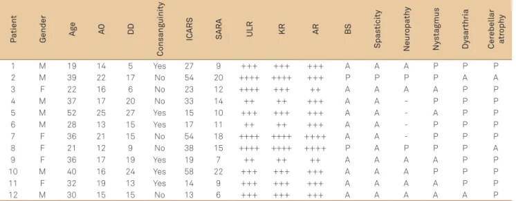
Table shows clinical and demographic features of the 12 patients with EOCA. Among patients with suspected EOCA, there was a slight male predominance (58.33%). he mean
age was 32.67±9.41, mean age at onset was 17.25±3.86, and
mean disease duration, 15.42±6.66 years. Regarding clinical
features, the mean score on ICARS was 31.09±17.50 and on
SARA was 12.82±5.38. Genetic test for FA was negative in all
patients. No correlation was found between ataxia severity (SARA and ICARS) and disease duration. Dysarthria and cer-ebellar atrophy were the most common features and were present in 10 of 12 patients (Fig 1).
FA was diagnosed in 37 patients and was the most common ARCA in our Ataxia Unit. EOCA was the second
P
a
tient
Gender A
ge AO DD
Consang
uinity
IC
ARS
S
ARA ULR KR AR BS
Spas
ticity
Neur
opa
th
y
Ny
s
tagmus
Dy
sar
thria
Cer
ebellar
a
tr
oph
y
1 M 19 14 5 Yes 27 9 +++ +++ +++ A A A P P P
2 M 39 22 17 No 54 20 ++++ ++++ +++ P P P P A A
3 F 22 16 6 No 23 12 ++++ +++ ++ A A A A P P
4 M 37 17 20 No 33 14 ++ ++ +++ A A - P P P
5 M 52 25 27 Yes 15 10 +++ +++ +++ A A - A P P
6 M 28 13 15 Yes 17 11 ++ ++ +++ A A - P P P
7 F 36 21 15 No 54 18 ++++ ++++ ++++ A A - P P P
8 F 21 12 9 No 38 15 ++++ ++++ ++++ P A P P P A
9 F 36 17 19 Yes 19 7 ++ ++ ++ A A A A P P
10 M 40 16 24 Yes 58 22 +++ +++ +++ A A A P P P
11 F 32 19 13 Yes 14 9 +++ +++ +++ A A A A P P
12 M 30 15 15 No 13 6 +++ +++ +++ A A A A A P
Table. Clinical features found in early onset ataxia with retained relexes.
347
José Luiz Pedroso et al. Early onset cerebellar ataxia with retained tendon relexes
most common cause of ARCA (12 patients), followed by ataxia with oculomotor apraxia type 2 (7 patients), atax-ia telangiectasatax-ia (4 patients), ARSACS (4 patients), ataxatax-ia with vitamin E deficiency (3 patients) and cerebrotendin-eous xanthomatosis (1 patient). There are several patients under investigation for ataxia with oculomotor apraxia type 1 (3 patients), coenzyme Q10 deficiency (2 patients), mithocondrial ataxia (3 patients) and Marinesco-Sjögren syndrome (1 patient). No patients with Refsun disease and abetalipoproteinemia were confirmed. Congenital ataxias were not included in this list.
DISCUSSION
Our study highlights how relevant is to consider EOCA in the differential diagnosis of ARCA, since it might cor-respond to its second most common cause. An autosomal recessive inherited disorder is suggested, as half of the pa-tients had consanguineous parents. Additionally, dysar-thria was presented in almost all patients and might be considered a frequent neurological finding. We also ob-served that almost all patients presented with cerebellar atrophy on brain magnetic resonance imaging (MRI) stud-ies, in opposite to FA patients. Conversely, spasticity was an unusual clinical feature. No correlation was found be-tween ataxia severity and disease duration, which express that EOCA has a heterogeneous progression.
Although the phenotype of FA has classical findings, such as loss of tendon reflexes, sensory loss, scoliosis, foot deformity, diabetes mellitus and cardiac abnormali-ties, there are variations in the clinical presentation. These include the late onset Friedreich ataxia (LOFA) and also
Friedreich ataxia with retained reflexes (FARR)5,6. Thus,
the presence of retained reflexes does not exclude the
diagnosis of FA, and a genetic test is recommended
de-spite an atypical ARCA presentation7. No patient with
FARR phenotype was identified in our study. A normal brain magnetic resonance imaging (MRI) without cere-bellar atrophy is also a remarkable finding in FA patients
(Fig 2)8,9.
One of the largest series of EOCA described by Chio et al. evaluated patients for 50 years. They concluded that EOCA was the third most common cause of ARCA, after
FA and ataxia telangiectasia10. Klockgether et al.
evalu-ated 14 patients with EOCA and compared clinical, elec-trophysiological and MRI observations with FA patients. Sensory disturbances, foot deformity and scoliosis were encountered less frequently in EOCA than in FA patients. Electrophysiological findings in EOCA were variable and pointed out to an axonal degeneration in peripher-al nerves. Our study demonstrated that axonperipher-al neuropa-thy was present in only 25% of patients. Cerebellar atrophy was a frequent neuroimaging feature in all EOCA series, in
opposite to FA9. The demonstration of cerebellar atrophy
in the majority of EOCA patients supported the view that
EOCA was distinct from FA9. Additionally, some data also
has demonstrated that EOCA is characterized by a
hetero-geneous progression11,12.
Although past decade has seen great advances in un-raveling the biological basis of hereditary ataxias, knowl-edge on the genetic features of EOCA is still extremely restricted. Sporadic reports have demonstrated new mu-tations in recessive ataxias. For instance, SYNE1 mutation is a gene responsible for a recessive inherited pure
cerebel-lar ataxia13. Concerning that genetic test is not available
for the diagnosis of EOCA, we strongly recommend a bio-marker investigation, including albumin, alphafetoprotein and vitamin E, and genetic test for FA, in order to exclude
other potential ARCA14.
Fig 1. (A) Sagittal T1-weighted and (B) axial T1-weighted brain MRI disclosing marked global cerebellar atrophy in a patient with early onset ataxia with retained relexes.
348 Arq Neuropsiquiatr 2013;71(6):345-348
1. Embiruçu EK, Martyn ML, Schlesinger D, Kok F. Autosomal
recessive ataxias: 20 types, and counting. Arq Neuropsiquiatr 2009;67:1143-1156.
2. Harding AE. Early onset cerebellar ataxia with retained tendon relexes: a clinical and genetic study of a disorder distinct from Friedreich’s ataxia. J Neurol Neurosurg Psychiatry 1981;44:503-508.
3. Harding AE. Friedreich’s ataxia: a clinical and genetic study of 90 families with an analysis of early diagnostic criteria and intrafamilial clustering of clinical features. Brain 1981;104:589-620.
4. Durr A, Cossee M, Agid Y, et al. Clinical and genetic abnormalities in patients with Friedreich’s ataxia. N Engl J Med 1996;335:1169-1175.
5. Armani M, Zortea M, Pastorello E, et al. Friedreich’s ataxia: clinical heterogeneity in two sisters. Neurol Sci 2006;27:140-142.
6. Palau F, Espinós C. Autosomal recessive cerebellar ataxias. Orphanet J Rare Dis 2006;1:47.
7. Schöls L, Amoiridis G, Przuntek H, Frank G, Epplen JT, Epplen C. Friedreich’s ataxia. Revision of the phenotype according to molecular genetics. Brain 1997;120:2131-2140.
8. Fogel BL, Perlman S. Clinical features and molecular genetics of autosomal recessive cerebellar ataxias. Lancet Neurol 2007;6:245-257.
9. Klockgether T, Petersen D, Grodd W, Dichgans J. Early onset cerebellar ataxia with retained tendon relexes. Clinical, electrophysiological and MRI observations in comparison with Friedreich’s ataxia. Brain 1991;114:1559-1573.
10. Chio A, Orsi L, Mortara P, Schiffer D. Early onset cerebellar ataxia with retained tendon relexes: prevalence and gene frequency in an Italian population. Clin Genet 1993;43:207-211.
11. De Castro M, Cruz-Martínez A, Vílchez JJ, et al. Early onset cerebellar ataxia and preservation of tendon relexes: clinical phenotypes associated with GAA trinucleotide repeat expanded and non-expanded genotypes. J Peripher Nerv Syst 1999;4:58-62.
12. Filla A, De Michele G, Cavalcanti F, et al. Clinical and genetic heterogeneity in early onset cerebellar ataxia with retained tendon relexes. J Neurol Neurosurg Psychiatry 1990;53:667-670.
13. Gros-Louis F, Dupré N, Dion P, et al. Mutations in SYNE1 lead to a newly discovered form of autosomal recessive cerebellar ataxia. Nat Genet 2007;39:80-85.
14. Braga-Neto P, Dutra LA, Pedroso JL, Barsottini OG. Alpha-fetoprotein as a biomarker for recessive ataxias. Arq Neuropsiquiatr 2010;68:953-955.
15. Ozeren A, Arac N, Ulku A. Early-onset cerebellar ataxia with retained tendon reflexes. Acta Neurol Scand 1989;80:593-597.
16. de Bot ST, Willemsen MA, Vermeer S, Kremer HP, van de Warrenburg BP. Reviewing the genetic causes of spastic-ataxias. Neurology 2012;79:1507-1514.
17. Pedroso JL, Braga-Neto P, Abrahão A, et al. Autosomal recessive spastic ataxia of Charlevoix-Saguenay (ARSACS): typical clinical and neuroimaging features in a Brazilian family. Arq Neuropsiquiatr 2011;69:288-291.
References
Interestingly, although patients described in our sample mostly had brisk relexes, spasticity was present only in one
patient, similarly to other series15. his is a crucial issue, since
spasticity in a context of ARCA (also called hereditary spastic-ataxias) might suggest other rare neurological conditions, such as ARSACS, hereditary spastic paraplegia and autosomal
reces-sive spastic ataxia16,17. herefore, spasticity should be considered
a neurological hallmark against the diagnosis of EOCA.
On the whole, this article highlighted the importance to consider EOCA as the second most common ARCA. he lack of a genetic marker should not be a limitation to consider this syndrome. Also, we reinforce the relevance to exclude other ARCA, by performing a biochemical investigation and genet-ic test for FA in order to exclude FARR. Based on this data, the clinical spectrum of EOCA might include autosomal cessive inherited cerebellar ataxia, dysarthria, retained re-lexes associated or not with neuropathy and cerebellar atro-phy on brain MRI. It is uncertain, however, whether EOCA is a homogeneous disease or a group of phenotypically similar syndromes represented by diferent genetic entities. Further molecular studies are required to provide deinitive answers to the questions that remain regarding EOCA.
Fig 2. (A) Sagittal T1-weighted and axial T2-weighted brain MRI disclosing normal cerebellar volume, without atrophy, in a patient with Friedreich ataxia; (C) sagittal T1-weighted thoracic spine MRI of the same patient, showing atrophy of the spinal cord.