CLINICAL SCIENCE
Spinocerebellar ataxias – genotype-phenotype
correlations in 104 Brazilian families
He´lio A. G. Teive,IRenato P. Munhoz,IWalter O. Arruda,IIscia Lopes-Cendes,II Salmo Raskin,IIILineu C. Werneck,I Tetsuo AshizawaIV
IHospital de Clı´nicas, Federal University of Parana´, Internal Medicine Department, Neurology Service, Movement Disorders Unit, Curitiba/PR, Brazil. IIFaculty of Medical Sciences, Department of Medical Genetics, (UNICAMP), Campinas/SP, Brazil.IIIGenetika Laboratory, Curitiba/PR, Brazil.IVUniversity of
Florida, Department of Neurology, Gainesville, FL, USA.
OBJECTIVE:Spinocerebellar ataxias are neurodegenerative disorders involving the cerebellum and its connections. There are more than 30 distinct subtypes, 16 of which are associated with an identified gene. The aim of the current study was to evaluate a large group of patients from 104 Brazilian families with spinocerebellar ataxias.
METHODS: We studied 150 patients from 104 families with spinocerebellar ataxias who had received molecular genetic testing for spinocerebellar ataxia types 1, 2, 3, 6, 7, 8, 10, 12, 17, and dentatorubral-pallidoluysian atrophy. A statistical analysis of the results was performed using basic descriptive statistics and the correlation coefficient (r), Student’s t-test, chi-square test, and Yates’ correction. The statistical significance level was established forp-values
,0.05.
RESULTS:The results show that the most common subtype was spinocerebellar ataxia 3, which was followed by spinocerebellar ataxia 10. Moreover, the comparison between patients with spinocerebellar ataxia 3, spinocerebellar ataxia 10, and other types of spinocerebellar ataxia revealed distinct clinical features for each type. In patients with spinocerebellar ataxia 3, the phenotype was highly pleomorphic, although the most common signs of disease included cerebellar ataxia (CA), ophthalmoplegia, diplopia, eyelid retraction, facial fasciculation, pyramidal signs, and peripheral neuropathy. In patients with spinocerebellar ataxia 10, the phenotype was also rather distinct and consisted of pure cerebellar ataxia and abnormal saccadic eye movement as well as ocular dysmetria. Patients with spinocerebellar ataxias 2 and 7 presented highly suggestive features of cerebellar ataxia, including slow saccadic ocular movements and areflexia in spinocerebellar ataxia 2 and visual loss in spinocerebellar ataxia 7.
CONCLUSIONS: Spinocerebellar ataxia 3 was the most common subtype examined, followed by spinocerebellar ataxia 10. Patients with spinocerebellar ataxia 2 and 7 demonstrated highly suggestive features, whereas the phenotype of spinocerebellar ataxia 3 patients was highly pleomorphic and spinocerebellar ataxia 10 patients exhibited pure cerebellar ataxia. Epilepsy was absent in all of the patients with spinocerebellar ataxia 10 in this series.
KEYWORDS: Spinocerebellar Ataxias; Cerebellar Ataxia; Cerebellar Atrophy; SCA3; SCA10.
Teive HA, Munhoz RP, Arruda WO, Lopes-Cendes I, Raskin S, Werneck LC, Ashizawa T. Spinocerebellar ataxias – genotype-phenotype correlations in 104 Brazilian families. Clinics. 2012;67(5):443-449.
Received for publication onOctober 22, 2011;First review completed onNovember 21, 2011;Accepted for publication onJanuary 16, 2012
E-mail: [email protected]
Tel.: 55 41 3019-5060
INTRODUCTION
Spinocerebellar ataxias (SCA) represents a large and complex group of heterogeneous autosomal dominant degenerative diseases characterized by progressive degen-eration of the cerebellum and its afferent and efferent connections. Other nervous system structures are typically affected, including the basal ganglia, brainstem nuclei,
pyramidal tracts, the posterior column and anterior horn of the spinal cord, and the peripheral nerves (1-6).
SCA are clinically characterized by the presence of cerebellar gait and limb ataxia (with dysmetria, dysdiado-chokinesia, intention tremor, dysarthria, and nystagmus), which may be accompanied by extracerebellar signs, such as ophthalmoplegia, pyramidal signs, movement disorders (including parkinsonism, dystonia, myoclonus, and chorea), dementia, epilepsy, visual disorders (including pigmentary retinopathy), and peripheral neuropathy (1-6).
SCA have a prevalence ranging from 1 to 5 cases per 100,000 individuals (7,8), and disease onset typically occurs between 30 and 50 years of age, although cases developing before the age of 20 and after the age of 60 have also been Copyrightß2012CLINICS– This is an Open Access article distributed under
described (2-10). The degenerative neuropathological pro-cess has been studied in depth in transgenic mice and Drosophila models (2-4,6,8). Neuroimaging, particularly magnetic resonance imaging, typically reveals cerebellar atrophy with or without brainstem involvement (olivopon-tocerebellar atrophy) (2,3,4,6,9,10). Initially defined as autosomal dominant cerebellar ataxias, SCA was subse-quently classified by Harding into the following four basic types: type 1, which is characterized by cerebellar ataxia (CA) with optic atrophy, ophthalmoplegia, dementia, amyotrophy, and extrapyramidal signs; type 2, involving retinal degeneration and accompanied by ophthalmoplegia and extrapyramidal signs; type 3, which is considered a ‘‘pure’’ type of CA; and type 4, which may present as deafness and myoclonia in addition to CA (5).
With recent advances in molecular genetics, several SCA genetic loci and genes have been identified on different chromosomes, and these findings have enabled the applica-tion of an improved classificaapplica-tion system based on clinical as well as genetic data (2,3,4,6,9-12).
Thirty-two different types of SCA have been identified to date, and these are designated SCA1 to SCA36. Dentatorubral-pallidoluysian atrophy (DRPLA) has also been included in this group of disorders. The particular gene responsible for each type of disease has been identified for SCA types 1-3, 5-8, 10-15, 17, 27, 31, and DRPLA. The remaining types (SCA 4, 18-23, 25, 26, 28-30, 32, 33-35, and 36) have been defined by linkage studies, as the associated genes and mutations have not yet been identified (2,3,4,6,9,10,11,12). Finally, it should be mentioned that SCA types 9 and 24 remain undefined, and these two types have been reserved for disorders yet to be described in the literature. Additionally, SCA16 appears to be identical to SCA15, and SCAs 29 and 15, as well as SCAs 22 and 19, may represent different allelic forms of the same gene (2,4,6,11,12). SCA3 is the most common form of the disease worldwide, whereas the prevalence of types 1, 2, 6, 7, and 8 is varied depending upon the ethnic background of the population (1,2,3,4,9-12).
The objective of this study was to evaluate the genotype-phenotype correlations in 104 Brazilian families with SCA.
METHODS
Patients
We studied 150 patients from 104 families with SCA, which had been extracted from a large Brazilian series of 190 SCA families (382 clinically affected family members and 296 patients evaluated by molecular genetic testing) and had received positive test results at the Hospital de Clı´nicas of the Federal University of Parana´ in Curitiba, Brazil between 1989 and 2009. The inclusion criteria included the following: 1) a progressive clinical phenotype in which ataxia was the prominent symptom; and 2) a positive familial history compatible with autosomal dominant inheritance. All of the patients were evaluated by at least one neurologist (HT) using a standard protocol that assessed gender, age of onset, duration of the disease, mean CAG or ATTCT polynucleo-tide expansion, clinical manifestations (such as CA, ocular movement disorders, visual loss, movement disorders, pyramidal signs, peripheral nerve signs, cognitive dysfunc-tion, or epilepsy), neuroimaging findings (brain CT and/or MRI), and any additional findings. All of the patients underwent routine laboratory tests, including CSF studies,
electroencephalography (EEG), nerve conduction velocity with electromyography (NCV/EMG), and neuropsycholo-gical tests (NPT) in selected cases. Signed informed consents were obtained following a protocol approved by the Institutional Ethics Committee of the Federal University of Parana´.
Genetic Analysis
Molecular diagnostic testing for SCA types 1, 2, 3, 6, 7, and DRPLA was performed at the Centre for Research in Neuroscience at the Montreal General Hospital Research Institute of McGill University in Montreal, Quebec, Canada between 1994 and 1998 (Prof. Guy Roleau, Dr. Izabel Silveira, and Dr. Iscia Lopes-Cendes) and subsequently (from 1998 to 2000) at the Medical Genetic Department of UNICAMP (Prof. Iscia Lopes-Cendes). Molecular genetic tests were performed from 2003 to the present at the Molecular Biology Laboratory (Neurology Service, Hospital de Clı´nicas, Federal University of Parana´: Prof. Lineu C. Werneck) and Genetika Laboratory (Prof. Salmo Raskin) in Curitiba/PR (SCA 1, 2, 3, 6, 7, 8, 12, and 17). Molecular diagnostic tests for SCA10 were performed at the Baylor DNA Diagnostic Laboratory (Baylor College of Medicine, Houston, Texas, USA) (Prof. T. Ashisawa’s group) and subsequently at the Galveston Department of Neurology (University of Texas Medical Branch) (Prof. T. Ashizawa).
Peripheral blood was collected from patients and rela-tives, and genomic DNA was isolated from peripheral blood leukocytes using the standard technique (Sambrock et al. 1989, Cold Spring Harbor Laboratory Press, NY, USA) (13). Expanded triplet repeats in the genes responsible for SCA1, SCA2, SCA3, SCA6, SCA7, SCA8, SCA12, SCA17, and DRPLA were amplified using the primer pairs Rep-1/Rep-2 (Orr et al. 1993), F-1/F-2 (Sanpei et al. 1996; Pulst et al. 1996), MJD25/MJD52 (Kawaguchi et al. 1994), S-5-F1/S-5-R1 (Zhunchenko et al. 1997), 4U1024/4U716 (David et al. 1997), SCA8-F3/SCA8-R4 (Koob et al. 1999), PPP2R2B-A/ PPP2R2B-B (Holmes et al. 1999), TBP-F/TBP-R (Nakamura et al. 2001) and CTG-B37 (699)/(840) (Koide et al. 1994), respectively (2,3,4,6,9,10,11,12). The analysis of the ATTCT repeat region within the SCA10 gene was performed by polymerase chain reaction (PCR) amplification using the primers attct-L (5’-AGAAAACAGATGGCAGAATGA-3’) and attct-R (5’-GCCTGGGCAACATAGAGAGA-3’), as previously described (11,14). Patient DNA samples that showed a single, normal SCA10 allele by PCR underwent Southern blot analysis to assess large expansion (14).
consisting of 800-4,500 repeats (normal alleles have 10-21 repeats) (14).
Statistical Analysis
The statistical analysis of the results was performed using basic descriptive statistics and the correlation coefficient (r), Student’s t-test, chi-square test, and Yates’ correction. The level of statistical significance was set top-values,0.05.
RESULTS
Mutations were identified in 66.3% of the families and were most frequently identified for SCA3 (72.46%). SCA10 represented the second most common type of SCA (11.60%), followed by SCA2 (7.25%), SCA7 (4.34%), SCA1 (2.90%), and SCA6 (1.45%).
Of the affected patients examined, the male to female ratio was 1:1.2, and the age at onset ranged from 22 to 60 years with a mean onset at 35 years¡6.2 years of age. Also,
the average length of time with disease for these patients was 13.6¡10.7 years (variance of 1-32 years).
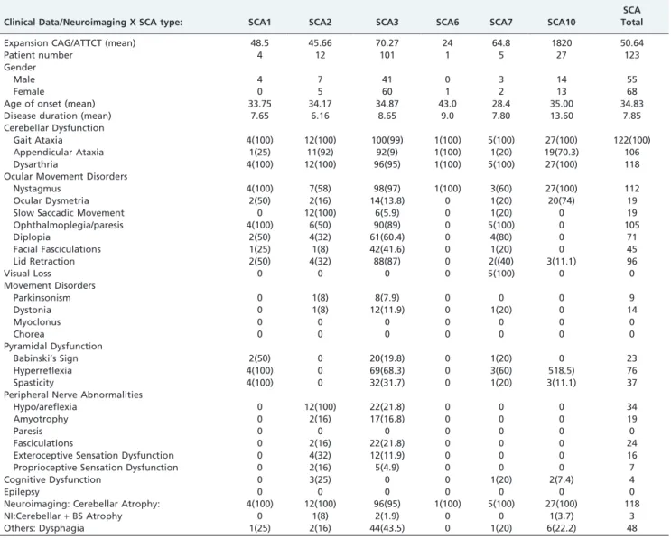
Table 1 shows a brief summary of the clinical data and the neuroimaging findings for SCA types 1,2,3,6,7,10 as well as overall SCA, excluding SCA10. This table also includes the demographic, clinical, and molecular data for each form of SCA.
In comparison to patients with SCA10, those with SCA3 presented more frequently with appendicular ataxia, ophthalmoplegia, diplopia, facial fasciculations, eyelid retraction (bulging eyes), severe hyperreflexia, severe hypo/areflexia, and muscular fasciculations. Significant differences (p,0.05) were also observed between these two SCA types regarding the presence of Babinski’s sign, spasticity, amyotrophy, and cognitive dysfunction. In contrast, ocular dysmetria was significantly more frequently seen in patients with SCA10.
In addition, when comparing patients with SCA10 to those with all other forms of SCA (excluding SCA3), this specific subtype presented more frequently with nystagmus and ocular dysmetria, whereas ophthalmoplegia/paresis, diplopia, slow saccadic movements, and hyporeflexia were observed less commonly in patients with SCA10 than other
Table 1 -SCA Types 1, 2, 3, 6, 7, 10 - Clinical Data/Neuroimaging.
Clinical Data/Neuroimaging X SCA type: SCA1 SCA2 SCA3 SCA6 SCA7 SCA10
SCA Total
Expansion CAG/ATTCT (mean) 48.5 45.66 70.27 24 64.8 1820 50.64
Patient number 4 12 101 1 5 27 123
Gender
Male 4 7 41 0 3 14 55
Female 0 5 60 1 2 13 68
Age of onset (mean) 33.75 34.17 34.87 43.0 28.4 35.00 34.83
Disease duration (mean) 7.65 6.16 8.65 9.0 7.80 13.60 7.85
Cerebellar Dysfunction
Gait Ataxia 4(100) 12(100) 100(99) 1(100) 5(100) 27(100) 122(100)
Appendicular Ataxia 1(25) 11(92) 92(9) 1(100) 1(20) 19(70.3) 106
Dysarthria 4(100) 12(100) 96(95) 1(100) 5(100) 27(100) 118
Ocular Movement Disorders
Nystagmus 4(100) 7(58) 98(97) 1(100) 3(60) 27(100) 112
Ocular Dysmetria 2(50) 2(16) 14(13.8) 0 1(20) 20(74) 19
Slow Saccadic Movement 0 12(100) 6(5.9) 0 1(20) 0 19
Ophthalmoplegia/paresis 4(100) 6(50) 90(89) 0 5(100) 0 105
Diplopia 2(50) 4(32) 61(60.4) 0 4(80) 0 71
Facial Fasciculations 1(25) 1(8) 42(41.6) 0 1(20) 0 45
Lid Retraction 2(50) 4(32) 88(87) 0 2((40) 3(11.1) 96
Visual Loss 0 0 0 0 5(100) 0 0
Movement Disorders
Parkinsonism 0 1(8) 8(7.9) 0 0 0 9
Dystonia 0 1(8) 12(11.9) 0 1(20) 0 14
Myoclonus 0 0 0 0 0 0 0
Chorea 0 0 0 0 0 0 0
Pyramidal Dysfunction
Babinski’s Sign 2(50) 0 20(19.8) 0 1(20) 0 23
Hyperreflexia 4(100) 0 69(68.3) 0 3(60) 518.5) 76
Spasticity 4(100) 0 32(31.7) 0 1(20) 3(11.1) 37
Peripheral Nerve Abnormalities
Hypo/areflexia 0 12(100) 22(21.8) 0 0 0 34
Amyotrophy 0 2(16) 17(16.8) 0 0 0 19
Paresis 0 0 0 0 0 0 0
Fasciculations 0 2(16) 22(21.8) 0 0 0 24
Exteroceptive Sensation Dysfunction 0 4(32) 12(11.9) 0 0 0 16
Proprioceptive Sensation Dysfunction 0 2(16) 5(4.9) 0 0 0 7
Cognitive Dysfunction 0 3(25) 0 0 1(20) 2(7.4) 4
Epilepsy 0 0 0 0 0 0 0
Neuroimaging: Cerebellar Atrophy: 4(100) 12(100) 96(95) 1(100) 5(100) 27(100) 118
NI:Cerebellar+BS Atrophy 0 1(8) 2(1.9) 0 0 1(3.7) 3
Others: Dysphagia 1(25) 2(16) 44(43.5) 0 1(20) 6(22.2) 48
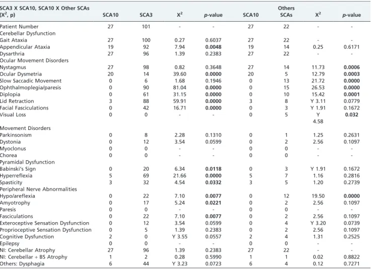
forms of SCA (SCA 1, 2, and 7). Slow saccadic eye movements associated with hypo/areflexia are typically suggestive of SCA2. As only one case of SCA6 was detected in our patient sample, no comparisons between this subtype and the other SCA subtypes could be made. The overall results are presented in Table 2.
The neurological signs most commonly found in patients with SCA3, SCA10, and other forms of SCA (SCA types 1, 2, 6, and 7) are summarized in Table 3.
DISCUSSION
In this sample of Brazilian patients with SCA, we observed that SCA3, which is also known as Machado-Joseph disease, was the most commonly identified subtype. In fact, SCA3 is also the most commonly encountered SCA worldwide, despite certain regional variations (SCA1 in Italy, SCA2 in India and Cuba, and SCA7 in Sweden, Finland, and South Africa) (2,3,4,6,9-12). In Brazil, several previously published studies of patients with SCA have identified SCA3 as occurring at the highest frequency (9,10,12,15).
Surprisingly, in our patient series, SCA10 was the second most frequently encountered SCA, accounting for approxi-mately 12% of the cases. As the causative genetic mutation
for this subtype was discovered fairly recently, SCA10 was not investigated prior to the year 2000 and was therefore was not reported in the previously published SCA patient series. Several other investigators (6,11,16) evaluated the
Table 2 -A comparative statistical analysis between the clinical data and neuroimaging results of patients with SCA3 x SCA10 and SCA10 x other SCAs.
SCA3 X SCA10, SCA10 X Other SCAs
(X2, p) SCA10 SCA3 X2 p-value SCA10
Others
SCAs X2 p-value
Patient Number 27 101 - - 27 22 -
-Cerebellar Dysfunction
Gait Ataxia 27 100 0.27 0.6037 27 22 -
-Appendicular Ataxia 19 92 7.94 0.0048 19 14 0.25 0.6171
Dysarthria 27 96 1.39 0.2383 27 22 -
-Ocular Movement Disorders
Nystagmus 27 98 0.82 0.3648 27 14 11.73 0.0006
Ocular Dysmetria 20 14 39.60 0.0000 20 5 12.79 0.0003
Slow Saccadic Movement 0 6 1.68 0.1946 0 13 21.72 0.0000
Ophthalmoplegia/paresis 0 90 81.04 0.0000 0 15 26.53 0.0000
Diplopia 0 61 31.15 0.0000 0 10 15.42 0.0001
Lid Retraction 3 88 59.91 0.0000 3 8 Y 3.11 0.0779
Facial Fasciculations 0 42 16.71 0.0000 0 3 Y 1.91 0.1672
Visual Loss 0 0 - - 0 5 Y
4.58
0.032
Movement Disorders
Parkinsonism 0 8 2.28 0.1310 0 1 1.25 0.2631
Dystonia 0 12 3.54 0.0599 0 2 2.56 0.1097
Myoclonus 0 0 - - 0 0 -
-Chorea 0 0 - - 0 0 -
-Pyramidal Dysfunction
Babinski’s Sign 0 20 6.34 0.0118 0 3 Y 1.91 0.1672
Hyperreflexia 5 69 21.66 0.0000 5 7 1.16 0.2816
Spasticity 3 32 4.54 0.0332 3 5 1.20 0.2739
Peripheral Nerve Abnormalities
Hypo/areflexia 0 22 7.10 0.0077 0 12 19.50 0.0000
Amyotrophy 0 17 5.24 0.0221 0 2 2.56 0.1097
Paresis 0 0 - - 0 0 -
-Fasciculations 0 22 7.10 0.0077 0 2 2.56 0.1097
Exteroceptive Sensation Dysfunction 0 12 3.54 0.0599 0 4 Y 3.20 0.0739
Proprioceptive Sensation Dysfunction 0 5 1.39 0.2383 0 2 2.56 0.1097
Cognitive Dysfunction 2 0 Y 3.55 0.0557 2 4 1.31 0.2525
Epilepsy 0 0 - - 0 0 -
-NI: Cerebellar Atrophy 27 96 1.39 0.2383 27 22 -
-NI: Cerebellar+BS Atrophy 1 2 0.28 0.5990 1 1 0.02 0.8822
Others: Dysphagia 6 44 Y 3.23 0.0723 6 4 0.12 0.7271
X2 = Chi-Square Test; Y = Yates’s Correction; NI = Neuroimaging; BS = Brainstem; P = Probability (
p,0.05).
Table 3 -The neurological signs most commonly identified in patients with SCA10, SCA3, or other types SCA.
SCA10 SCA3 Other SCAs
Neurological Signs
Appendicular Ataxia - +
-Nystagmus + - +
Ocular Dysmetria + -
-Slow Saccadic Movement - - +
Ophthalmoplegia/paresis - + +
Diplopia - + +
Lid Retraction - +
-Facial Fasciculations - +
-Babinski’s Sign - +
-Hyperreflexia - +
-Spasticity - +
-Hypo/Areflexia - + +
Amyotrophy - +
-occurrence of SCA10 in European, Asian, and North American patients, although no cases were identified.
SCA10 was originally described in Mexican patients and subsequently in Brazilian patients, and SCA10 represents the second most common type of SCA after SCA2 (in Mexico) and SCA3 (in Brazil) (6,11,17-19). Moreover, cases of SCA10 have been detected in other Latin American countries, including Argentina and Venezuela (19).
The other forms of SCA found in this patient series consisted of SCA2, SCA7, SCA1, and SCA6. No cases of DRPLA were found.
We initially compared the two most commonly identified forms of SCA and found that several neurological signs could be significantly correlated with either SCA3 or SCA10. In general, clinical experience has demonstrated that SCA3 presents great phenotypic heterogeneity with large inter-familial variation. However, the neurological signs identi-fied in the current study (including ophthalmoplegia, diplopia, facial fasciculations, eyelid retraction (bulging eyes), spasticity, severe hyperreflexia, Babinski’s sign, severe hypo/areflexia, amyotrophy, and muscular fascicu-lations) reflect a more common phenotype among patients with SCA3.
The genotype-phenotype correlation in SCA3 has been the focus of several studies, and many of these published results are identical to those reported in the present study (20,21). Since the original description of SCA3 by Coutinho and Andrade in 1978, which was followed by additional studies from the same group, most investigators have emphasized the presence of a characteristic phenotype consisting of ophthalmoplegia, bulging eyes, facial fascicu-lations, pyramidal signs, and peripheral neuropathy, as well as dystonia in younger patients (20-22). More recently, the observation of variant phenotypes for SCA3 has led to the description of the following sub-phenotypes: subtype I (with a predominance of extrapyramidal signs and dysto-nia); subtype II (with CA and pyramidal tract signs); subtype III (with CA and peripheral neuropathy signs); subtype 4 (with parkinsonism); and subtype 5 (resembling spastic paraplegia) (21). In contrast, the study by Scho¨ls et al. (23) describing German patients did not report the typical signs described for Portuguese SCA3 cases, such as bulging eyes, dystonia, and rigidity. In addition, Klockgether et al. as well as other groups have suggested that significant overlap exists between different SCAs, which impedes the utility of purely clinical-based diagnoses (4,24). Finally, other authors have attempted to describe oculomotor phenotypes suggestive of several forms of SCA, although these finding have no significant impact in regards to daily clinical practice (25).
With respect to movement disorders in our patient series, we observed that 15.3% of patients presented signs of dystonia and parkinsonism (nine patients), and the majority of these symptoms were presented by patients with SCA3. Similar results have also been reported by Schols et al. and Garcia Ruiz et al. (26,27).
The majority of patients with SCA10 in the current study presented a phenotype of ‘‘pure’’ CA with occasional slight pyramidal tract signs (hyperreflexia and discrete spasticity). This presentation without epilepsy or peripheral neuropathy differs from the phenotype described in Mexican patients, who were shown to present with epilepsy in 72.2% of cases and peripheral nephropathy in 66% of cases (28-32). These observations have led to debates regarding the reason for these
different phenotypic manifestations in Mexican and Brazilian SCA10 patients. The first explanation for these differences correlated the observed phenotype with the expansion of the ATTCT pentanucleotide repeat size (greater on average in Mexicans) but was refuted in 2004 (11). Later, Matsuura et al. (33) showed that the presence of a complex interruption pattern composed of two different repeat interruptions, ATTTTCT and ATATTCT, could explain the presence of a phenotype consisting of epilepsy associated with CA.
More recently, Teive at al. evaluated the frequency of epilepsy in a group of 80 patients from 10 Brazilian families with SCA10 and identified the presence of epilepsy in only 3.75% of the cases, whereas case reports from Argentina and Venezuela have presented a phenotype resembling those of Mexican origin (18). Another interesting clinical aspect of patients with SCA10 is that they generally do not present ophthalmoplegia/paresis, although they do commonly present nystagmus in lateral views and ocular dysmetria (11,19,34).
Finally, we also evaluated the genotype-phenotype correlation between patients with SCA10 and all other types of SCA (excluding SCA3). These results demonstrated that ophthalmoplegia/paresis and diplopia were the most frequently observed signs in patients with other types of SCA, including SCA 1, 2, and 7. However, the presence of slow saccadic ocular movements and hypo/areflexia were more common in patients with SCA2. These findings have been repeatedly reported in the literature since the initial description of SCA2 (3,4,6,35). Also, we should highlight that the association between slow saccadic eye movements and signs of peripheral neuropathy are highly suggestive, at least from a clinical standpoint, of SCA2 (3,4,6,35). The same is also true for the association between CA and vision loss due to retinopathy in the case of SCA7 (3,4,6,35).
In our clinical setting, neither patients with SCA1 nor SCA6 presented a characteristic phenotype, which was likely due to the small number of cases identified.
SCA represents an extensive and complex group of autosomal dominant neurodegenerative diseases, and to date, SCA3, SCA1, SCA2, SCA6 and SCA7 are the most frequently identified types of SCA worldwide. In this series of Brazilian SCA patients, SCA3 was the most commonly identified type, followed by SCA10. Upon comparisons between SCA3, SCA10 and other SCAs, patients with each of these conditions revealed several clinical features that may be useful for the clinical classification of an affected patient or family. In SCA3 patients, the phenotype was highly pleomorphic, although CA, ophthalmoplegia, diplopia, eye-lid retraction, facial fasciculation, pyramidal signs and peripheral neuropathy were the most frequently observed signs of disease. In SCA10 patients, the phenotype was rather peculiar and consisted of pure CA, saccadic eye movement, and ocular dysmetria. For the other SCAs, SCA2 and SCA7 exhibited highly suggestive phenotypes, presenting as CA with slow saccadic ocular movements and profound areflexia in SCA2 and cerebellar ataxia and visual loss in SCA7. The phenotypes of patients with SCA1, SCA6, and SCA10 were commonly nonspecific (e.g., pure cerebellar ataxia).
ACKNOWLEDGMENTS
Clı´nicas, Federal University of Parana´), Dr. Ben Roa, Dr. Ping Fang (Baylor DNA Diagnostic Laboratory, Baylor College of Medicine, Houston, Texas, USA), Dr. Rui Gao (University of Texas Medical Branch, Galveston, Texas, USA), Jilin Liu (University of Florida, Gainesville, Florida, USA), and Ms. Misti C. White (Baylor DNA Diagnostic Laboratory, Baylor College of Medicine, Houston, Texas, USA), for their collaboration in performing the molecular genetic exams of SCA patients. The authors also thank Prof. Rosana H. Scola (Electroneuromyography Unit, Neurology Service, Hospital de Clı´nicas, Federal University of Parana´) and Dr. Alexandre L. Longo, Dr. Carla C. Moro, Dr. Norberto Cabral (Clı´nica Neurolo´gica de Joinville, Santa Catarina), Prof. Ylmar Correa Neto (Internal Medicine Department, Federal University of Santa Catarina), Prof. Paulo N. D. Sa´, and Prof. Paulo C. T. Bittencourt (Neurology Service, Federal University of Santa Catarina) for their help with this study. Support by NIH NS041547 (TA).
Conflicts of interests:Drs. Arruda, Munhoz, Raskin, Lopes-Cendes,
Werneck, and Ashizawa have nothing to report. Dr. Teive has received speaking honoraria from Allergan, Boeringher-Ingelheim, Ipsen, Roche, and Novartis.
AUTHOR CONTRIBUTIONS
Teive HA conceived and designed the study and was also responsible for the acquisition, analysis and interpretation of data, manuscript drafting, critical revision of the manuscript for important intellectual content and study supervision. Werneck LC conceived and designed the study and was also responsible for the analysis and interpretation of data, critical revision of the manuscript for important intellectual content, study supervision and administrative, technical, and material support. Arruda WO was responsible for the data acquisition, analysis and interpretation, manuscript drafting and critical revision of the manuscript for important intellectual content. Munhoz RP was responsible for the data acquisition, analysis and interpretation, critical revision of the manuscript for important intellectual content, study supervision, and administrative, technical and material support. Raskin S was responsible for the data analysis and interpretation, critical revision of the manuscript for important intellectual content, and administrative, technical, and material support. Lopes-Cendes I was responsible for the data analysis and interpretation, critical revision of the manuscript for important intellectual content, study supervision, admin-istrative, technical, and material support. Ashizawa T was responsible for the critical revision of the manuscript for important intellectual content, study supervision;, and contributed with administrative, technical and material support.
REFERENCES
1. Teive HA. Spinocerebellar Degenerations in Japan. New insights from an epidemiological study. Neuroepidemiology. 2009;32(3):184-5. 2. Durr A. Autosomal dominant cerebellar ataxias: polyglutamine
expan-sions and beyond. Lancet Neurology. 2010;9(9):885-94, http:// dx.doi.org/10.1016/S1474-4422(10)70183-6.
3. Scho¨ls L, Bauer P, Schmidt T, Schulte T, Riess O. Autosomal dominant cerebellar ataxias: clinical features, genetics, and pathogenesis. Lancet Neurol. 2004;3(5):291-304, http://dx.doi.org/10.1016/S1474-4422(04) 00737-9.
4. Klockgether T, Lu¨dtke R, Kramer B, Abele M, Bu¨rk K, Scho¨ls L, et al. The natural history of degenerative ataxia: a retrospective study of 466 patients. Brain. 1998;121(4):589-600, http://dx.doi.org/10.1093/brain/ 121.4.589.
5. Harding AE. The hereditary ataxias and related disorders. Churchill Livingstone, Edimburgh, 1984;129-65.
6. Teive HAG. Spinocerebellar Ataxias. Arq Neuropsiquiatr. 2009;67(4):1133-42, http://dx.doi.org/10.1590/S0004-282X2009000600035.
7. van de Warremburg BP, Sinke RJ, Verschuuren-Bemelmans CC. Spinocerebelllar ataxias in the Netherlands: prevalence and age at onset variance analysis. Neurology. 2002;58(5):702-8.
8. Erichsen AK, Koht J, Stray-Pedersen A, Abdelnoor M, Tallaksen ME. Prevalence of hereditary ataxia and spastic paraplegia in southeast Norway: a population-based study. Brain. 2009;132(6):1577-88, http:// dx.doi.org/10.1093/brain/awp056.
9. Silveira I, Lopes-Cendes I, Kish S, Maciel P, Gaspar C, Coutinho P, et al. Frequency of spinocerebellar ataxia type 1, dentatorubropallidoluysian atrophy, and Machado-Joseph disease mutations in a large group of spinocerebellar ataxia patients. Neurology. 1996;46(1):214-8.
10. Lopes-Cendes I, Teive HAG, Calcagnotto ME, Da Costa JC, Cardoso F, Viana E, et al. Frequency of the different mutations causing spinocerebellar
ataxia (SCA 1, SCA 2, SCA 3/MJD and DRPLA) in a large group of brazilian patients. Arq Neuropsiquiatr. 1997;55(3B):519-29, http://dx.doi.org/ 10.1590/S0004-282X1997000400001.
11. Teive HAG, Roa B, Raskin S, Fang P, Arruda WO, Neto YC, et al. Clinical phenotype of Brazilian patients with spinocerebellar ataxia 10. Neurology. 2004;63(8):1509-12.
12. Jardim LB, Silveira I, Pereira ML, Ferro A, Alonso I, do Ce´u Moreira M, et al. A survey of spinocerebellar ataxia in South Brazil - 66 new cases with Machado-Joseph disease, SCA 7, SCA 8, or unidentified disease-causing mutations. J Neurol. 2001; 248(10):870-6, http://dx.doi.org/ 10.1007/s004150170072.
13. Sambrock J, Fritsh EF, Maniatis T. Molecular Cloning: A Laboratory Manual 2nd ed., Cold Spring Harbor, NY (1989), 1.8-1.9, 1.86. 14. Matsuura T, Yamagata T, Burgess DL, Rasmussen A, Grewal RP, Watase
K, et al. Large expansions of the ATTCT pentanucleotide repeat in spinocerebellar ataxia type 10. Nat Genet. 2000;26(2):191-4, http:// dx.doi.org/10.1038/79911.
15. Jardim LB, Pereira ML, Silveira I, Ferro A, Sequeiros J, Giugliani R. Neurologic findings in Machado-Joseph disease. Relation with disease duration, sybtypes, and (CAG)n. Arch Neurol. 2001;58(4):899-904. 16. Matsuura T, Ranum LPW, Volpini V, Pandolfo M, Sasaki H, Tashiro K,
et al. Spinocerebellar ataxia type 10 is rare in populations other than Mexicans. Neurology. 2002;58(6):983-4.
17. Raskin S, Ashizawa T, Teive HAG, Arruda WO, Fang P, Gao R, et al. Reduced penetrance in a Brazilian family with spinocerebellar ataxia type 10. Arch Neurol. 2007;64(4):591-4, http://dx.doi.org/10.1001/ archneur.64.4.591.
18. Teive HAG, Munhoz RP, Raskin S, Arruda WO, de Paola L, Werneck LC, et al. Spinocerebellar ataxia type 10: Frequency of epilepsy in a large sample of Brazilian patients. Mov Disord. 2010;25(16):2875-8, http:// dx.doi.org/10.1002/mds.23324.
19. Teive HAG, Munhoz RP, Arruda WO, Raskin S, Werneck LC, Ashizawa T. Spinocerebellar Ataxia Type 10 - A Review. Parkinsonism Relat Disord 2011;17(9):655-61.
20. Coutinho P, Andrade C. Autosomal dominant system degeneration in Portuguese families of the Azorean islands: a new genetic disorder involving cerebellar, pyramidal, extrapyramidal and spinal cord motor functions. Neurology. 1978;28(7):703-9.
21. Lima L, Coutinho P. Clinical criteria for diagnosis of Machado-Joseph disease: report of a non-azorean Portuguese family. Neurology. 1980;30(3):319-22.
22. Silveira I, Miranda C, Guimara˜es L, Moreira M, Alonso I, Mendonc¸a P, et al. Trinucleotide repeats in 202 families with ataxia: a small expanded (CAG)n allele at the SCA17 locus. Arch Neurol. 2002; 59(4):623-9, http:// dx.doi.org/10.1001/archneur.59.4.623.
23. Scho¨ls L, Amoiridis G, Epplen JT, Langkafel M, Przuntek H, Riess O. Relations between genotype and phenotype in German patients with the Machado-Joseph disease mutation. J Neurol Neurosurgery & Psychiatry. 1996;61(5):466-70, http://dx.doi.org/10.1136/jnnp.61.5.466.
24. Lopes-Cendes I, Silveira I, Maciel P, Gaspar C, Radvany J, Chitayat D, et al. Limits of clinical assessment in the acurate diagnosis of Machado-Joseph disease. Arch Neurol. 1996;53(11):1168-74, http://dx.doi.org/ 10.1001/archneur.1996.00550110120020.
25. Buttner N, Geschwind D, Jen JC, Perlman S, Pulst SM, Baloh RW. Oculomotor phenotypes in autosomal dominant ataxias. Arch Neurol 1998;55:1353-57.
26. Schols L, Peters S, Szymanski S, Kruger R, Lange S, Hardt C, et al. Extrapiramidal motor signs in degenerative ataxias. Arch Neurol. 2000;57(10):1495-1500, http://dx.doi.org/10.1001/archneur.57.10.1495. 27. Garcia Ruiz PJ, Mayo D, Hernandez J, Cantarero S, Ayuso C. Movement
disorders in hereditary ataxias. J Neurol Sci. 2002;202(1-2):59-64, http:// dx.doi.org/10.1016/S0022-510X(02)00211-3.
28. Matsuura T, Achari M, Khakavi M, Bachinski LL, Huda ZY, Ashizawa T. Mapping of the gene for a novel spinocerebellar ataxia with pure cerebellar signs and epilepsy. Ann Neurol. 1999;45(3):407-11, http://dx.doi.org/ 10.1002/1531-8249(199903)45:3,407::AID-ANA21.3.0.CO;2-D.
29. Zu L, Figueroa KP, Grewal R, Pulst SM. Mapping of a new autosomal dominant spinocerebellar ataxia to chromosome 22. Am J Hum Genet. 1999;64(2):594-9, http://dx.doi.org/10.1086/302247.
30. Grewal RP, Tayag E, Figueroa KP, Zu L, Durazo A, Nunez C, et al. Clinical and genetic analysis of a distinct autosomal dominant spinocerebellar ataxia. Neurology. 1998;51(5):1423-26.
31. Rasmunssen A, Matsuura T, Ruano L, Yescas P, Ochoa A, Ashizawa T, et al. Clinical and Genetic analysis of four Mexican families with spinocerebellar ataxia type 10. Ann Neurol. 2001;50(2):234-9, http:// dx.doi.org/10.1002/ana.1081.
32. Grewal RP, Achari M, Matsuura T, Durazo A, Tayag E, Zu L, et al. Clinical features and ATTCT repeat expansion in spinocerebellar ataxia type 10. Arch Neurol. 2002;59(8):1285-90, http://dx.doi.org/10.1001/ archneur.59.8.1285.
34. Teive HAG, Arruda WO, Raskin S, Munhoz RP, Zavala J, Werneck LC, et al. Symptom onset of spinocerebellar ataxia type 10 in pregnancy and puerperium. J Clin Neurosci. 2011;18(3):437-8, http://dx.doi.org/ 10.1016/j.jocn.2010.07.102.