Submitted30 May 2016
Accepted 4 November 2016
Published20 December 2016
Corresponding authors
Kun Wang, kunwang@mospital.com Likwang Chen, likwang@nhri.org.tw
Academic editor
Yeong Yeh Lee
Additional Information and Declarations can be found on page 9
DOI10.7717/peerj.2753
Copyright
2016 Hsu et al.
Distributed under
Creative Commons CC-BY 4.0
OPEN ACCESS
Risk of lung cancer in patients with
gastro-esophageal reflux disease: a
population-based cohort study
Chi-Kuei Hsu1, Chih-Cheng Lai2, Kun Wang3,*and Likwang Chen4,*
1Department of Internal Medicine, E-Da Hospital, Kaohsiung, Taiwan
2Department of Intensive Care Medicine, Chi Mei Medical Center, Liouying, Tainan, Taiwan 3Department of Internal Medicine, Cardinal Tien Hospital, New Taipei City, Taiwan 4National Health Research Institutes, Miaoli, Taiwan
*These authors contributed equally to this work.
ABSTRACT
This large-scale, controlled cohort study estimated the risks of lung cancer in pa-tients with gastro-esophageal reflux disease (GERD) in Taiwan. We conducted this population-based study using data from the National Health Insurance Research Database of Taiwan during the period from 1997 to 2010. Patients with GERD were diagnosed using endoscopy, and controls were matched to patients with GERD at a ratio of 1:4. We identified 15,412 patients with GERD and 60,957 controls. Compared with the controls, the patients with GERD had higher rates of osteoporosis, diabetes mellitus, asthma, chronic obstructive pulmonary disease, pneumonia, bronchiectasis, depression, anxiety, hypertension, dyslipidemia, chronic liver disease, congestive heart failure, atrial fibrillation, stroke, chronic kidney disease, and coronary artery disease (all
P< .05). A total of 85 patients had lung cancer among patients with GERD during the
follow-up of 42,555 years, and the rate of lung cancer was 0.0020 per person-year. By contrast, 232 patients had lung cancer among patients without GERD during the follow-up of 175,319 person-years, and the rate of lung cancer was 0.0013 per person-year. By using stepwise Cox regression model, the overall incidence of lung cancer remained significantly higher in the patients with GERD than in the controls (hazard ratio, 1.53; 95% CI [1.19–1.98]). The cumulative incidence of lung cancer was higher in the patients with GERD than in the controls (P=.0012). In conclusion, our
large population-based cohort study provides evidence that GERD may increase the risk of lung cancer in Asians.
SubjectsEpidemiology, Gastroenterology and Hepatology, Oncology
Keywords Lung cancer, GERD, Risk factors
INTRODUCTION
of GERD are esophageal symptoms, including heartburn, dysphagia, and regurgitation, and it can cause extra-esophageal presentation such as bronchospasm, laryngitis, and chronic cough. Because it may cause lung injury from recurrent microaspiration, GERD is associated with the risk of several lung diseases, such as idiopathic pulmonary fibrosis, cystic fibrosis, connective tissue disease, asthma, chronic obstructive pulmonary disease (COPD), and interstitial lung disease (Blondeau et al., 2008;D’Ovidio et al., 2005;Mise et al., 2010;Morehead, 2009;Pacheco-Galvan, Hart & Morice, 2011;Pashinsky, Jaffin & Litle, 2009;Salvioli et al., 2006;Sweet et al., 2009;Patti et al., 2008).
Recently,Vereczkei et al. (2008)investigated the association between GERD and non-small cell lung cancer (NSCLC) and found that a considerably higher proportion of patients with NSCLC had GERD than the general population, irrespective of cell type. Therefore, a study proposed that GERD-associated chronic lung injury may be one element of lung can-cer promotion (Herbella et al., 2015). However, it enrolled only 25 patients with surgically treated adenocarcinoma and squamous cell carcinoma, and the relationship between GERD and lung cancer remains unclear (Herbella et al., 2015;Vereczkei et al., 2008). Therefore, whether GERD is associated with an increased risk of lung cancer should be determined. Hence, we performed a large-scale, controlled cohort study to estimate the hazard rates of lung cancer in patients with GERD by using a nationwide, population-based database in Taiwan.
MATERIALS & METHODS
Data source
The National Health Insurance (NHI) program of Taiwan is a nationwide insurance program that covers outpatient visits, hospital admissions, prescriptions, interventional procedures, and disease profiles for >99% of the population of Taiwan (23.12 million people in 2009) (Chen et al., 2011). Taiwan’s National Health Research Institute (NHRI) used the original data from the NHI program to construct a longitudinal database of patients admitted between 1997 and 2010. This cohort includes 2,619,534 hospitalized patients, rep-resenting 10% of all NHI enrollees. This sampled fraction (a 3.4:1 ratio) is based on a regula-tion that limits the maximal amount of NHI data that can be extracted for research purposes. The National Health Insurance Research Database (NHIRD) is one of the largest and most comprehensive databases worldwide and has been used extensively in various studies of prescription use, diagnoses, and hospitalizations. This study was approved by the Institutional Review Board of Cardinal Tien Hospital (Number: EC1011008-E-R1).
Identification of patients with GERD and without GERD
without GERD) to patients with GERD by age, sex, and the index date at a ratio of 1:4. In the non-GERD group, patients with a history of lung cancer or peptic ulcer disease were excluded.
Baseline variables
We collected data on demographic and clinical characteristics of the study population, including age, sex, and comorbidities. Comorbidities were defined according to the ICD-9-CM and procedure codes within 1 year before index admission. We used a relatively strict criterion to define comorbidities: coding one morbidity required at least one admission or 3 outpatient clinic visits for disease treatment during the year before index admission.
Definition of outcome
We followed up each patient until December 31, 2010, to observe for the development of de novo lung cancer. In Taiwan, patients with cancer can apply for a catastrophic illness certificate that exempts them from any out-of-pocket expenses for cancer evaluation and care. The development of de novo lung cancer was identified by the ICD-9-CM code 162 having been noted on the catastrophic illness certificate as a previous study (Jian et al., 2015). The follow-up duration was calculated from the date of GERD diagnosis (index date) to the date of the first recorded cancer code.
Statistical analysis
All data were analyzed using SAS Version 9.3 software (SAS Institute). Categorical variables are expressed as numbers or percentages and were compared using the chi-square test. Inci-dence rates of lung cancer in both GERD and non-GERD groups were calculated by Poisson regression. The Kaplan–Meier method was used to estimate the cumulative incidence rate of lung cancer in patients with or without GERD. The cumulative incidence curves of both groups were compared using the log-rank test. We used Log-Minus-Log survival plots to evaluate proportional hazard assumption. To assess the risk of lung cancer, a list of potential risk factors associated both with admission of lung cancer and with GERD status was considered in the Cox regression model. Univariable and multivariable Cox regression models (stepwise selection) were performed to examine the association of lung cancer with potential confounding factors such as osteoporosis, diabetes mellitus (DM), asthma, COPD, pneumonia, anxiety, hypertension, dyslipidemia, chronic liver disease, congestive heart failure (CHF), atrial fibrillation, stroke, chronic kidney disease (CKD), and coronary artery disease (CAD). Two-sidedPvalues < .05 were considered statistically significant.
RESULTS
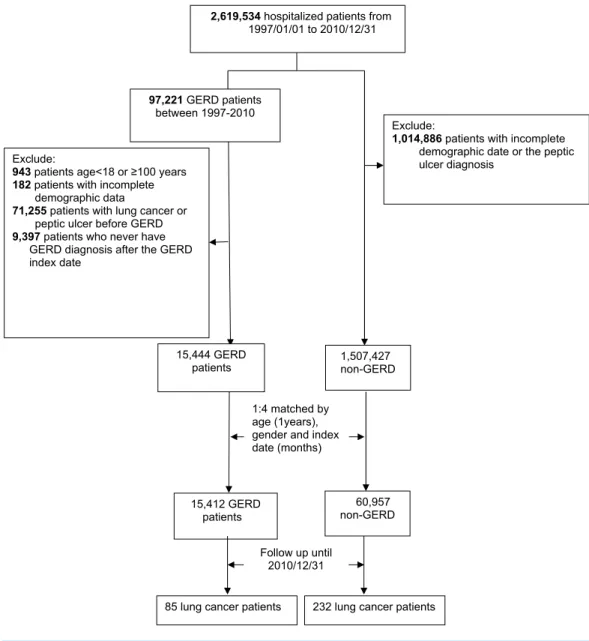
2,619,534 hospitalized patients from 1997/01/01 to 2010/12/31
97,221 GERD patients between 19972010
15,444 GERD patients Exclude:
943 patients age<18 or ≥100 years 182 patients with incomplete
demographic data
71,255 patients with lung cancer or peptic ulcer before GERD 9,397 patients who never have
GERD diagnosis after the GERD index date
1,507,427 nonGERD
Exclude:
1,014,886 patients with incomplete demographic date or the peptic ulcer diagnosis
1:4 matched by age (1years), gender and index date (months)
15,412 GERD
patients nonGERD60,957
Follow up until 2010/12/31
85 lung cancer patients 232 lung cancer patients
Figure 1 Study algorithm for patient enrollment.
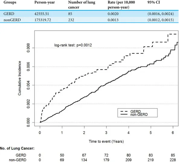
A total of 85 patients had lung cancer among patients with GERD during the follow-up of 42,555 person-years, and the rate of lung cancer was 0.0020 per person-year. By contrast, 232 patients without GERD had lung cancer during the follow-up of 175,319 person-years, and the rate of lung cancer was 0.0013 per person-year (Table 1). The baseline characteristics and comorbidities are listed inTable 2. Compared with the controls, the patients with GERD displayed higher rates of osteoporosis, asthma, COPD, pneumonia, bronchiectasis, depression, anxiety, hypertension, dyslipidemia, chronic liver disease, CHF, atrial fibrilla-tion, stroke, CKD, and CAD (allP< .05). There is no violation of the proportional hazard
Table 1 Incidence rates of lung cancer events per 10,000 person-year among gastro-esophageal reflux disease (GERD) and non-GERD group.
Groups Person-year Number of lung
cancer
Rate (per 10,000 person-year)
95% CI
GERD 42555.51 85 0.0020 (0.0016, 0.0024)
nonGERD 175319.72 232 0.0013 (0.0012, 0.0015)
Figure 2 Cumulative incidence rate of lung cancer for patients with or without GERD.
CI [1.19–1.98];Table 3). As shown inFig. 2, the cumulative incidence of lung cancer was higher in the patients with GERD than in the controls (P=.0012).
DISCUSSION
Table 2 Baseline characteristics of study population stratified by gastro-esophageal reflux disease (GERD) and non-GERD group.
Variables Number (%) of patients
with GERDN=15,412
Number (%) of patients without GERDN=60,957
χ2 df p-value
Gender 0.326(1) 0.568
Female 7,849 (50.9) 31,201 (51.2)
Male 7,563 (49.1) 29,756 (48.8)
Age group 4.203(2) 0.122
18–54 years 6,503 (42.2) 26,049 (42.7)
54–64 5,961 (38.7) 23,677 (38.8)
≥65 years 2,948 (19.1) 11,231 (18.4)
Underlying diseases/conditions
Osteoporosis 15.366(1) <.001
No 15,173 (98.5) 60,250 (98.84)
Yes 239 (1.6) 707 (1.2)
Diabetes mellitus 16.298(1) <.001
No 14,078 (91.3) 55,030 (90.3)
Yes 1,334 (8.7) 5,927 (9.7)
Tuberculosis 0.930(1) 0.335
No 15,342 (99.6) 60,714 (99.6)
Yes 70 (0.5) 243 (0.4)
Asthma 95.058(1) <.001
No 14,881 (96.6) 59,673 (97.9)
Yes 531 (3.5) 1,284 (2.1)
Chronic obstructive pulmonary diseases 259.590(1) <.001
No 14,158 (91.9) 58,015 (95.2)
Yes 1,254 (8.4) 2,942 (4.8)
Pneumonia 247.106(1) <.001
No 14,929 (96.9) 60,157 (98.7)
Yes 483 (3.1) 800 (1.3)
Pneumoconiosis 0.031(1) 0.861
No 15,401 (99.9) 60,916 (99.9)
Yes 11 (0.1) 41 (0.1)
Bronchiectasis 28.373(1) <.001
No 15,349 (99.6) 60,847 (99.8)
Yes 63 (0.4) 110 (0.2)
Depression 37.239(1) <.001
No 15,242 (98.9) 60,570 (99.4)
Yes 170 (1.1) 387 (0.6)
Anxiety 365.826(1) <.001
No 14,167 (91.9) 58,337 (95.7)
Yes 1,245 (8.1) 2,620 (4.3)
Hypertension 9.954(1) 0.002
No 12,031 (78.1) 48,291 (79.2)
Yes 3,381 (22.0) 12,666 (20.8)
Table 2(continued)
Variables Number (%) of patients
with GERDN=15,412
Number (%) of patients without GERDN=60,957
χ2 df p-value
Dyslipidemia 11.772(1) <.001
No 13,993 (90.8) 55,871 (91.7)
Yes 1,419 (9.2) 5,086 (8.3)
Chronic liver disease 75.039(1) <.001
No 14,865 (96.5) 59,546 (97.7)
Yes 547 (3.6) 1,411 (2.3)
Congestive heart failure 21.013(1) <.001
No 15,118 (98.1) 60,101 (98.6)
Yes 294 (2.0) 856 (1.4)
Atrial fibrillation 4.635(1) 0.031
No 15,274 (99.1) 60,514 (99.3)
Yes 138 (0.9) 443 (0.7)
Myocardial infarction 0.289(1) 0.591
No 15,316 (99.4) 60,600 (99.4)
Yes 96 (0.62) 357 (0.6)
Stroke 19.921(1) <.001
No 14,581 (94.6) 58,190 (95.5)
Yes 831 (5.4) 2,767 (4.6)
Peripheral vascular disease 0.720(1) 0.720
No 15,350 (99.6) 60,724 (99.6)
Yes 62 (0.4) 233 (0.7)
Chronic kidney diseases 32.318(1) <.001
No 15,083 (97.9) 60,050 (98.5)
Yes 329 (2.1) 907 (1.5)
Coronary artery diseases 32.782(1) <.001
No 15,051 (97.7) 59,947 (98.3)
Yes 361 (2.3) 1,010 (1.7)
refluxate, no study has assessed the possible relationship between GERD and lung cancer. Our study is the first to demonstrate a significant positive association between GERD and lung cancer. This finding was supported by the increased risk of lung cancer in comparison with age- and sex-matched controls (crude HR, 1.53; 95% CI [1.19–1.98]). Our findings have some clinical implications. After confirming this significant association between GERD and lung cancer, it was suggested that aggressive treatment of GERD possibly prevents the development of lung cancer. However, further studies should be warranted to prove the possible chemopreventive role of antacid use in patients with GERD.
Table 3 Crude hazard ratios (HR) among gastro-esophageal reflux disease (GERD) and non-GERD group.
Variables Beta value Crude HR (95%CI) pvalue
GERD 0.43 1.53 (1.19–1.98) 0.001
Osteoporosis 0.59 1.80 (0.88–3.69) 0.110
Diabetes mellitus 0.05 1.05 (0.76–1.46) 0.752
Asthma 0.20 1.22 (0.65–2.29) 0.538
Chronic obstructive pulmonary diseases 0.30 1.34 (0.92–1.96) 0.125
Pneumonia 0.40 1.49 (0.76–2.90) 0.245
Bronchiectasis 0.69 2.00 (0.18–22.06) 0.571
Depression 0.84 2.31 (0.55–9.69) 0.251
Anxiety −0.08 0.92 (0.53–1.60) 0.765
Hypertension −0.11 0.90 (0.69–1.17) 0.429
Dyslipidemia −0.02 0.98 (0.65–1.46) 0.913
Chronic liver disease −0.25 0.78 (0.36–1.71) 0.539
Congestive heart failure −0.88 0.42 (0.15–1.19) 0.102
Atrial fibrillation −1.23 0.29 (0.07–1.25) 0.098
Stroke −0.42 0.66 (0.41–1.05) 0.079
Chronic kidney diseases −0.31 0.73 (0.33–1.60) 0.433
Coronary artery diseases −0.28 0.76 (0.36–1.58) 0.458
the likelihood of non-response and loss of follow-up to a minimum. Besides, there were some variables during the multivariable analysis. We controlled them by statistic methods (Table 3). Most important of all, we used a nationwide and population-based database– Taiwan NHIRD. Thus, the findings in the present work can be generalized in the real world.
Several mechanisms can help explain the significant relationship between GERD and lung cancer. First, several studies have shown that the refluxate can destroy the epithelium of the larynx or pharynx by means of introducing chronic inflammation (Rees et al., 2008) or activating proliferative signaling pathways (Dvorak et al., 2011;Johnston et al., 2012;
Sung et al., 2003) and further result in malignant transformation. In addition, based on the studies investigating the pathogenesis of Barrett’s esophagus and esophageal carcinoma, both acid and bile can promate carcinogenesis through the induction of DNA damage and the influence of cell proliferation and apoptosis (Denlinger & Thompson, 2012;Fang et al., 2013). These pathogenesis may happen in the respiratory tract, and contribute to the development of lung cancer. Second, the trend of the predominance of lung adeno-carcinoma among all cell type is similar with the distribution trend of esophageal cancer (Etzel et al., 2006;Liam et al., 2006). Third, the origin of central lung adenocarcinoma is dif-ferent from peripheral lung cancer’s. Lung cancer at a central site is more prone to be affected by gastric refluxate than at a peripheral site. Thus, lung adenocarcinoma at a central site is more likely to arise in the glandular epithelium in contrast to lung cancer at a peripheral site which possibly originates from type II pneumocytes and Clara cells (Fukui et al., 2013).
CAD, or COPD to minimize the influence of smoking. In addition, the data regarding the type of lung cancer was not available. Therefore, we cannot further analyze the association between GERD and the specific type of lung cancer. Second, patients with GERD may more often visit physicians than patients without GERD and this difference may cause possible surveillance bias. Finally, we did not collect the data about the use of anti-GERD treatments such as proton pump inhibitors or histamine-2-receptor antagonist.
CONCLUSIONS
Our large, population-based cohort study provides evidence that GERD may increase the risk of lung cancer.
ADDITIONAL INFORMATION AND DECLARATIONS
Funding
This study was supported by grants from National Health Research Institutes (intramural funding). The funders had no role in study design, data collection and analysis, decision to publish, or preparation of the manuscript.
Grant Disclosures
The following grant information was disclosed by the authors: National Health Research Institutes.
Competing Interests
The authors declare there are no competing interests.
Author Contributions
• Chi-Kuei Hsu and Chih-Cheng Lai conceived and designed the experiments, wrote the
paper.
• Kun Wang prepared figures and/or tables, reviewed drafts of the paper.
• Likwang Chen analyzed the data, prepared figures and/or tables, reviewed drafts of the
paper.
Ethics
The following information was supplied relating to ethical approvals (i.e., approving body and any reference numbers):
Cardinal Tien Hospital (Approval number: EC1011008-E-R1).
Data Availability
The following information was supplied regarding data availability:
REFERENCES
Bacciu A, Mercante G, Ingegnoli A, Ferri T, Muzzetto P, Leandro G, Di Mario F, Bacciu S. 2004.Effects of gastroesophageal reflux disease in laryngeal carcinoma.Clinical Otolaryngology and Allied Sciences29(5):545–548
DOI 10.1111/j.1365-2273.2004.00851.x.
Blondeau K, Dupont LJ, Mertens V, Verleden G, Malfroot A, Vandenplas Y, Hauser B, Sifrim D. 2008.Gastro-oesophageal reflux and aspiration of gastric contents in adult patients with cystic fibrosis.Gut 57(8):1049–1055DOI 10.1136/gut.2007.146134.
Bredenoord AJ, Pandolfino JE, Smout AJ. 2013.Gastro-oesophageal reflux disease.
Lancet 381(9881):1933–1942DOI 10.1016/s0140-6736(12)62171-0.
Chen PC, Muo CH, Lee YT, Yu YH, Sung FC. 2011.Lung cancer and incidence of stroke: a population-based cohort study.Stroke42(11):3034–3039
DOI 10.1161/strokeaha.111.615534.
Denlinger CE, Thompson RK. 2012.Molecular basis of esophageal cancer development and progression.Surgical Clinics of North America92(5):1089–1103
DOI 10.1016/j.suc.2012.07.002.
D’Ovidio F, Singer LG, Hadjiliadis D, Pierre A, Waddell TK, De Perrot M, Hutcheon M, Miller L, Darling G, Keshavjee S. 2005.Prevalence of gastroesophageal reflux in end-stage lung disease candidates for lung transplant.Annals of Thoracic Surgery 80(4):1254–1260DOI 10.1016/j.athoracsur.2005.03.106.
Dvorak K, Goldman A, Kong J, Lynch JP, Hutchinson L, Houghton JM, Chen H, Chen X, Krishnadath KK, Westra WM. 2011.Molecular mechanisms of Barrett’s esopha-gus and adenocarcinoma.Annals of the New York Academy of Sciences1232:381–391
DOI 10.1111/j.1749-6632.2011.06062.x.
El-Serag HB. 2007.Time trends of gastroesophageal reflux disease: a systematic review.
Clinical Gastroenterology and Hepatology5(1):17–26DOI 10.1016/j.cgh.2006.09.016.
El-Serag HB, Sweet S, Winchester CC, Dent J. 2014.Update on the epidemiology of gastro-oesophageal reflux disease: a systematic review.Gut63(6):871–880
DOI 10.1136/gutjnl-2012-304269.
Etzel CJ, Lu M, Merriman K, Liu M, Vaporciyan A, Spitz MR. 2006.An epidemiologic study of early onset lung cancer.Lung Cancer52(2):129–134
DOI 10.1016/j.lungcan.2005.11.018.
Fang Y, Chen X, Bajpai M, Verma A, Das KM, Souza RF, Garman KS, Donohoe CL, O’Farrell NJ, Reynolds JV, Dvorak K. 2013.Cellular origins and molecular mechanisms of Barrett’s esophagus and esophageal adenocarcinoma.Annals of the New York Academy of Sciences1300:187–199
DOI 10.1111/nyas.12249.
Fukui T, Shaykhiev R, Agosto-Perez F, Mezey JG, Downey RJ, Travis WD, Crystal RG. 2013.Lung adenocarcinoma subtypes based on expression of human airway basal cell genes.European Respiratory Journal42(5):1332–1344
Herbella FA, Neto SP, Santoro IL, Figueiredo LC. 2015.Gastroesophageal reflux disease and non-esophageal cancer.World Journal of Gastroenterology 21(3):815–819
DOI 10.3748/wjg.v21.i3.815.
Jian ZH, Huang JY, Ko PC, Jan SR, Nfor ON, Lung CC, Ku WY, Ho CC, Pan HH, Liaw YP. 2015.Impact of coexisting pulmonary diseases on survival of patients with lung adenocarcinoma: a STROBE-compliant article.Medicine94(4):e443
DOI 10.1097/MD.0000000000000443.
Johnston N, Yan JC, Hoekzema CR, Samuels TL, Stoner GD, Blumin JH, Bock JM. 2012.Pepsin promotes proliferation of laryngeal and pharyngeal epithelial cells.
Laryngoscope122(6):1317–1325DOI 10.1002/lary.23307.
Langevin SM, Michaud DS, Marsit CJ, Nelson HH, Birnbaum AE, Eliot M, Christensen BC, McClean MD, Kelsey KT. 2013.Gastric reflux is an independent risk factor for laryngopharyngeal carcinoma.Cancer Epidemiology, Biomarkers & Prevention 22(6):1061–1068DOI 10.1158/1055-9965.EPI-13-0183.
Lee YL, Hu HY, Yang NP, Chou P, Chu D. 2014.Dental prophylaxis decreases the risk of esophageal cancer in males; a nationwide population-based study in Taiwan.PLoS ONE9(10):e109444DOI 10.1371/journal.pone.0109444.
Liam CK, Pang YK, Leow CH, Poosparajah S, Menon A. 2006.Changes in the dis-tribution of lung cancer cell types and patient demography in a developing mul-tiracial Asian country: experience of a university teaching hospital.Lung Cancer 53(1):23–30DOI 10.1016/j.lungcan.2006.03.009.
Mise K, Capkun V, Jurcev-Savicevic A, Sundov Z, Bradaric A, Mladinov S. 2010.The influence of gastroesophageal reflux in the lung: a case-control study.Respirology 15(5):837–842DOI 10.1111/j.1440-1843.2010.01777.x.
Moayyedi P, Talley NJ. 2006.Gastro-oesophageal reflux disease.Lancet 367(9528): 2086–2100DOI 10.1016/s0140-6736(06)68932-0.
Morehead RS. 2009.Gastro-oesophageal reflux disease and non-asthma lung disease.
European Respiratory Review18(114):233–243DOI 10.1183/09059180.00002509.
Pacheco-Galvan A, Hart SP, Morice AH. 2011.Relationship between gastro-oesophageal reflux and airway diseases: the airway reflux paradigm.Archivos de Bronconeumologia 47(4):195–203DOI 10.1016/j.arbres.2011.02.001.
Pashinsky YY, Jaffin BW, Litle VR. 2009.Gastroesophageal reflux disease and idiopathic pulmonary fibrosis.Mount Sinai Journal of Medicine76(1):24–29
DOI 10.1002/msj.20088.
Patti MG, Gasper WJ, Fisichella PM, Nipomnick I, Palazzo F. 2008.Gastroesophageal reflux disease and connective tissue disorders: pathophysiology and implications for treatment.Journal of Gastrointestinal Surgery12(11):1900–1906
DOI 10.1007/s11605-008-0674-9.
Qadeer MA, Colabianchi N, Vaezi MF. 2005.Is GERD a risk factor for laryngeal cancer?
Laryngoscope115(3):486–491DOI 10.1097/01.mlg.0000157851.24272.41.
response to laryngopharyngeal reflux.American Journal of Respiratory and Critical Care Medicine 177(11):1187–1193DOI 10.1164/rccm.200706-895OC.
Salvioli B, Belmonte G, Stanghellini V, Baldi E, Fasano L, Pacilli AM, De Giorgio R, Barbara G, Bini L, Cogliandro R, Fabbri M, Corinaldesi R. 2006. Gastro-oesophageal reflux and interstitial lung disease.Digestive and Liver Disease 38(12):879–884DOI 10.1016/j.dld.2006.05.012.
Sung MW, Roh JL, Park BJ, Park SW, Kwon TK, Lee SJ, Kim KH. 2003.Bile acid induces cyclo-oxygenase-2 expression in cultured human pharyngeal cells: a possible mechanism of carcinogenesis in the upper aerodigestive tract by laryngopharyngeal reflux.Laryngoscope113(6):1059–1063DOI 10.1097/00005537-200306000-00027.
Sweet MP, Patti MG, Hoopes C, Hays SR, Golden JA. 2009.Gastro-oesophageal reflux and aspiration in patients with advanced lung disease.Thorax64(2):167–173
DOI 10.1136/thx.2007.082719.
Vaezi MF, Qadeer MA, Lopez R, Colabianchi N. 2006.Laryngeal cancer and gas-troesophageal reflux disease: a case-control study.American Journal of Medicine 119(9):768–776DOI 10.1016/j.amjmed.2006.01.019.
Vakil N. 2010.Disease definition, clinical manifestations, epidemiology and natural history of GERD.Best Practice & Research: Clinical Gastroenterology 24(6):759–764
DOI 10.1016/j.bpg.2010.09.009.
Vakil N, Van Zanten SV, Kahrilas P, Dent J, Jones R. 2006.The Montreal definition and classification of gastroesophageal reflux disease: a global evidence-based consensus.
American Journal of Gastroenterology101(8):1900–1920
DOI 10.1111/j.1572-0241.2006.00630.x.