Submitted 19 May 2015 Accepted 1 August 2015 Published20 August 2015
Corresponding author Vytautas Smirnovas, [email protected]
Academic editor Vladimir Uversky
Additional Information and Declarations can be found on page 9
DOI10.7717/peerj.1207
Copyright 2015 Sneideris et al.
Distributed under
Creative Commons CC-BY 4.0
OPEN ACCESS
Polymorphism of amyloid-like fibrils can
be defined by the concentration of seeds
Tomas Sneideris∗, Kataˇzyna Milto∗and Vytautas Smirnovas
Department of Biothermodynamics and Drug Design, Vilnius University, Institute of Biotechnology, Vilnius, Lithuania
∗These authors contributed equally to this work.
ABSTRACT
Prions are infectious proteins where the same protein may express distinct strains. The strains are enciphered by different misfolded conformations. Strain-like phenomena have also been reported in a number of other amyloid-forming proteins. One of the features of amyloid strains is the ability to self-propagate, maintaining a constant set of physical properties despite being propagated under conditions different from those that allowed initial formation of the strain. Here we report a cross-seeding experiment using strains formed under different conditions. Using high concentrations of seeds results in rapid elongation and new fibrils preserve the properties of the seeding fibrils. At low seed concentrations, secondary nucleation plays the major role and new fibrils gain properties predicted by the environment rather than the structure of the seeds. Our findings could explain conformational switching between amyloid strains observed in a wide variety ofin vivoandin vitro
experiments.
Subjects Biochemistry, Biophysics
Keywords Amyloid, Prion, Protein misfolding, Protein aggregation, Amyloid-like fibrils, Prion strain, Polymorphism, Elongation, Nucleation
INTRODUCTION
A lot of information on possible mechanisms of amyloid-like fibril formation comes fromin vitrostudies of the aggregation kinetics (Knowles et al., 2009;Arosio et al., 2014; Meisl et al., 2014). It is thought that four major steps are involved in fibril formation (Meisl et al., 2014). In the case of spontaneous aggregation, everything starts from primary nucleation. It takes time for a group of soluble protein molecules to get together and misfold into an amyloid-like structure, which serves as a nucleus for fibrillation. Once nuclei are formed, they start elongation into fibrils by attaching soluble protein at the ends and refolding it into an amyloid-like structure. Although nucleation and elongation could be sufficient for describing fibrillation, in many cases secondary processes, such as fibril fragmentation and secondary nucleation are extremely important (Knowles et al., 2009;Meisl et al., 2014). Fibril fragmentation increases the number of fibril ends, which leads to faster elongation. The presence of fibrils can induce formation of new nuclei with much shorter lag times compared to primary nucleation; this is referred to as secondary nucleation (Meisl et al., 2014).
How would such a mechanism of fibril formation work in the case of different amyloid strains? Strain-like structural polymorphism was observed in a number of different amyloid-forming proteins (Tanaka et al., 2004;Tanaka et al., 2005;Yamaguchi et al., 2004;Dzwolak et al., 2004;Petkova et al., 2005;Jones & Surewicz, 2005;Heise et al., 2005; Paravastu et al., 2008;Makarava et al., 2009;Colby et al., 2009;Dinkel et al., 2011;Jones et al., 2011;Chatani et al., 2012;Bousset et al., 2013;Ghaemmaghami et al., 2013;Cobb et al., 2014;Tycko, 2014;Surmacz-Chwedoruk, Babenko & Dzwolak, 2014). To form different amyloid strainsde novousing the same protein, different environmental conditions, such as temperature (Tanaka et al., 2005), shear forces (Makarava et al., 2009), concentration of denaturants (Cobb et al., 2014) or co-solvents (Dzwolak et al., 2004) are involved. Once nuclei are formed, they are able to carry strain-specific properties even in unfavorable environments (Dzwolak et al., 2004;Petkova et al., 2005;Makarava et al., 2009;Cobb et al., 2014;Surmacz-Chwedoruk, Babenko & Dzwolak, 2014). This indicates that environment defines different strains during primary nucleation, but affects only kinetics, not the structure, of fibrils formed via elongation. In the case of secondary nucleation, formation of new nuclei is induced by existing fibrils, but there is no experimental evidence if the structure of these nuclei is determined by the environment conditions, or by structure of the fibrils. Or in other words, can secondary nucleation be responsible for conformational switching in amyloid-like fibril strains?
MATERIALS AND METHODS
Recombinant mouse prion protein fragment (rMoPrP(89-230)) used in this study was purified and stored as described previously (Milto, Michailova & Smirnovas, 2014). Protein grade guanidine hydrochloride (GuHCl) was purchased from Carl Roth GmbH, guanidine thiocyanate (GuSCN) and other chemicals were purchased from Fisher Scientific UK.
IKA KS 4000i). For seeding experiments rPrP-A4Mfibrils were treated for 10 min using Bandelin Sonopuls 3100 ultrasonic homogenizer equipped with MS72 tip (using 20% power, cycles of 30 s/30 s sonication/rest, total energy applied to the sample per cycle— 0.36 kJ). The sample was kept on ice during the sonication. Right after the treatment, fibrils were mixed with 0.5 mg/ml of mouse prion solution in 2 M GuHCl in 50 mM phosphate buffer, pH 6, containing 50µM ThT. Elongation kinetics at 60◦C temperature was
moni-tored by ThT fluorescence assay (excitation at 470 nm, emission at 510 nm) using Qiagen Rotor-Gene Q real-time analyzer (Milto, Michailova & Smirnovas, 2014). ThT fluorescence curves were normalized by dividing each point by the maximum intensity of the curve.
For denaturation assays, amyloid fibrils were resuspended to a concentration of 25µM
in 50 mM phosphate buffer, pH 6, containing 0.5 M GuSCN and homogenized by sonication (same way as in preparation of seeds). These solutions were diluted 1:4 in a buffer containing varying concentrations of GuSCN, and incubated for 60 min at 25◦C in
Maxymum RecoveryTMmicrotubes (Axygen Scientific, Inc., Union City, California, USA). 150µL of samples were mixed with 850µL of 100 mM phosphate buffer, pH 7, containing
ThT (final concentration after dilution was 50µM), then each mixture was sonicated for
15 s (same conditions as described above). Fluorescence was measured at 480 nm using the excitation wavelength of 440 nm. Denaturation curves were normalized by dividing each point by the average intensity of the points in the plateau region. Fractional loss of signal at increasing denaturant concentrations corresponds to the fraction of rPrP dissociated from amyloid fibrils.
For AFM experiments, 30µL of the sample were deposited on freshly cleaved mica and
left to adsorb for 1 min, the sample was rinsed with several mL of water and dried gently using airflow. AFM images were recorded in the Tapping-in-Air mode at a drive frequency of approximately 300 kHz, using a Dimension Icon (Bruker, Santa Barbara, California, USA) scanning probe microscope system. Aluminium-coated silicon tips (RTESPA-300) from Bruker were used as a probe.
To prepare samples for the FTIR measurements, rMoPrP aggregates were separated from the buffer by centrifugation (30 min, 15,000 g), and resuspended in D2O, sedimen-tation and resuspension was repeated three times to minimize the amount of GuHCl and H2O. After resuspension samples were homogenized by 1 min sonication (same conditions as described above). The FTIR spectra were recorded using Bruker Alpha spectrometer equipped with deuterium triglycine sulfate (DTGS) detector. For all measurements, CaF2 transmission windows and 0.1 mm Teflon spacers were used. Spectra were recorded at room temperature. For each spectrum, 256 interferograms of 2 cm−1resolution were
co-added. A corresponding buffer spectrum was subtracted from each sample spectrum. All the spectra were normalized to the same area of amide I/I’ band. All data processing was performed using GRAMS software.
RESULTS
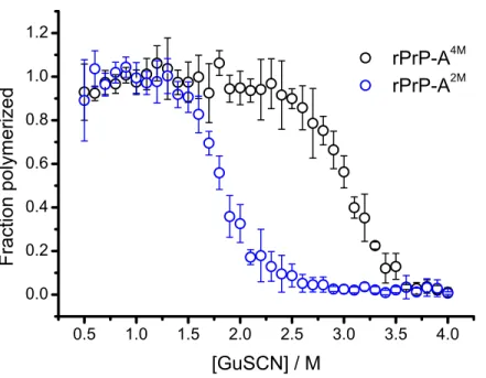
Figure 1 Denaturation profiles of rPrP-A2Mand rPrP-A4Mfibrils in GuSCN reveal different confor-mational stabilities.Standard errors calculated from 6 measurements using Student’st-distribution at P=0.05.
2009). Different strains of recombinant mammalian prion protein amyloid-like fibrils made in 2 and 4 M guanidine hydrochloride (rPrP-A2Mand rPrP-A4M, respectively) were thoroughly characterized by Surewicz group (Cobb et al., 2014). We used recombinant N-terminally truncated mouse prion protein (rMoPrP(89-230)) to create rPrP-A2Mand rPrP-A4Mstrains of amyloid-like fibrils. Similar to recent data on recombinant human PrP (Cobb et al., 2014), rMoPrP fibrils formed in 2 and 4 M guanidine hydrochloride (GuHCl) have different conformational stability (Fig. 1). Due to the fact that rPrP-A4Mfibrils could not be fully denatured using even 7.5 M GuHCl (Cobb et al., 2014), a denaturation assay using a more strongly chaotropic salt, guanidine thiocynate (GuSCN) was performed. Midpoint of denaturation of rPrP-A2Mis at∼1.8 M GuSCN and rPrP-A4Mis at∼3 M GuSCN, respectively. This difference served as a simple, unbiased marker of different strains in further experiments.
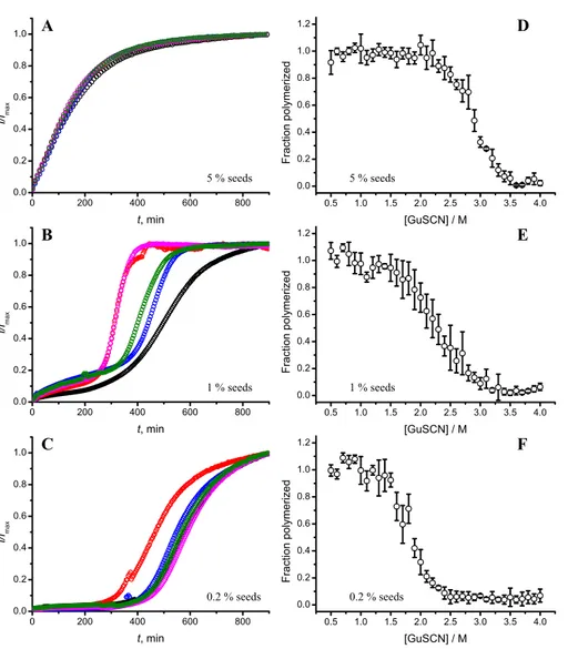
Figure 2 Concentration of seeds determines the mechanism of aggregation and stability of the final strain.Different amounts of rPrP-A4Mfibrils (sonicated for 300 s) were added to the solution of rMoPrP, prepared in 2 M GuHCl, 50 mM phosphate buffer, pH6. The kinetics was followed at 60 ◦C using Thioflavin T (ThT) fluorescence assay, five data repeats at each seed concentration plotted in (A–C). No change of ThT fluorescence was observed in samples without seeds. Denaturation profiles in GuSCN reveal different conformational stabilities of formed fibrils (D–F). Standard errors calculated from 6 measurements using Student’st-distribution atP=0.05.
attributed to fibril-induced secondary nucleation (seeSupplemental Information). The fibril denaturation assay (Fig. 2D) revealed that stability of fibrils formed in the presence of 5% seeds (midpoint at∼2.9 M GuSCN) is very similar to rPrP-A4Mstrain, which was used as a seed. At 1% seed volume (Fig. 2E), stability of fibrils is lower (midpoint at∼2.2 M GuSCN), and at 0.2% of seeds (Fig. 2F) it is the same (midpoint at∼1.8 M GuSCN) as the rPrP-A2Mstrain. This allows hypothesizing that fibrils initiated by secondary nucleation do not follow the seeding template, despite using template fibrils as nucleation sites.
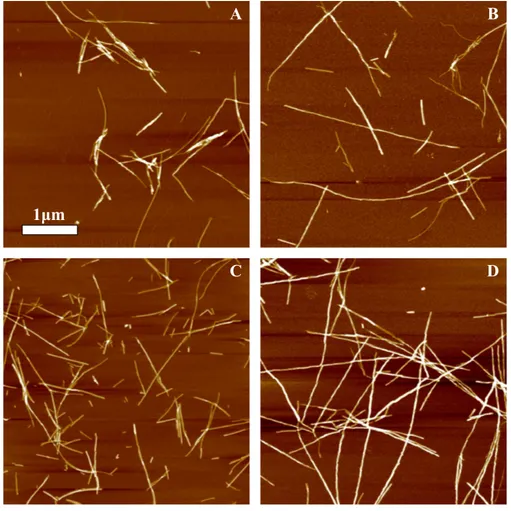
Figure 3 AFM images of rMoPrP amyloid-like aggregates.(A) and (B) show fibrils of rPrP-A4Mand rPrP-A2Mstrains, (C) and (D) show fibrils formed during cross-seeding in the presence of 5% and 0.2% seeds, respectively.
obvious when comparing fibrils formed in presence of 5% and 0.2% seeds (Figs. 3C and3D). The majority of fibrils formed in presence of high amount of seeds are 4–8 nm in diameter, while these formed at low seed concentration are usually 8–16 nm.
FTIR spectra of rMoPrP amyloid-like fibrils display major band in the amide I/I’ region, corresponding to beta-sheet structure with subtle difference in band frequencies between rPrP-A4Mand rPrP-A2Mstrains (Fig. 4). The spectrum of rPrP-A4Mstrain is very similar to the spectrum of fibrils, prepared in the presence of 5% seeds; both show peak maxima at∼1,620 cm−1. The spectrum of rPrP-A2Mstrain and the spectrum of fibrils, prepared in the presence of 0.2% seeds show peak maxima at∼1,624 cm−1. This data serve as additional confirmation that propagation of the strain-specific structure depends on the amount of seeds and possibly on the mechanism of aggregation.
Figure 4 FTIR spectra of rPrP amyloid-like fibrils.
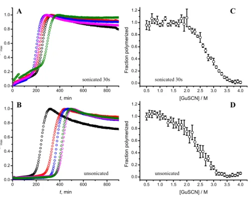
Figure 5 AFM images of rPrP-A4Mfibrils after 300 s (A) and 30 s (B) sonication.
Figure 6 Effect of sonication on the kinetics of aggregation (A–B) and stability of formed fibrils (C–D).The same amount of seeds (5%) was used in all experiments. Five data repeats plotted in (A) and (B). Standard errors calculated from 6 measurements using Student’st-distribution atP=0.05.
DISCUSSION
Taken together the data with different seed concentrations (Fig. 2), and sonication times (Fig. 6), show that stability of fibrils is dependent on the kinetics. Different processes in fibril formation leads to the mixture of rPrP-A4Mand rPrP-A2Mfibril populations in all samples, and different proportions of two strains determine their denaturation profiles. In-crease of fibril ends leads to shorter lag times and faster elongation rates, and to the bigger proportion of more stable fibrils. In fact, we cannot exclude the impact of fibril surface as a catalyzer of secondary nucleation. Larger super-structures can be disrupted by sonication thus releasing more fibril surface. We haven’t found large clumps using microscopy, but the possibility of larger aggregates is suggested by the decline of final fluorescence in samples where mild or no sonication was used. Larger aggregates used as seeds grow further and may settle out of solution leading to the decrese of ThT fluorescence.
Amyloid strain switching has been observed in animal studies (Bartz et al., 2000;Asante et al., 2002;Lloyd et al., 2004;Ghaemmaghami et al., 2013), cell culture (Li et al., 2010), and experimentsin vitro(Castilla et al., 2008;Makarava et al., 2009;Surmacz-Chwedoruk, Babenko & Dzwolak, 2014). Two possibilities are suggested to explain this phenomenon (Collinge & Clarke, 2007;Cobb & Surewicz, 2009). The first one describes coexistence of multiple structures in the infective material, when only the dominant type would be recognized experimentally; however, upon transmission to different host, the minor population may self-propagate much better and become dominant, reflected in the change of strain properties. Recently this way of amyloid strain switching was demonstrated for insulin fibrilsin vitro(Surmacz-Chwedoruk, Babenko & Dzwolak, 2014). The second possibility suggested that sometimes host protein can adopt amyloid conformations distinct from the heterologous template. The Baskakov group demonstrated adaptive conformational switching within individual fibrils as a possible mechanism for such change (Makarava et al., 2009). Our data suggests a possibility of strain switching via secondary nucleation pathways. Moreover, secondary nucleation could explain switching of strains in absence of species barrier, for example in case of recently described Darwinian evolution of prions in cell culture, which showed strain mutations within a single host protein (Li et al., 2010) or in case of protein misfolding cyclic amplification (PMCA) of recombinant PrP (Smirnovas et al., 2009). In summary, we hypothesize that continuous propagation or switching between amyloid strains may be determined by the mechanism of replication in addition to the environment. In cases when a species barrier or environmental barrier stops or slows down fibril elongation, there is the possibility of secondary nucleation events to seed the formation of different strains. The mechanism is dependent on the concentration of fibrils, which opens up a new dimension in cross-species and cross-environment seeding/infection experiments.
We would like to acknowledge that part of the described kinetic profiles differs from the general fibrillation kinetics, normally observed in the field. Thus, in the absence of supporting investigations of different systems, we would like to stress that all our findings may be limited to the described system and any extrapolation to other amyloid proteins and/or other conditions of fibrillation needs an additional experimental evidence.
ACKNOWLEDGEMENTS
The authors thank Prof. Witold Surewicz for sharing MoPrP(89-230) plasmid, Dr. Marija Jankunec for help with AFM, and Dr. Jonathan G. Cannon for critical reading of the manuscript.
ADDITIONAL INFORMATION AND DECLARATIONS
Funding
Grant Disclosures
The following grant information was disclosed by the authors: Research Council of Lithuania: MIP-030/2012.
Marie Curie Career Integration Grant: 293476.
Competing Interests
The authors declare there are no competing interests.
Author Contributions
• Tomas Sneideris and Kataˇzyna Milto conceived and designed the experiments, performed the experiments, analyzed the data, prepared figures and/or tables, reviewed drafts of the paper.
• Vytautas Smirnovas conceived and designed the experiments, analyzed the data, contributed reagents/materials/analysis tools, wrote the paper, prepared figures and/or tables, reviewed drafts of the paper.
Supplemental Information
Supplemental information for this article can be found online athttp://dx.doi.org/ 10.7717/peerj.1207#supplemental-information.
REFERENCES
Aguzzi A. 2014.Neurodegeneration: alzheimer’s disease under strain.Nature512:32–34
DOI 10.1038/512032a.
Angot E, Steiner JA, Hansen C, Li JY, Brundin P. 2010.Are synucleinopathies prion-like disorders?The Lancet Neurology9:1128–1138DOI 10.1016/S1474-4422(10)70213-1.
Arosio P, Cukalevski R, Frohm B, Knowles TPJ, Linse S. 2014. Quantification of the
concentration of Abeta42 propagons during the lag phase by an amyloid chain reaction assay.
Journal of the American Chemical Society136:219–225DOI 10.1021/ja408765u.
Asante EA, Linehan JM, Desbruslais M, Joiner S, Gowland I, Wood AL, Welch J, Hill AF, Lloyd SE, Wadsworth JDF, Collinge J. 2002.BSE prions propagate as either variant CJD-like or sporadic CJD-like prion strains in transgenic mice expressing human prion protein.EMBO Journal21:6358–6366DOI 10.1093/emboj/cdf653.
Bartz JC, Bessen RA, McKenzie D, Marsh RF, Aiken JM. 2000.Adaptation and selection of prion protein strain conformations following interspecies transmission of transmissible mink encephalopathy.Journal of Virology74:5542–5547DOI 10.1128/JVI.74.12.5542-5547.2000.
Bousset L, Pieri L, Ruiz-Arlandis G, Gath J, Jensen PH, Habenstein B, Madiona K, Olieric V, B¨ockmann A, Meier BH, Melki R. 2013.Structural and functional characterization of two alpha-synuclein strains.Nature Communications4:Article 2575DOI 10.1038/ncomms3575.
Brundin P, Melki R, Kopito R. 2010. Prion-like transmission of protein aggregates in neurodegenerative diseases. Nature Reviews. Molecular Cell Biology 11:301–307
DOI 10.1038/nrm2873.
Castilla J, Gonzalez-Romero D, Sa´a P, Morales R, De Castro J, Soto C. 2008.Crossing the species barrier by PrPSc replicationin vitrogenerates unique infectious prions.Cell134:757–768
Chatani E, Yagi H, Naiki H, Goto Y. 2012.Polymorphism of beta 2-microglobulin amyloid fibrils manifested by ultrasonication-enhanced fibril formation in trifluoroethanol.Journal of Biological Chemistry287:22827–22837DOI 10.1074/jbc.M111.333310.
Cobb NJ, Apostol MI, Chen S, Smirnovas V, Surewicz WK. 2014.Conformational stability of mammalian prion protein amyloid fibrils is dictated by a packing polymorphism within the core region.Journal of Biological Chemistry289:2643–2650DOI 10.1074/jbc.M113.520718.
Cobb NJ, Surewicz WK. 2009.Prion diseases and their biochemical mechanisms.Biochemistry
48:2574–2585DOI 10.1021/bi900108v.
Colby DW, Giles K, Legname G, Wille H, Baskakov IV, DeArmond SJ, Prusiner SB. 2009.Design and construction of diverse mammalian prion strains.Proceedings of the National Academy of Sciences of the United States of America106:20417–20422DOI 10.1073/pnas.0910350106.
Colby DW, Prusiner SB. 2011.Prions. Cold Spring Harbor Perspectives in Biology3:1–22
DOI 10.1101/cshperspect.a006833.
Collinge J. 2001.Prion diseases of humans and animals: their causes and molecular basis.Annual Review of Neuroscience24:519–550DOI 10.1146/annurev.neuro.24.1.519.
Collinge J, Clarke AR. 2007.A general model of prion strains and their pathogenicity.Science
318:930–936DOI 10.1126/science.1138718.
Dinkel PD, Siddiqua A, Huynh H, Shah M, Margittai M. 2011. Variations in filament conformation dictate seeding barrier between three- and four-repeat tau.Biochemistry
50:4330–4336DOI 10.1021/bi2004685.
Dzwolak W, Smirnovas V, Jansen R, Winter R. 2004.Insulin forms amyloid in a strain-dependent manner: an FT-IR spectroscopic study.Protein Science: a Publication of the Protein Society
13:1927–1932DOI 10.1110/ps.03607204.
Eisele YS, Oberm¨uller U, Heilbronner G, Baumann F, Kaeser SA, Wolburg H, Walker LC, Staufenbiel M, Heikenwalder M, Jucker M. 2010.Peripherally applied Abeta-containing in-oculates induce cerebral beta-amyloidosis.Science330:980–982DOI 10.1126/science.1194516.
Eisele YS. 2013.From soluble Abeta to progressive Abeta aggregation: could prion-like templated misfolding play a role?Brain Pathology23:333–341DOI 10.1111/bpa.12049.
Frost B, Diamond MI. 2010.Prion-like mechanisms in neurodegenerative diseases. Nature Reviews. Neuroscience11:155–159DOI 10.1038/nrn2786.
Ghaemmaghami S, Colby DW, Nguyen HOB, Hayashi S, Oehler A, Dearmond SJ, Prusiner SB. 2013.Convergent replication of mouse synthetic prion strains.American Journal of Pathology
182:866–874DOI 10.1016/j.ajpath.2012.11.038.
Goedert M, Falcon B, Clavaguera F, Tolnay M. 2014.Prion-like mechanisms in the pathogenesis of tauopathies and synucleinopathies.Current Neurology and Neuroscience Reports14:Article
495DOI 10.1007/s11910-014-0495-z.
Heise H, Hoyer W, Becker S, Andronesi OC, Riedel D, Baldus M. 2005. Molecular-level secondary structure, polymorphism, and dynamics of full-length alpha-synuclein fibrils studied by solid-state NMR.Proceedings of the National Academy of Sciences of the United States of America102:15871–15876DOI 10.1073/pnas.0506109102.
Jones EM, Surewicz WK. 2005. Fibril conformation as the basis of species- and strain-dependent seeding specificity of mammalian prion amyloids. Cell121:63–72
DOI 10.1016/j.cell.2005.01.034.
Jones EM, Wu B, Surewicz K, Nadaud PS, Helmus JJ, Chen S, Jaroniec CP, Surewicz WK. 2011.
Knowles TPJ, Waudby CA, Devlin GL, Cohen SIA, Aguzzi A, Vendruscolo M, Terentjev EM, Welland ME, Dobson CM. 2009.An analytical solution to the kinetics of breakable filament assembly.Science326:1533–1537DOI 10.1126/science.1178250.
Li J, Browning S, Mahal SP, Oelschlegel AM, Weissmann C. 2010.Darwinian evolution of prions in cell culture.Science327:869–872DOI 10.1126/science.1183218.
Lloyd SE, Linehan JM, Desbruslais M, Joiner S, Buckell J, Brandner S, Wadsworth JDF,
Collinge J. 2004.Characterization of two distinct prion strains derived from bovine spongiform encephalopathy transmissions to inbred mice.Journal of General Virology85:2471–2478
DOI 10.1099/vir.0.79889-0.
Lundmark K, Westermark GT, Nystr¨om S, Murphy CL, Solomon A, Westermark P. 2002.
Transmissibility of systemic amyloidosis by a prion-like mechanism.Proceedings of the National Academy of Sciences of the United States of America99:6979–6984DOI 10.1073/pnas.092205999.
Makarava N, Ostapchenko VG, Savtchenko R, Baskakov IV. 2009.Conformational switching within individual amyloid fibrils. Journal of Biological Chemistry284:14386–14395
DOI 10.1074/jbc.M900533200.
Masuda-Suzukake M, Nonaka T, Hosokawa M, Oikawa T, Arai T, Akiyama H, Mann DMA, Hasegawa M. 2013. Prion-like spreading of pathologicalα-synuclein in brain.Brain
136:1128–1138DOI 10.1093/brain/awt037.
Meisl G, Yang X, Hellstrand E, Frohm B, Kirkegaard JB, Cohen SIA, Dobson CM, Linse S, Knowles TPJ. 2014.Differences in nucleation behavior underlie the contrasting aggregation kinetics of the Aβ40 and Aβ42 peptides.Proceedings of the National Academy of Sciences of the United States of America111:9384–9389DOI 10.1073/pnas.1401564111.
Milto K, Michailova K, Smirnovas V. 2014.Elongation of mouse prion protein amyloid-like fibrils: effect of temperature and denaturant concentration. PLoS ONE9:e94469
DOI 10.1371/journal.pone.0094469.
Paravastu AK, Leapman RD, Yau W-M, Tycko R. 2008. Molecular structural basis for polymorphism in Alzheimer’s beta-amyloid fibrils.Proceedings of the National Academy of Sciences of the United States of America105:18349–18354DOI 10.1073/pnas.0806270105.
Petkova AT, Leapman RD, Guo Z, Yau W-M, Mattson MP, Tycko R. 2005.Self-propagating, molecular-level polymorphism in alzheimer’s beta-amyloid fibrils.Science307:262–265
DOI 10.1126/science.1105850.
Prusiner SB. 1998.Prions.Proceedings of the National Academy of Sciences of the United States of America95:13363–13383DOI 10.1073/pnas.95.23.13363.
Safar J, Wille H, Itri V, Groth D, Serban H, Torchia M, Cohen FE, Prusiner SB. 1998.Eight prion strains have PrP(Sc) molecules with different conformations.Nature Medicine4:1157–1165
DOI 10.1038/2654.
Sim VL, Caughey B. 2009.Ultrastructures and strain comparison of under-glycosylated scrapie prion fibrils.Neurobiology of Aging30:2031–2042DOI 10.1016/j.neurobiolaging.2008.02.016.
Smirnovas V, Kim J Il, Lu X, Atarashi R, Caughey B, Surewicz WK. 2009.Distinct structures of scrapie prion protein (PrPSc)-seeded versus spontaneous recombinant prion protein fibrils revealed by hydrogen/deuterium exchange.Journal of Biological Chemistry284:24233–24241
DOI 10.1074/jbc.M109.036558.
Soto C, Estrada L, Castilla J. 2006. Amyloids, prions and the inherent infectious nature of misfolded protein aggregates. Trends in Biochemical Sciences 31:150–155
DOI 10.1016/j.tibs.2006.01.002.
Prusiner SB. 2014.Distinct synthetic Aβ prion strains producing different amyloid deposits in bigenic mice.Proceedings of the National Academy of Sciences of the United States of America
111:2–7DOI 10.1073/pnas.1408968111.
Surmacz-Chwedoruk W, Babenko V, Dzwolak W. 2014.Master and slave relationship between two types of self-propagating insulin amyloid fibrils.The Journal of Physical Chemistry B
118:13582–13589DOI 10.1021/jp510980b.
Tanaka M, Chien P, Naber N, Cooke R, Weissman JS. 2004. Conformational variations in an infectious protein determine prion strain differences. Nature 428:323–328
DOI 10.1038/nature02392.
Tanaka M, Chien P, Yonekura K, Weissman JS. 2005.Mechanism of cross-species prion transmission: an infectious conformation compatible with two highly divergent yeast prion proteins.Cell121:49–62DOI 10.1016/j.cell.2005.03.008.
Tycko R. 2014.Physical and structural basis for polymorphism in amyloid fibrils.Protein Science: a Publication of the Protein Society23:1528–1539DOI 10.1002/pro.2544.
Watts JC, Condello C, St¨ohr J, Oehler A, Lee J, DeArmond SJ, Lannfelt L, Ingelsson M, Giles K, Prusiner SB. 2014. Serial propagation of distinct strains of Aβ prions from alzheimer’s disease patients.Proceedings of the National Academy of Sciences111:10323–10328
DOI 10.1073/pnas.1408900111.
Westermark GT, Westermark P. 2010.Prion-like aggregates: infectious agents in human disease.
Trends in Molecular Medicine16:501–507DOI 10.1016/j.molmed.2010.08.004.
Yamaguchi KI, Katou H, Hoshino M, Hasegawa K, Naiki H, Goto Y. 2004. Core and