Clinical Features Indicating Nigrostriatal
Dopaminergic Degeneration in Drug-Induced
Parkinsonism
Seung Ha Lee,1 Han Kyeol Kim,1 Young Gun Lee,1 Chul Hyoung Lyoo,1 Sung Jun Ahn,2 Myung Sik Lee1
1Departments of Neurology and 2Radiology, Yonsei University College of Medicine, Gangnam Severance Hospital, Seoul, Korea
ABSTRACT
ObjectiveaaPatients with drug-induced parkinsonism (DIP) may have nigrostriatal dopaminergic degeneration. We studied the clinical features that may indicate nigrostriatal dopaminergic degeneration in patients with DIP.
MethodsaaForty-one DIP patients were classiied into normal and abnormal [18F] FP-CIT scan groups. Diferences in 32 clini-cal features and drug withdrawal efects were studied.
ResultsaaTwenty-eight patients had normal (Group I) and 13 patients had abnormal (Group II) scans. Eight patients of Group I, but none of Group II, had taken calcium channel blockers (p = 0.040). hree patients of Group I and six of Group II had hy-posmia (p = 0.018). Ater drug withdrawal, Group I showed greater improvement in Uniied Parkinson’s Disease Rating Scale total motor scores and subscores for bradykinesia and tremors than Group II. Only hyposmia was an independent factor associ-ated with abnormal scans, but it had suboptimal sensitivity.
ConclusionaaNone of the clinical features were practical indicators of nigrostriatal dopaminergic degeneration in patients with DIP.
Key WordsaaDrug-induced parkinsonism; dopamine transporter; positron-emission tomography; hyposmia.
ORIGINAL ARTICLE
https://doi.org/10.14802/jmd.16045 / J Mov Disord 2017;10(1):35-39 pISSN 2005-940X / eISSN 2093-4939
Received: September 27, 2016 Revised: November 9, 2016 Accepted: November 10, 2016
Corresponding author: Myung Sik Lee, MD, PhD, Department of Neurology, Yonsei University College of Medicine, Gangnam Severance Hospital, 211 Eonju-ro, Gangnam-gu, Seoul 06273, Korea / Tel: +82-2-2019-3322 / Fax: +82-2-3462-5904 / E-mail: [email protected]
cc his is an Open Access article distributed under the terms of the Creative Commons Attribution Non-Commercial License (http://creativecommons.org/ licenses/by-nc/3.0) which permits unrestricted non-commercial use, distribution, and reproduction in any medium, provided the original work is properly cited.
JMD
Nigrostriatal dopaminergic degeneration is found in 30 to 75% of patients clinically diagnosed with drug-induced parkin-sonism (DIP).1-4 Dopamine transporter (DAT) single photon emission computed tomography (SPECT) and photon emission tomography (PET) studies of DIP showed that patients with normal scans have dopa-resistant parkinsonism that may not progress even if the ofending drug treatment is continued. Con-versely, patients with reduced striatal uptake have dopa-respon-sive parkinsonism that may deteriorate even ater drug withdraw-al.3-6 hus, clinical features must be deined to help diferentiate DIP patients with normal and abnormal scans. DAT imaging evaluates the striatal presynaptic dopaminergic function and is useful for the diferential diagnosis of parkinsonism and the
as-sessment of severity and progression.7,8 Additionally, [18F] N-(3-fluoropropyl)-2β-carbon ethoxy-3β-(4-iodophenyl) nortropane (FP-CIT) is superior to SPECT because of its better spatial reso-lution.8
J Mov Disord 2017;10(1):35-39
JMD
However, those four studies included only 13 to 22 patients with DIP.
In 41 patients with DIP, we studied demographic and clinical parameters that may differentiate DIP patients with normal and abnormal [18F] FP-CIT PET scans. In 31 of the 41 patients, we studied wheth-er DIP patients with normal and abnormal scans could be diferentiated by changes in Uniied Parkin-son’s Disease Rating Scale (UPDRS) scores ater drug withdrawal.
MATERIALS & METHODS
Patients
We reviewed the medical records of 41 DIP pa-tients who underwent brain MRI and DAT PET studies. DIP was diagnosed according to parameters described in a previous report: the presence of two or more cardinal symptoms of parkinsonism, no his-tory of parkinsonism before use of the offending drug and onset of parkinsonism symptoms during use of the ofending drug.13 Patients were excluded if they had signs compatible with atypical parkinson-ism and other causes of secondary parkinsonparkinson-ism. In 31 of the 41 patients, we evaluated changes in par-kinsonian motor deicits ater withdrawal of the of-fending drug. he present study was approved by the Institutional Review Board of the Gangnam Sever-ance Hospital, Yonsei University College of Medicine (IRB # 3-2016-0018).
Methods
We recorded the demographics, duration of par-kinsonism, family history of parpar-kinsonism, smoking history, and offending drug histories. We collected data about falls, aspiration, dysarthria, and BLM dyskinesia. We registered initial parkinsonian symp-toms including asymmetry. We collected data on non-motor symptoms and Mini-Mental State Ex-amination (MMSE) scores.
We measured UPDRS motor scores and counted patients who presented with asymmetric parkinso-nian motor deficits (≥ 4 side to side difference in UPDRS motor score).9 We also computed indexes of asymmetric parkinsonian motor deficits (sum of UPDRS motor scores of the more affected side di-vided by that of the less afected side) and dispropor-tionate arm involvement (sum of UPDRS motor scores of the arms divided by that of the legs). In 31
of 41 patients, changes in UPDRS total motor scores and subscores for bradykinesia, rigidity, tremors, and axial motor deicits were measured ater withdrawal of the drug for at least 2 months.
Quantitative measurements of PET images were performed as reported previously by our study group.14 We considered a scan as abnormal when the mean bilateral putamen DAT uptake was reduced more than two standard deviations from that of 36 age-matched healthy controls who had no history of neurological illness and no abnormal signs on neu-rological examination. We classified patients into normal and abnormal scan groups.
Statistics
Using chi-square tests (Fisher’s exact tests) for caegorical variables and independent two sample t-tests for continuous variables, we examined the group diferences for all the parameters. For the parameters with significant group differences, we performed univariate and multivariate backward binary logistic regression analyses controlled for age and sex to identify independent factors in distinguishing DIP with normal and abnormal scans. For the use of cal-cium channel blockers (CCBs), exact binary logistic regression analysis was performed using Statistical Analysis System sotware (SAS version 9.1; SAS Inc., Cary, NC, USA). All other statistical analyses were performed using SPSS (version 23.0; SPSS Inc., Chi-cago, IL, USA). he results were considered statisti-cally signiicant when the p-value was < 0.05.
RESULTS
Twenty-eight patients (Group I; 68.3%) had nor-mal scans and 13 (Group II; 31.7%) had abnornor-mal scans. The mean age at onset was similar between Group I and II. Women were more prevalent than men in both groups. here was no signiicant group difference in female predominance. The means of the duration of parkinsonism were not signiicantly diferent (Table 1).
Dopamine PET in Drug-Induced Parkinsonism Lee SH, et al.
was not signiicant (Table 1).
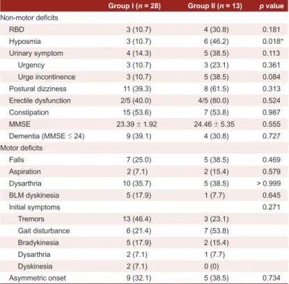
The frequencies of various non-motor deficits were not signiicantly diferent between groups. How-ever, compared to Group I, Group II patients had more frequent hyposmia (p = 0.018). here was no significant group difference in the mean MMSE scores or the frequency of dementia (MMSE score ≤ 24) (Table 2).
Compared to Group I, Group II patients more of-ten had a history of falls, aspiration, and dysarthria, but the diferences were not signiicant. BLM dyski-nesia was uncommon in both groups. In both groups, tremors and gait disturbances were common initial symptoms. here was no signiicant diference in the frequency of asymmetric onset (Table 2).
he means of UPDRS total motor scores of Group I and II were similar. In addition, there were no sig-niicant diferences in subscores for bradykinesia, ri-gidity, tremor or axial motor impairments. Four pa-tients (14.3%) of Group I and three papa-tients (23.1%) of Group II had asymmetric parkinsonism. There were no signiicant diferences in the UPDRS index-es of asymmetry or disproportionate upper-to-lower involvement of the extremities (Supplementary Table 2 in the only online Data Supplement).
In 31 patients (Group I = 20 patients; Group II = 11 patients), changes in UPDRS scores were mea-sured after a mean of 4.03 months of drug with-drawal. Compared to Group II, Group I showed greater reductions in UPDRS total motor scores (-8.75 ± 9.16 vs. 0.18 ± 7.76; p = 0.011) and subscores for bradykinesia (p = 0.029), total tremors (p = 0.019), and resting tremors (p = 0.021). However, there were no diferences in residual parkinsonian motor dei-cits and changes in the indexes of asymmetry or dis-proportionate upper-to-lower involvement of the ex-tremities (Supplementary Table 2 in the only online Data Supplement).
Univariate regression analyses showed that the fre-quency of hyposmia (p = 0.017) and greater reduc-tions in total motor scores (p = 0.040) and subscores for bradykinesia (p = 0.047) after drug withdrawal were associated with abnormal scans (Supplementary Table 3 in the only online Data Supplement). Using multivariate regression analysis, only the frequency of hyposmia was an independent factor associated with abnormal scans (p = 0.030) (Supplementary Table 3 in the only online Data Supplement). Hyposmia had relatively high speciicity (89.3%) and negative
pre-dictive value (78.1%) but had low sensitivity (46.2%) and positive predictive value (66.7%) for abnormal scans.
DISCUSSION
In the present study, we performed a systematic analysis of 32 clinical parameters between patients with normal and abnormal DAT scans. Compared to DIP patients with normal scans, those with ab-normal scans had more frequent hyposmia. No
pa-Table 1. Demographics and clinical characteristics of 41 patients with drug-induced parkinsonism with normal and reduced putamen FP-CIT uptake
Group I (n = 28) Group II (n = 13) p value
Age at onset (yrs) 71.29 ± 9.34 67.38 ± 8.88 0.214
Age at examination (yrs) 72.72 ± 9.68 68.46 ± 8.84 0.187
Gender (male: female) 5:23 (82.1) 5:8 (61.5) 0.241
Duration of drug exposure (mo)* 4.66 ± 3.79 3.04 ± 2.96 0.182
Duration of parkinsonism (mo)† 14.16 ± 14.54 15.54 ± 15.44 0.783
Family history of parkinsonism 2 (7.1) 1 (7.7) > 0.999
Smoking history 1 (3.6) 2 (15.4) 0.232
Group I: normal putamen FP-CIT uptake, Group II: reduced putamen FP-CIT uptake. Mean
± standard deviation, numbers in parentheses = %. *duration of drug exposure prior to symptom onset (mo), †duration of parkinsonism before diagnosis (mo).
Table 2. Comparisons of non-motor and motor deicits between 41 drug-induced par -kinsonism patients with normal and reduced putamen FP-CIT uptake
Group I (n = 28) Group II (n = 13) p value
Non-motor deicits
RBD 3 (10.7) 4 (30.8) 0.181
Hyposmia 3 (10.7) 6 (46.2) 0.018*
Urinary symptom 4 (14.3) 5 (38.5) 0.113
Urgency 3 (10.7) 3 (23.1) 0.361
Urge incontinence 3 (10.7) 5 (38.5) 0.084
Postural dizziness 11 (39.3) 8 (61.5) 0.313
Erectile dysfunction 2/5 (40.0) 4/5 (80.0) 0.524
Constipation 15 (53.6) 7 (53.8) 0.987
MMSE 23.39 ± 1.92 24.46 ± 5.35 0.555
Dementia (MMSE ≤ 24) 9 (39.1) 4 (30.8) 0.727
Motor deicits
Falls 7 (25.0) 5 (38.5) 0.469
Aspiration 2 (7.1) 2 (15.4) 0.579
Dysarthria 10 (35.7) 5 (38.5) > 0.999
BLM dyskinesia 5 (17.9) 1 (7.7) 0.645
Initial symptoms 0.271
Tremors 13 (46.4) 3 (23.1)
Gait disturbance 6 (21.4) 7 (53.8)
Bradykinesia 5 (17.9) 2 (15.4)
Dysarthria 2 (7.1) 1 (7.7)
Dyskinesia 2 (7.1) 0 (0)
Asymmetric onset 9 (32.1) 5 (38.5) 0.734
Group I: normal putamen FP-CIT uptake, Group II: reduced putamen FP-CIT uptake. Mean
J Mov Disord 2017;10(1):35-39
JMD
tients with reduced putamen DAT uptake had been exposed to CCBs. hey showed deterioration or less improvement in UPDRS total motor scores and sub-scores for bradykinesia and tremors ater drug with-drawal. However, only hyposmia was associated in-dependently with abnormal scans at baseline.
A DAT SPECT study reported that DIP patients with abnormal scans were significantly older than those with normal scans.9 However, the present study and other studies have shown no signiicant difer-ence in age of onset.1-4,10,15
An Italian group reported no female predomi-nance in DIP patients with normal and reduced stri-atal DAT uptake.4,9 However, two other studies from the same group and the present study found similar female predominance in DIP patients with normal and abnormal scans.2,3 These findings suggest the high vulnerability of females to DIP, even in females with reduced nigrostriatal dopaminergic innerva-tion.16
An FP-CIT PET study reported lower symmetric frequency of parkinsonism in the abnormal scan than the normal scan.10 However, the present study showed no signiicant diference in asymmetric par-kinsonism as measured by frequency, UPDRS score and asymmetry index. he previous study also showed that the interval from drug intake to the onset of par-kinsonian symptoms was longer in patients with an abnormal scan than a normal scan. However, the pres-ent study showed no significant difference between the two groups.
he drug exposure period before the onset of par-kinsonism was longer in patients treated with CCBs than those treated with dopamine blockers.17 Tremor was the main symptom induced by CCBs, whereas ri-gidity was the most common feature induced by other ofending drugs.17 In the present study, eight patients with normal scans had taken CCBs, while none with abnormal scans had taken CCBs (p = 0.040). Al-though the difference was not significant using re-gression analyses, large studies are needed to deter-mine whether CCBs can rarely uncover subclinical nigrostriatal dopaminergic degeneration.
A DAT SPECT study of DIP reported that patients with abnormal scans had abnormal odor threshold, discrimination and identification, while patients with normal scans had abnormal odor threshold.18 In the present study, 46% of patients with abnormal scans reported hyposmia compared to 11% of those
with normal scans. Although a history of hyposmia was an independent factor, its suboptimal sensitivity for the detection of abnormal scans in DIP limits its use in clinical practice.
In the present study, 39% of patients with abnor-mal scans had urge incontinence. heir mean dura-tion of parkinsonism was only one year. Postmortem pathological studies are needed to deine how many of these patients have multiple system atrophy.
A DAT SPECT study of DIP reported that BLM dyskinesia is more frequent in patients with normal scans than in those with abnormal scans. Most pa-tients included in that study had schizophrenia and were being treated with neuroleptics.2 The present study of patients with diferent illnesses treated using a variety of drugs did not show diferences in the fre-quency of BLM dyskinesia.15
No previous dopaminergic brain imaging studies compared diferences in the initial symptoms of DIP patients with normal and abnormal scans. Only one PET study compared the frequency of tremors at rest and orolingual dyskinesia.10 However, they did not compare other initial symptoms. In the present study, the most common initial symptoms were tremors in patients with normal scans and gait disturbances in patients with abnormal scans. However, there were no signiicant group diferences in the frequencies of initial symptoms, including asymmetric onset.
Two SPECT studies and one PET study reported that DIP patients with abnormal scans had more se-vere and asymmetric parkinsonism.2,4,10 However, in the present study, patients with abnormal scans had no significant differences in UPDRS motor scores nor the frequencies and severities of asymmetric parkinsonism.1,9 The present study also showed no significant group differences in UPDRS subscores for axial motor deficits and indexes for dispropor-tionate upper-to-lower involvement of the extremi-ties.
Complete recovery ater drug withdrawal has been considered an indicator of normal nigrostriatal do-paminergic function in patients with DIP.15,19 In the present study, none of the UPDRS changes indepen-dently indicated an abnormal scan. In addition, pa-tients with normal and abnormal scans showed a similar degree of residual parkinsonian motor dei-cits.
with-Dopamine PET in Drug-Induced Parkinsonism Lee SH, et al.
drawal, only hyposmia was independently associated with nigrostriatal dopaminergic degeneration in pa-tients with DIP. However, hyposmia does not appear to be a practical indicator of abnormal scans. Dopa-minergic functional imaging studies are needed to detect nigrostriatal degeneration in patients clinically diagnosed with DIP.
Supplementary Materials
he online-only Data Supplement is available with this arti-cle at https://doi.org/10.14802/jmd.16045.
Conflicts of Interest
he authors have no inancial conlicts of interest.
REFERENCES
1. Lorberboym M, Treves TA, Melamed E, Lampl Y, Hell-mann M, Djaldetti R. [123I]-FP/CIT SPECT imaging for distinguishing drug-induced parkinsonism from Parkin-son’s disease. Mov Disord 2006;21:510-514.
2. Tinazzi M, Ottaviani S, Isaias IU, Pasquin I, Steinmayr M, Vampini C, et al. [123I]FP-CIT SPET imaging in drug-in-duced Parkinsonism. Mov Disord 2008;23:1825-1829. 3. Tinazzi M, Antonini A, Bovi T, Pasquin I, Steinmayr M,
Moretto G, et al. Clinical and [123I]FP-CIT SPET imaging follow-up in patients with drug-induced parkinsonism. J Neurol 2009;256:910-915.
4. Tinazzi M, Morgante F, Matinella A, Bovi T, Cannas A, Solla P, et al. Imaging of the dopamine transporter predicts pattern of disease progression and response to levodopa in patients with schizophrenia and parkinsonism: a 2-year follow-up multicenter study. Schizophr Res 2014;152:344-349.
5. Burn DJ, Brooks DJ. Nigral dysfunction in drug-induced parkinsonism: an 18F-dopa PET study. Neurology 1993; 43(3 Pt 1):552-556.
6. Hong JY, Sunwoo MK, Oh JS, Kim JS, Sohn YH, Lee PH. Persistent drug-induced parkinsonism in patients with normal dopamine transporter imaging. PLoS One 2016; 11:e0157410.
7. Marshall V, Grosset D. Role of dopamine transporter im-aging in routine clinical practice. Mov Disord 2003;18:1415-1423.
8. Park E, Hwang YM, Lee CN, Kim S, Oh SY, Kim YC, et al. Diferential diagnosis of patients with inconclusive parkin-sonian features using [(18)F]FP-CIT PET/CT. Nucl Med Mol Imaging 2014;48:106-113.
9. Tinazzi M, Cipriani A, Matinella A, Cannas A, Solla P, Ni-coletti A, et al. [¹²³I]FP-CIT single photon emission com-puted tomography findings in drug-induced Parkinson-ism. Schizophr Res 2012;139:40-45.
10. Shin HW, Kim JS, Oh M, You S, Kim YJ, Kim J, et al. Clini-cal features of drug-induced parkinsonism based on [18F] FP-CIT positron emission tomography. Neurol Sci 2015; 36:269-274.
11. Hambÿe AS, Vervaet A, Dethy S. FP-CIT SPECT in clini-cally inconclusive Parkinsonian syndrome during amioda-rone treatment: a study with follow-up. Nucl Med Com-mun 2010;31:583-589.
12. Olivares Romero J, Arjona Padillo A. Diagnostic accuracy of 123 I-FP-CIT SPECT in diagnosing drug-induced par-kinsonism: a prospective study. Neurologia 2013;28:276-282.
13. Jiménez-Jiménez FJ, Ortí-Pareja M, Ayuso-Peralta L, Gas-alla T, Cabrera-Valdivia F, Vaquero A, et al. Drug-induced parkinsonism in a movement disorders unit: a four-year survey. Parkinsonism Relat Disord 1996;2:145-149. 14. Kim JS, Cho H, Choi JY, Lee SH, Ryu YH, Lyoo CH, et al.
Feasibility of computed tomography-guided methods for spatial normalization of dopamine transporter positron emission tomography image. PLoS One 2015;10:e0132585. 15. Diaz-Corrales FJ, Sanz-Viedma S, Garcia-Solis D, Escobar-Delgado T, Mir P. Clinical features and 123I-FP-CIT SPECT imaging in drug-induced parkinsonism and Parkinson’s disease. Eur J Nucl Med Mol Imaging 2010;37:556-564. 16. Ayd FJ Jr. A survey of drug-induced extrapyramidal
reac-tions. JAMA 1961;175:1054-1060.
17. Bondon-Guitton E, Perez-Lloret S, Bagheri H, Brefel C, Rascol O, Montastruc JL. Drug-induced parkinsonism: a review of 17 years’ experience in a regional pharmacovigi-lance center in France. Mov Disord 2011;26:2226-2231. 18. Bovi T, Antonini A, Ottaviani S, Antonioli A, Cecchini MP,
Di Francesco V, et al. he status of olfactory function and the striatal dopaminergic system in drug-induced parkin-sonism. J Neurol 2010;257:1882-1889.