https://doi.org/10.1590/0004-282X20180076
ARTICLE
Blepharospasm with elevated
anti-acetylcholine receptor antibody titer
Blefaroespasmo com título elevado de anticorpos antirreceptores de acetilcolina
Min Tang
1, Wu Li
2, Ping Liu
1, Fangping He
1, Fang Ji
1, Fanxia Meng
1Blepharospasm (BSP) is a focal dystonia involving the
orbicularis oculi and accessory muscles, leading to
involun-tary, inappropriate eyelid closure
1. The majority of patients
with myasthenia gravis (MG), a nerve-muscle junction dis
-ease, have generalized fluctuating weakness and ocular
muscle fatigue. An increase of the anti-acetylcholine
recep-tor antibody (anti-AChR-Abs) titer is one of the specific tests
for MG. Anti-AChR-Abs are highly disease-specific and are
detected in approximately 80–85% of patients with
general-ized MG and 50% of patients with ocular MG
2. However,
ele-vated anti-AChR Abs have been found in patients who have
both MG and another disease such as Guillain-Barré
syn-drome
3, amyotrophic lateral sclerosis
4and myotonic
dystro-phy type 2
5. BSP and MG are two distinct diseases. The for
-mer is distinguished from typical conditions of MG in which
eyelid closure is due to weakness of the levator palpebrae
muscles. However, Kurlan et al.
6reported five patients with
coexistent Meige’s syndrome and MG. In the current study,
we found that anti-AChR Abs levels were elevated in patients
with BSP who did not have MG. The aim of this study was to
evaluate whether the level of anti-AChR Abs is linked to the
clinical parameters of BSP.
1Department of Neurology,First Affiliated Hospital, Zhejiang University School of Medicine, 79 Qingchun Road, Hangzhou 310003, China. 2Tongde Hospital of Zhejiang Province, Hangzhou, Zhejiang, Province, P.R. China.
Correspondence: Min Tang; Department of Neurology,First Affiliated Hospital,School of Medicine, Zhejiang University,79 Qingchun Road, Zhejiang, Hangzhou 310003, China; E-mail: [email protected]
Conflict of interest: There is no conflict of interest to declare.
Support: Natural Science Fund of Zhejiang Province [grant no. LQ14H090002] and the Traditional Chinese Medicine Science and Technology Plan of Zhejiang Province [grant no.2015ZQ023].
Received 26 November 2017; Received in final form 10 April 2018; Accepted 04 May 2018.
ABSTRACT
Objective:
To determine whether serum levels of anti-acetylcholine receptor antibody (anti-AChR-Abs) are related to clinical parameters of
blepharospasm (BSP).
Methods:
Eighty-three adults with BSP, 60 outpatients with hemifacial spasm (HFS) and 58 controls were recruited.
Personal history, demographic factors, response to botulinum toxin type A (BoNT-A) and other neurological conditions were recorded.
Anti-AChR-Abs levels were quantified using an enzyme-linked immunosorbent assay.
Results:
The anti-AChR Abs levels were 0.237 ± 0.022
optical density units in the BSP group, which was significantly different from the HFS group (0.160 ± 0.064) and control group (0.126 ± 0.038).
The anti-AChR Abs level was correlated with age and the duration of response to the BoNT-A injection.
Conclusion:
Patients with BSP had
an elevated anti-AChR Abs titer, which suggests that dysimmunity plays a role in the onset of BSP. An increased anti-AChR Abs titer may be
a predictor for poor response to BoNT-A in BSP.
Keywords:
Blepharospasm, hemifacial spasm, botulinum toxin type A.
RESUMO
Objetivo:
Determinar se os níveis séricos do anticorpo antirreceptor de acetilcolina (anti-AChR-Abs) estão relacionados aos parâmetros
clínicos do blefaroespasmo (BSP).
Métodos:
Fora recrutados 83 adultos com BSP, 60 pacientes ambulatoriais com espasmo hemifacial
(HFS) e 58 controles. Foi aplicado um questionário para registrar história pessoal, fatores demográficos, resposta à toxina botulínica
tipo A (BoNT-A) e outras condições neurológicas. Os níveis de anti-AChR-Abs foram quantificados usando um ensaio imunoenzimático.
Resultados:
O nível de anti-AChR-Abs foi de 0,237 ± 0,022 unidades de densidade óptica (OD) no grupo BSP, significativamente diferente
em comparação com o grupo HFS (0,160 ± 0,064) e o grupo controle (0,126 ± 0,038). O nível de anti-AChR-Abs se correlacionou com a idade e
a duração da resposta à injeção de BoNT-A.
Conclusão:
Pacientes com BSP apresentaram títulos elevados de anti-AChR-Abs, o que sugere
que a desimunidade desempenha um papel no surgimento de BSP. O aumento do título de anti-AChR-Abs pode ser um preditor de resposta
insuficiente à BoNT-A em BSP.
METHODS
Study population
Eighty-three patients with BSP were enrolled from June
2013 to June 2016 in the Department of Neurology of the First
Affiliated Hospital of Zhejiang University. BSP was diagnosed
according to published criteria
7. These cases were age- and
sex-matched to 60 outpatients with hemifacial spasm (HFS;
hospital controls) and 58 healthy people (population
con-trols). This study was approved by the ethics committee of
the hospital. Written informed consent was obtained from
all participants. Clinical outcomes were evaluated in terms
of latency, duration of effect, and clinical response, which was
evaluated by means of a modified Jankovic Rating Scale
8. We
assessed the patients before receiving a botulinum toxin type
A (BoNT-A) injection and one month after the injections,
using the Jankovic Rating Scale. We defined poor respond
-ers (treatment failures) as those who, subjectively or from
the doctor’s observation, did not achieve adequate symptom
relief (primary or secondary non-responders).
Anti-AChR-Abs levels measurement by
enzyme-linked immunosorbent assays (ELISA)
Plasma samples from the three groups were collected.
All patient samples were tested for antibody binding to
human muscle AChR using an ELISA kit (Cusabio; Barksdale,
Delaware; USA), following the manufacturer’s instructions
9.
Samples were placed in a serum separator tube and stored
overnight at 4°C to clot. The samples were centrifuged at
approximately 3,000 rpm for 15 minutes. Serum was decanted
and diluted 101-fold and assayed immediately. Wells were
filled with 100 µL of sample. Phosphate buffered saline (pH
7.0–7.2) was used as a blank control. A microplate reader
(Biomad, USA) was used to measure the optical density of
each sample at 450 nm. Samples were measured in triplicate.
Statistical analysis
Data are expressed as mean ± SD. ANOVA was used to
compare averages. Anti-AChR-Abs levels were correlated
with other parameters using Pearson’s parametric
correla-tion analysis. A value of p < 0.05 was considered statistically
significant.
RESULTS
Demographics of the study subjects.
Sixty women (72%) and 23 men (28%) were enrolled
in this study. The mean age of BSP onset was 55 ± 11 years
(range, 23–79 years). The percentage of patients with BSP
onset varied with age: five (6%) before or at age 40 years, 36
(43.4%) between ages 41 and 50 years, 23 (27.7%) between
ages 51 and 60 years, and 19 (22.9%) older than 60 years.
For diagnoses, 53 (64%) patients had BSP and 30 (36%) had
Meige’s syndrome. Of the 83 patients with BPS, 47 (56.6%)
had sensory tricks (Figure 1). The mean duration of the
disease was 31 ± 27 months (0.5–180 months). Thirty-five
patients, who had abnormal anti-AChR Abs levels (higher
than the normal reference range), received a single or a
combination of tests related to MG. These tests included a
repetitive nerve stimulation test in 34 patients, a
neostig-mine test in two patients, chest computed-tomography in 30
patients and single-fiber electromyography in 22 patients.
All 35 of the patients who underwent the tests related to
MG had negative results. The mean duration of the BoNT-A
injection treatment was 2.1 ± 0.7 years (range, 0–5 years).
The mean number of injections was 1.6 ± 0.3 (range, 0–8).
The mean dose per treatment was 36 ± 3 units (range, 28–80
units). The mean duration of response to BoNT-A was 1.6 ±
0.3 months (range, 0.5–8 months).
Serum anti-AChR Abs levels in patients with BSP,
HFS and healthy controls
The serum level of anti-AChR Abs was significantly higher
(p < 0.001) in patients with BSP compared with patients with
HFS and the healthy controls (Table). There was no signifi
-cant difference (p > 0.05) in serum anti-AChR Abs levels
between the BSP group with and without sensory tricks, and
the left HFS and right HFS groups (Table).
Correlation between serum anti-AChR Abs levels
and sex, age, duration of disease, duration of
response to BoNT-A and Jankovic Rating Scale
scores in patients with BSP
The level of serum anti-AChR Abs increased with age
(Figure 2B), and decreased with the duration of time after
the BoNT-A treatment (Figure 2E; r
2= 0.2365), which
sug-gests that high levels of anti-AChR Abs indicate a poor
response to BoNT-A treatment. One female patient, who
had an increased anti-AChR Abs level as high as three
times that of the controls, had only two to three weeks of
relief after the BoNT-A treatment. This patient had a repeti
-tive nerve stimulation test
10and single-fiber electromyog
-raphy. The results were normal, and the neostigmine test
was negative. The chest computed tomography, free thyrox
-ine, free triiodothyron-ine, total thyrox-ine, total
triiodothy-ronine, thyrotropin, the thyroid peroxidase antibody and
antinuclear antibody levels were all normal. She
under-went an electromyography test of the orbicularis oculi
mus-cle, which revealed continuous motor unit potentials. The
patient did not complain of weakness in other muscles,
and all the results confirmed the diagnosis of BSP rather
than of MG. The patient accepted an injection of BoNT-A
at the total dose 50 U. There was no obvious change after
the injection.
DISCUSSION
Our major finding of this study was that the level of
anti-AChR Abs in BSP patients was higher than the levels in patients
with HFS and healthy controls. Anti-AChR antibodies are
typi-cally IgG1 or IgG3, with a high affinity for AChR, and a dissocia
-tion constant of 10
-10M anti-AChR Abs are specific for patients
with MG
11;
and MG and BSP coexist in some patients
6,12,13,
sug-gesting a strong association between BSP and MG. The com
-mon features for both MG and BSP include predominant eyelid
involvement, ptosis with unequal palpebral fissures, rest benefit,
diurnal change, aggravation from reading and fatigue, excessive
blinking, and photophobia. The presence of symptoms of ocular
surface irritation and concomitant lower facial or cervical
dys-tonia are more common in patients with BSP. One hypothesized
mechanism for the coexistence of BSP and MG is that
myas-thenic ocular muscle weakness produces abnormal feedback
to the central nervous system in patients with BSP and MG
14,15.
Abnormalities in peripheral afferent inputs, such as continual
ptosis and eye blinking, may interfere with motor program exe
-cution. Another hypothesis is that eye blinking compensates for
eye positional errors resulting from a fatigued extraocular
mus-cle. Frequent compensatory blinking may produce abnormal
feedback to the central nervous system.
In this study, the BSP patients had increased anti-AChR
Abs levels only and had no other clinical signs of MG. None of
our patients developed MG or a weakness of other muscles.
As well, in this study, some of the BSP patients with high
anti-AChR Abs levels had sensory tricks, which are seldom pres
-ent in MG. Electromyography of the BSP pati-ents’ orbicularis
showed spasm potentials (Figure 2). Therefore, the AChR
antibody was a ‘false-positive’ test for MG. The presence of
autoantibodies against AChR indicate a possible pathogenic
relationship with the development of BSP.
Rate: 26.8Hz
Trig:
Off
10/11s
1
100 uV
Amp 4: 20-3k, 50Hz
50 ms
A
B
C
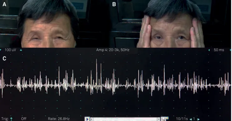
Figure 1.
Sensory tricks and orbicularis electromyography of a blepharospasm patient with high anti-AChR Abs level. (A) The
movements of eyelid closing particularities in a blepharospasm patient with a high anti-AChR Abs level. (B) Sensory trick in
this patient. When she put her hands beside her eyes, the symptoms of spasm were dramatically alleviated. (C) orbicularis
electromyography of the patient showing spasm potentials.
Table.
Levels of anti-AChR Abs in different groups.
Group
N
Anti-AChR-Abs
(optical density)
Blepharospasm
83
0.237 ± 0.022*
Blepharospasm with sensory trick
47
0.236 ± 0.014**
Blepharospasm without sensory trick
36
0.239 ± 0.021
Hemifacial spasm
60
0.160 ± 0.064
Left hemifacial spasm
30
0.173 ± 0.083***
Right hemifacial spasm
30
0.155 ± 0.083
Controls
58
0.126 ± 0.038
HFS is another common craniofacial movement disorder,
typically caused by vascular compression of cranial nerve VII,
leading to involuntary unilateral contractions of the muscles
used in facial expression
16. In our study, there was no difference
between the level of anti-AChR Abs in patients with HFS and that
in controls, suggesting that BSP and HFS have different etiologies.
Moore et al.
17, in their study, found that multiple sclerosis led to
BSP and dystonia in a sibling pair. There have been other reports
suggesting a relationship between dysimmunity and BSP
18,19,20. We
speculate that elevated anti-AChR Abs levels indicate a role for an
autoimmune component in the onset of BSP. Further longitudinal
studies are needed to confirm this hypothesis.
BSP is a focal dystonia that involves orbicularis oculi and
accessory muscles leading to involuntary and inappropriate
eyelid closure
21. An injection of BoNT-A is the first-choice
therapy for BSP. However, up to 10% of the patients
repeat-edly injected with BoNT-A lose response over time
20. This
treatment failure might be associated with several factors
22,23,
one of which is BoNT-A neutralizing antibodies
24,25. In this
study, we found that the levels of serum anti-AChR Abs were
closely related to the benefit duration after BoNT-A injection.
Some of the BSP patients always had a very poor response
to BoNT-A stating with the first injection, as opposed to
this developing over time, even after increasing the dose of
the BoNT-A (up to 80 U). We found that the anti-AChR Abs
level increased with age, which is consistent with a previous
report
26. No relationships were found with gender, disease
course, sensory trick, or severity of disease.
Gender
optical density
3
2
1
0
0
0.2
0.4
0.6
0.8
1
1.2
Age (years)
optical density
80
60
30
0
0
0.2
0.4
0.6
0.8
1
1.2
70
50
20
90
40
10
Duration of disease (months)
optical density
120
80
20
0
0
0.2
0.4
0.6
0.8
1
1.2
Jankovic Rating Scale score
optical density
8
6
3
0
0
0.2
0.4
0.6
0.8
1
1.2
7
5
2
9
4
1
100
60
140
40
Duration of response (months)
optical density
8
6
1
0
0
0.2
0.4
0.6
0.8
1
1.2
7
5
9
3
R
2= 0.0056
R
2= 0.0028
R
2= 0.2365
4
2
References
1. Malinovsky V. Benign essential blepharospasm. J Am Optom Assoc. 1987 Aug;58(8):646-51
2. Dalakas MC. B cells in the pathophysiology of autoimmune neurological disorders: a credible therapeutic
target. Pharmacol Ther. 2006 Oct;112(1):57-70. https://doi.org/10.1016/j.pharmthera.2006.03.005
3. Zhou C, Wu J, Zhang H. Myasthenia gravis and Guillain-Barré cooccurrence syndrome. Am J Emerg Med. 2013 Dec;31(12):1710. https://doi.org/10.1016/j.ajem.2013.08.030
4. Mehanna R, Patton EL Jr, Phan CL, Harati Y. Amyotrophic lateral sclerosis with positive anti-acetylcholine receptor antibodies. Case report and review of the literature. J Clin Neuromuscul Dis. 2012 Dec;14(2):82-5. https://doi.org/10.1097/CND.0b013e31824db163
5. Nikolic A, Rakocevic Stojanovic V, Romac S, Savic D, Basta I, Lavrnic D. The coexistence of myasthenia gravis and myotonic dystrophy type 2 in a single patient. J Clin Neurol. 2013 Apr;9(2):130-2. https://doi.org/10.3988/jcn.2013.9.2.130
6. Kurlan R, Jankovic J, Rubin A, Patten B, Griggs R, Shoulson I. Coexistent Meige’s syndrome and myasthenia gravis. A relationship between blinking and extraocular muscle fatigue? Arch Neurol. 1987 Oct;44(10):1057-60. https://doi.org/10.1001/archneur.1987.00520220055017
7. Fahn S. Concept and classification of dystonia. Adv Neurol. 1988;50:1-8.
8. Jankovic J, Kenney C, Grafe S, Goertelmeyer R, Comes G. Relationship between various clinical outcome assessments in patients with blepharospasm. Mov Disord. 2009 Feb;24(3):407-13. https://doi.org/10.1002/mds.22368
9. Yang H, Goluszko E, David C, Okita DK, Conti-Fine B, Chan TS et al. Mapping myasthenia gravis-associated T cell epitopes on human acetylcholine receptors in HLA transgenic mice. J Clin Invest. 2002 Apr;109(8):1111-20. https://doi.org/10.1172/JCI14255
10. Daroff RB, Fenichel GM, Jankovic J, Mazziotta JC. Bradley’s neurology in clinical practice. 6th ed. London: Elsevier; 2012. Bashar Katirji. Clinical Neurophysiology, Chapter 32B,:416-19.
11. Leite MI, Waters P, Vincent A. Diagnostic use of autoantibodies in myasthenia gravis. Autoimmunity. 2010 Aug;43(5-6):371-9. https://doi.org/10.3109/08916930903541208
12. Hara K, Matsuda A, Kitsukawa Y, Tanaka K, Nishizawa M, Tagawa A. Botulinum toxin treatment for blepharospasm associated with myasthenia gravis. Mov Disord. 2007 Jul;22(9):1363-4. https://doi.org/10.1002/mds.21558
13. Kamada M, Fukutake T, Hiroyoshi Y, Sato S, Shibayama H, Nishino H. [A case of myasthenia gravis associated with blephaloptosis mimicking blephalospasm]. Rinsho Shinkeigaku. 2004 Jan;44(1):54. Japanese.
14. Zee DS, Chu FC, Leigh RJ, Savino PJ, Schatz NJ, Reingold DB et al. Blink-saccade synkinesis. Neurology. 1983 Sep;33(9):1233-6. https://doi.org/10.1212/WNL.33.9.1233
15. Hain TC, Zee DS, Mordes M. Blink-induced saccadic oscillations. Ann Neurol. 1986 Mar;19(3):299-301. https://doi.org/10.1002/ana.410190315
16. Tan EK, Chan LL, Lim SH, Lim WE, Khoo JB, Tan KP. Role of magnetic resonance imaging and magnetic resonance angiography in patients with hemifacial spasm. Ann Acad Med Singapore. 1999 Mar;28(2):169-73.
17. Moore CE, Lees AJ, Schady W. Multiple sclerosis leading to blepharospasm and dystonia in a sibling pair. J Neurol. 1996 Sep;243(9):667-70. https://doi.org/10.1007/BF00878668
18. Kanzato N, Motomura M, Suehara M, Arimura K. Lambert-Eaton myasthenic syndrome with ophthalmoparesis and pseudoblepharospasm. Muscle Nerve. 1999 Dec;22(12):1727-30. https://doi.org/10.1002/(SICI)1097-4598(199912)22:12<1727::AID-MUS19>3.0.CO;2-K
19. Jankovic J, Patten BM. Blepharospasm and autoimmune diseases. Mov Disord. 1987;2(3):159-63. https://doi.org/10.1002/mds.870020303
20. Lange O, Bigalke H, Dengler R, Wegner F, Groot M, Wohlfarth K. Neutralizing antibodies and secondary therapy failure after treatment with botulinum toxin type A: much ado about nothing? Clin Neuropharmacol. 2009 Jul-Aug;32(4):213-8. https://doi.org/10.1097/WNF.0b013e3181914d0a
21. Naumann M, Boo LM, Ackerman AH, Gallagher CJ. Immunogenicity of botulinum toxins. J Neural Transm (Vienna). 2013 Feb;120(2):275-90. https://doi.org/10.1007/s00702-012-0893-9
22. Rana AQ, Shah R. Combination of blepharospasm and apraxia of eyelid opening: a condition resistant to treatment. Acta Neurol Belg. 2012 Mar;112(1):95-6. https://doi.org/10.1007/s13760-012-0019-z
23. Dolimbek BZ, Aoki KR, Steward LE, Jankovic J, Atassi MZ. Mapping of the regions on the heavy chain of botulinum neurotoxin A (BoNT/A) recognized by antibodies of cervical dystonia patients with immunoresistance to BoNT/A. Mol Immunol. 2007 Feb;44(5):1029-41. https://doi.org/10.1016/j.molimm.2006.03.011
24. Atassi MZ, Dolimbek BZ, Jankovic J, Steward LE, Aoki KR. Regions of botulinum neurotoxin A light chain recognized by human anti-toxin antibodies from cervical dystonia patients immunoresistant to toxin treatment. The antigenic structure of the active toxin recognized by human antibodies. Immunobiology. 2011 Jul;216(7):782-92. https://doi.org/10.1016/j.imbio.2010.12.009
25. Rajagopalan N, Humphrey PR, Bucknall RC. Torticollis and blepharospasm in systemic lupus erythematosus. Mov Disord. 1989;4(4):345-8. https://doi.org/10.1002/mds.870040410