opening had unexpected effects on ligand orientation, affinity, and ordered water structure. Some ligands lost over ten-fold in affinity and reoriented in the cavity, while others retained their geometries, formed new interactions with water networks, and improved affinity. To test our ability to discover new ligands against this opened site prospectively, a 534,000 fragment library was docked against the open cavity using two models of ligand solvation. Using an older solvation model that prioritized many neutral molecules, three such uncharged docking hits were tested, none of which was observed to bind; these molecules were not highly ranked by the new, context-dependent solvation score. Using this new method, another 15 highly-ranked molecules were tested for binding. In contrast to the previous result, 14 of these bound detectably, with affinities ranging from 8mM to 2 mM. In crystal structures, four of these new ligands superposed well with the docking
predictions but two did not, reflecting unanticipated interactions with newly ordered waters molecules. Comparing recognition between this open cavity and its buried analog begins to isolate the roles of ordered solvent in a system that lends itself readily to prospective testing and that may be broadly useful to the community.
Citation:Barelier S, Boyce SE, Fish I, Fischer M, Goodin DB, et al. (2013) Roles for Ordered and Bulk Solvent in Ligand Recognition and Docking in Two Related Cavities. PLoS ONE 8(7): e69153. doi:10.1371/journal.pone.0069153
Editor:Rafael Josef Najmanovich, Universite de Sherbrooke, Canada
ReceivedApril 25, 2013;AcceptedMay 30, 2013;PublishedJuly 18, 2013
Copyright:ß2013 Barelier et al. This is an open-access article distributed under the terms of the Creative Commons Attribution License, which permits unrestricted use, distribution, and reproduction in any medium, provided the original author and source are credited.
Funding:Funding provided by US NIH grant GM59957 (to BKS) (http://www.nigms.nih.gov/). The funder had no role in study design, data collection and analysis, decision to publish, or preparation of the manuscript.
Competing Interests:The authors have declared that no competing interests exist.
* E-mail: [email protected]
¤ Current address: Gilead Sciences, Inc., Foster City, California, United States of America
.These authors contributed equally to this work.
Introduction
Molecular docking is widely used to screen large libraries of molecules for those that will complement a site on a biological target. Whereas the technique has had important successes over the last decade [1–10], it retains several liabilities: it cannot predict binding affinities, nor even rank-order the affinities of diverse molecules. Consequently, docking is benchmarked for its ability to enrich ligands over non-binding decoy molecules [11] or, more compellingly, by prospective hit-rates (actives/tested). The retreat to these criteria reflects the entangled challenges that docking faces: it screens million-molecule libraries, and the molecules are diverse in chemotypes, topology, and physical properties. The diversity of these libraries negates one of the great equalizers of medicinal chemists: comparing differences in related series. Meanwhile, docking scoring functions must model ligand interac-tions in physically complicated binding sites with multiple residue types and strong, counter-balancing terms like electrostatic interactions, desolvation and hydrophobic burial, all in a condensed phase [12].
by ion-pair interactions with Asp235 [22,23]. Because of their simplicity, docking against these model cavities has revealed particular errors in our scoring functions and our representation of molecular properties, most often by the misprediction of molecules, which in these simple sites are often illuminating. Examples include the importance of using higher-level partial atomic charges for ligands [20], the challenges posed by decoy molecules when van der Waals repulsion terms are softened [24], the need to account for strain energy when modeling receptor flexibility [25], the trade-offs between optimizing geometric fidelity and ligand discovery [26], the consequences of neglecting ordered and especially bridging waters in the docking calculations [27], the challenges of correctly balancing van der Waals and electrostatic interaction terms in docking [21], and the opportunities and challenges for even the highest level of theory to predict binding affinities in these simple sites [28].
For all their advantages, the cavity sites leave important questions unaddressed, especially relating to the interaction with a bulk solvent interface, the higher dielectric boundary that it implies and, in many of the cavities, displacement of ordered waters – these are terms and challenges often encountered in biological targets. The failure to represent these terms owes to the buried nature of these cavities, which is typically a simplifying advantage of them, but does preclude a direct bulk water interface (though not an electrostatic interaction with it, of course [29]). We therefore looked for a cavity site that had an interface with bulk solvent but otherwise kept its qualities of simplicity, size, and dominance by a single interaction term.
We turned to a mutant of the CcP W191G cavity where the substitution Pro190RGly has been made and residues Gly192
and Ala193 have been deleted (P190G/W191G/D192-3) [30].
These residues do not themselves directly interact with ligands but form a capping loop that seals off the original W191G cavity; their deletion opens this cavity to solvent. In crystal structures of the apo- and of the 2 ligand complexes determined before this study, this opening sequesters a chimney of eight ordered water molecules from the center of the active site to the bulk (Figure 1). In this new ‘‘Gateless’’ cavity we wished to investigate the following questions. First, how would ligands of the closed W191G cavity be affected by the opening to bulk solvent? In the closed cavity, small aryl cations like N-methyl-pyridine and thiophene-amidinium, which ion-pair with Asp235 (Asp233 in the Gateless mutant), had bound two to three log-orders better than neutral molecules like phenol and catechol. In the Gateless mutant one could imagine that the proximity to the bulk would diminish the affinity for mono-cations by increasing the effective dielectric or the solvation of the anionic Asp233, thus increasing competition between ligands and water. Empirically, such a loss in affinity has in fact been observed among three cationic ligands known for this cavity [30]. Counter-balancing this, the penalty for ligand desolvation might also be reduced, actually strengthening some affinities. Second, we wondered if a docking screen would track these changes – whatever they were – in the identities of the ligands it would predict, and how different models of ligand solvation, imple-mented in the docking method, would perform. Because this Gateless cavity remains relatively small, at ,450 A˚3, we
anticipated many likely ligands in the ZINC library [31]. We therefore addressed these questions in a prospective docking screen, where the predictions were tested experimentally by binding affinity measurements and by X-ray crystallography.
Results
Comparison of Ligand Binding to the Closed and Open Cavities
Our first interest was to investigate the effect of opening the cavity to bulk solvent. Six known ligands of the buried W191G cavity were tested for binding to the Gateless cavity (P190G/ W191G/D192-3) [30] by UV-Vis titration or by Isothermal
Titration Calorimetry (ITC) (Figure 2, Table S1 and Figure S1). To investigate these effects at atomic resolution, the six ligands were then crystallized in complex with the new Gateless cavity, with resolutions ranging from 1.19 to 1.60 A˚ (Figure 3
and Table S2), and compared to their complexes with the W191G cavity (Figure 4).
In going from the W191G to Gateless cavities, three cationic compounds suffered a substantial loss in affinity: 2-amino-5-methylthiazole (1), 2,4-diaminopyrimidine (2) and 3-amino-1-methylpyridinium (4) (16-fold, 8-fold and 6-fold, respectively) (Figure 2). Conversely, the affinity of a fourth aryl cation, 2,6-diaminopyridine (3), was about 1.5-fold better in Gateless than in the closed W191G. The affinity of neutral phenol (5) was almost unchanged between the two cavities, while that of 3-fluorocatechol (6) improved 2-fold (from 7.7 mM in W191G to 3.1 mM in Gateless).
In the two cases where the affinity of the ligand increased upon opening the cavity to solvent (compounds3and6), we observed no change in ligand binding mode between the W191G and Gateless cavities (Figure 4 CandF). In both Gateless complexes, the ligands participated, without reorientation, in an extensive water network created by the opening of the cavity to solvent. This new water network occupies the vacant space between the small ligands and the interface with bulk solvent and connects Gly178 to Met228. Phenol (5)also adopted the same geometry in W191G and Gateless, although a second binding mode was observed in the Gateless complex that had not been observed in W191G (Figure 4 E). The new water network observed for compounds3and6was also observed for phenol. Conversely, compounds 1, 2 and 4
underwent gross changes in their orientations in the Gateless versus the W191G cavity, and suffered substantial losses in affinity (from 1 to 1.6 kcal/mol). In the W191G/1 complex, the ligand interacted with Asp235 and the backbone carbonyl of Met230 (Figure 4 A). The same pattern of interactions was observed for the W191G/2complex, with an extra contact with Leu177 and a conserved water molecule (Figure 4 B). In the Gateless/1
complex, however, the ligand flipped by almost 180 degrees away from the Asp235 (now Asp233, owing to the residue deletion), opening this residue to solvation and to a direct interaction with the new water network (Figure 4 A). Similarly, compound 2
bound to Gateless with 2 orientations, neither of which resembled the W191G binding mode (Figure 4 B). In both, the nitrogen that is formally charged on the pyrimidine ring pointed away from Asp235(233), interacting with the Leu177 backbone instead. Here again, the new orientation appears to maximize interactions with the new water network at the cost of interacting with the anchoring aspartate, which is now more accessible to solvent. Finally, compound 4, which ion-paired with Asp235 via its pyridinium nitrogen in W191G (Figure 4 D), flipped to appose this same group with His175 in Gateless. This flip leads to more extensive interactions with the new water network for both the ligand and the aspartate, which though it did not directly interact with the new waters, was closer to them (Figure 4 D).
their orientation when binding to the open cavity have weaker affinities. Naturally, these ligands do not bind weaker because they change orientations; rather, they change orientations because the geometry they had adopted in the closed cavity – where they typically form a direct salt-bridge to Asp235– has become higher in energy than an orientation that maximizes their interactions with the new water network. This is a point to which we return.
Prospective Docking against the Open Cavity
To investigate the ability of docking to predict new ligands of the Gateless cavity, 534,000 molecules with molecular weight between 30 and 250 Da were docked against its structure. Using a variation of an receiver operator curve (ROC) that plots log10of the percent of decoys found on the x-axis, which acts to up-weight early ligand enrichment, and that corrects for the enrichment seen at random, logAUC32, high enrichment of known ligands was
observed (logAUC 42.56), with the three sub-millimolar ligands (1,
2and3) in the top 1.4% of the database (Figure S2). Whereas the average molecular weight of the six known ligands for Gateless (Figure 2) is 112 Da, larger compounds dominated the top of the docking list, with an average molecular weight of 210 Da for the first 500 compounds (80% between 190 and 250 Da). Intriguingly, a substantial number of highly-ranked compounds were un-charged (.20% in the top 5000 molecules)(Figure S3).
We were skeptical of this result, given the very modest affinities of phenol (5) and 3-fluorocatechol (6) for this site, and so turned to a Solvent-Excluded Volume (SEV) method of accounting for ligand desolvation in docking [32]. This method calculates the amount of solvent dielectric excluded by the volume of low-dielectric protein for any given configuration of a ligand in a binding site, using this value to calculate the ligand desolvation through a version of the Born equation. In this treatment a ligand
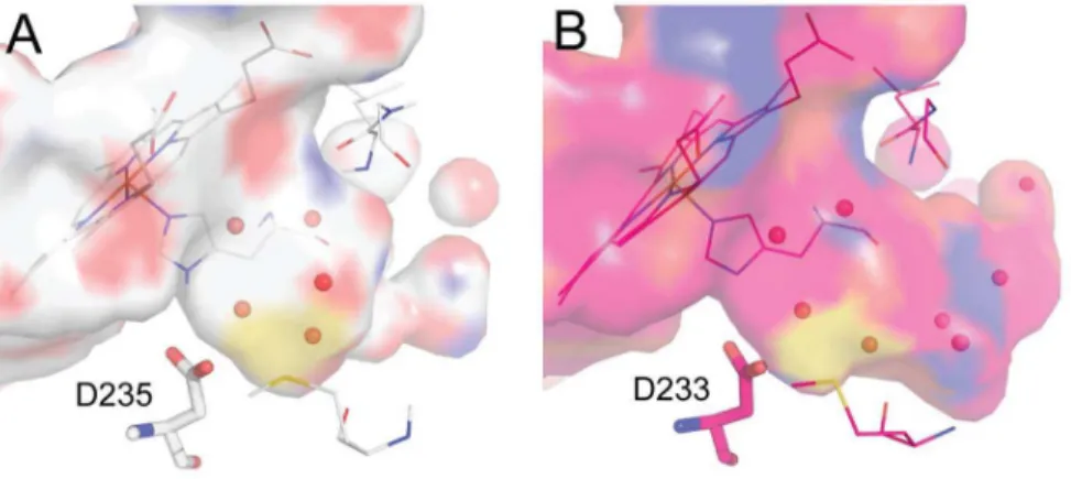
Figure 1. Comparison of the closed and open cavities.(A) The W191G cavity displays a closed and buried cavity that accommodates five ordered water molecules (PDB 1CMQ). (B) The Gateless cavity shows an open and larger pocket. Eight ordered water molecules extend from the back of the Gateless cavity, near Asp233, and out to solvent (PDB 1KXN).
doi:10.1371/journal.pone.0069153.g001
Figure 2. Comparison of binding affinities to the CcP W191G and Gateless cavities.{
Approximate KD determined by endpoint UV-Vis assay or partial ITC curves - assessment of these compounds was limited by solubility. ITC data for compounds1,2,3,4and6are available as supporting information (Table S1).
is more or less desolvated depending on the overall volume of protein that surrounds it, and even a fully buried ligand retains an interaction with the bulk, remaining partially solvated, as is physically correct. This SEV method provides a better model than either considering a ligand fully desolvated on docking to a site– which over-penalizes it–or not desolvating it at all, which under-penalizes it, and does so in a physics-based manner consistent with the rest of the DOCK3.6 scoring function [32]. In retrospective studies, this SEV method had improved enrichments for sites where the solvent interface plays an important role [32] and also appeared to do so in a prospective screen [33]; this study represents the first test of the method, in a model system that enables detailed analysis of the results. Redocking the ZINC library with the SEV method, the fraction of neutral compounds in the top 5000 molecules of the docking list dropped to 3% (Figure S3) and enrichment for active compounds improved slightly (logAUC 44.34). This fit with our expectation that this site, though opened to solvent, would still be dominated by cationic ligand recognition. To actually test this, three neutral molecules, compounds 7, 8 and 9, that were prioritized by the older full desolvation method (ranked 395, 493 and 500) and de-prioritized by the SEV method (ranked 2389, 2612 and 2950) were tested for binding; none showed measurable affinity for the Gateless cavity at up to 1 mM concentration (Figure 5).
The Gateless cavity offered the first chance to test the new SEV solvation method prospectively. A further fifteen molecules, in addition to the three neutral molecules from the previous hit list, were selected from among the top 500 compounds of the SEV solvation-based hit list, or top 0.1% of the database screened, and tested for affinity. In addition to the docking score that ranked them among the top 500 molecules, these compounds were selected for favorable interactions with key binding site residues, such as Asp233, for chemotype diversity, and for compounds that were unburdened by known problems of the DOCK3.6 protocol and scoring, pricipally incorrect ionization and tautomerization states of the docked molecules, and occasionally high-internal energy conformations, as previously described33. Binding was detected for 14 of these 15 with affinities ranging from 8 to 982mM (ligand efficiencies from 0.36 to 0.66) (Figure 6). Crystal structures of Gateless in complex with six of the new docking-predicted ligands were determined with resolutions ranging from 1.30 to 1.86 A˚ (Figure 7and Table S2). The structures of two ligand complexes, those of compounds10and17,superposed to within 0.5 A˚ of the docking prediction, three structures (14, 22
and24) did to within 1.4 A˚ of the docking prediction, and for one ligand (20) the docking pose was over 3 A˚ away from the crystallographic result (Figure 8). The crystallographic orienta-tions of compounds14and24differed mainly by a translational
Figure 3. Electron density difference maps.Initial Fo2Fcmaps contoured at 3sfor (A)1, (B)2, (C)3, (D)4, (E)5and (F)6. PDB codes are as follows:11AEN/4JM5;22EUN/4JM6;32ANZ/4JM8;42EUO/4JM9;52AS3/4JMW;62AS4/4JMA.
Figure 4. Crystallographic poses for six ligands in W191G (gray) and Gateless (pink). (A) 2-amino-5-methylthiazole (1). (B) 2,4-diaminopyrimidine (2). (C) 2,6-diaminopyridine (3). (D) 3-amino-1-methylpyridinium (4). (E) Phenol (5). (F) 3-fluorocatechol (6). PDB codes for the W191G/GA structures are as follows:11AEN/4JM5;22EUN/4JM6;32ANZ/4JM8;42EUO/4JM9;52AS3/4JMW;62AS4/4JMA.
doi:10.1371/journal.pone.0069153.g004
Figure 5. Three neutral compounds highly-ranked by the DOCK3.54 ligand solvation method[20,74]do not bind detectably to the Gateless cavity.These ligands were de-prioritized by the Solvent-Excluded Volume ligand solvation method used in DOCK3.6. [32].
shift, leading to a 1.1 A˚ r.m.s.d. between docking and crystallo-graphic poses in both cases (Figure 8 BandF). Intriguingly, the aldehyde in the docking pose of24was rotated by,90 degrees,
but this variation did not affect the pose prediction. For compound
22, the correspondence between docking and crystallographic pose was slightly worse (1.4 A˚ ) (Figure 8 E). In the docking pose, the ligand contacted Gly178 and Met228 via the benzimidazole nitrogens. In the crystal structure,22rotated away from Met228 to interact with Leu177 and Gly178 via one benzimidazole nitrogen and the methylamine tail. The second benzimidazole nitrogen hydrogen-bonded with two ordered waters; these water molecules were not modeled in the docking. Finally, the docking prediction for20 (Figure 8 D) was inconsistent with the crystal structure (r.m.s.d. 3.1 A˚ ). In the docking pose, the nitrogen on the imidazo ring interacted with Asp233 while the amine made contact with Gly178. This fragment had two configurations in the electron density, both modeled at 50% occupancy and neither of them resembling the docking pose.
Discussion
Both bulk and ordered solvent effects play crucial roles in ligand binding [34–37], and this has motivated the development of methods to model ordered water molecules in molecular design [38–46]. Disentangling bulk contributions from those of ordered water molecules, and from the other convoluted terms encoun-tered in biologically relevant target sites, has remained challeng-ing. Because of its simplicity, the ability to determine structures to high resolution, the ability to seek and test new molecules prospectively, and to compare results with an analogous site that
is closed to the bulk, the Gateless cavity seems well-suited to testing specific solvent-derived terms in protein ligand binding. Three key observations emerge from this study. First, and contrary to our own expectations, opening the cavity to bulk solvent has no general effect on the relative affinities of cationic and neutral ligands; the former continue to bind much more strongly, with the latter barely measurable. The effects on ligand affinity and binding geometry were context-dependent, and whereas several cationic ligands bound weaker to the opened Gateless cavity than to the analogous closed cavity (W191G), one cationic ligand had better affinity, as did one neutral ligand. The one common theme only emerges from the structures of the ligand-cavity complexes: those ligands that maintained their interactions with the anchoring aspartate and, at the same time, increased interactions with the new water channel, saw their affinity increase or stay the same. Ligands that could not do both changed their binding modes to favor a larger interface with the new water network, and saw their affinities drop (presumably they would have dropped further still had they maintained their closed-cavity geometries). Second, and again to our surprise, docking predictions broadly tracked these empirical trends, and as we moved to a more sophisticated physical model of ligand desolvation did so better still. In a prospective screen using this more sophisticated solvation term, the docking hit rate was high, with 14 of 15 ligands tested confirmed experimentally. The predicted and experimentally determined ligand geometries corresponded well for 4 of the 6 structures. Third, structures where we observed a substantial discrepancy between the predicted and experimental ligand poses
Figure 6. Binding affinities and DOCK ranks (Solvent-Excluded Volume solvation method) for compounds selected from a screen of 534,000 fragments against CcP Gateless cavity.{
saw the intervention of new ordered water molecules that we had not anticipated in the docking model.
The loss of affinity for compounds1,2, and4, with their flipped geometries in the site, versus the improved affinities for compounds 3 and 6, and their conserved geometries, points to two effects that in these matched cavities perhaps may be disentangled. The opening to bulk solvent and the appearance of the new water network may reflect an improved solvation of the cavity in the Gateless mutant relative to that of W191G, reducing the relative free energy of the apo Gateless site. Conversely, in the closed loop W191G, the waters that provide the ‘‘chimney’’ from the site to the bulk, with which the ligands interact in the Gateless cavity, cannot bind as they are sterically excluded by the loop itself. This effect, in itself, likely reduces the affinity of all the ligands, which must now compete with lower energy solvent. This may also explain why the weakened ligands, like compound1, flip away from the anchoring aspartate: this allows interactions with the new water network precluded by their interactions with the aspartate, which effectively buried their polar groups, and better solvates the aspartate itself. Ligands like 3 and 6, on the other hand, can adopt geometries that allow for extensive interface with the new water network, gaining interactions they did not make previously (or diminishing their own desolvation), while maintain-ing their original salt-bridge with the aspartate. Viewed another way, both ligands and the cavity may be optimizing their
interaction with bulk solvent, for which the ordered waters are simply a visible proxy. Because all six ligands adopt geometries that seem to optimize interactions with the water network, we ourselves favor the more atomistic explanation. In either case, these observations illustrate how interactions with water can both compete with ligand-protein interactions – weakening net affinities and changing binding geometries – or complement them, improving affinity, depending on the particular features of the ligands. It is also illuminating that despite orientations of cationic ligands that apparently disrupt a crucial salt-bridge, the electro-static interaction between the aspartate and the ligands is still maintained, and likely still the single most important contributor to recognition. This is reflected in the much higher affinity of cationic ligands over neutral ones, the failure of any newly docked neutral ligands to measurably bind to the cavity, and the high affinities of newly prioritized cationic ligands. Methods that base interaction energies on hydrogen-bond inventories, rather than overall electrostatics, may miss these contributions (DOCK 3.6 [32,47,48], like related physics-based approaches [49–55], uses a probe-charge model against an electrostatic potential map, and so does not depend on direct ligand-protein contacts, but rather electrostatic complementarity to an overall receptor potential).
In addition to asking how opening a cavity to solvent affects ligand recognition, a key part of this study was testing how docking would track the changes in site environment. In particular, we
Figure 7. Electron density difference maps.Initial Fo2Fcmaps contoured at 3sfor (A)10, (B)14, (C)17, (D)20, (E)22and (F)24. PDB codes are as follows:104JMB;144JMS;174JMT;20JMV;224JMZ;244JN0.
were interested in evaluating a new ‘‘Solvent-Excluded Volume’’ (SEV) method to treat ligand desolvation on docking [32]. This method should in principle better calculate partial ligand desolvation (i.e., retention of bulk solvent interaction) upon binding than our previous model, though neither considers ordered waters. In a large library screen, the older, less physical desolvation model highly-ranked mono-cations as likely molecules for the closed cavity, as is appropriate. However, against the open cavity about 20% of the top-ranked molecules were neutral; this reflects the high de-solvation penalty for cations, which are modeled as being almost entirely desolvated even in the open cavity. Conversely, with the new SEV method mono-cations entirely dominated the hit list (Figure S3); this reflects their substantially lower desolvation costs in the new method, owing to
their retention of a substantial bulk solvation energy (see above), and consequent relative advantages over neutral molecules. We prospectively tested its performance in three ways: by testing three neutral molecules that the older method had prioritized (ranked 395, 493 and 500 out of 534,000 screened) but that the SEV method had deprioritized (ranked 2389, 2612 and 2950) (Figure 5), by testing 15 new molecules highly-ranked by the SEV method against the open cavity (Figure 6), and by determining X-ray crystal structures of six new ligands.
Consistent with the new solvation treatment, and inconsistent with the older method, the three neutral molecules were not observed to bind to the cavity at concentrations up to 1 mM. Conversely, 14 of the 15 new molecules highly ranked in the docking screen by the SEV solvation model, all cations, were
Figure 8. Superposition of the docking pose (green) and the crystallographic pose (pink) for six ligands prioritized by docking against the open Gateless cavity.Compounds (A)10. (B)14. (C)17. (D)20. (E)22. (F)24. All waters shown are from the co-complexed crystal structures. PDB codes are as follows:104JMB;144JMS;174JMT;20JMV;224JMZ;244JN0.
docking and crystallographic poses clearly disagreed (20,
Figure 8 D). In both of these last two cases, the presence of ordered water molecules in the binding site may explain reduced fidelity of the docking. Compound22interacts with two ordered waters that were not modeled in the docking and the ligand is rotated as compared to the dock pose. Compound 20does not interact directly with water molecules, but the charged moiety on the ligand interacts with Gly178, which is itself involved in the new ordered water network. Here again, the role of the ordered waters, which we did not explicitly model, is highlighted.
The theoretical basis of ordered and bulk water effects on binding have been previously explored [34,35,39,56–62], and there is a substantial body of empirical observations in this area [63–67]; what is new here is the engineering of two simple cavities, one a perturbation of the other, where these effects can be at least partially isolated. The closed (W191G) and opened Gateless (P190G/W191G/D192-3) cavities in Cytochrome c Peroxidase
conserve most features that dominate ligand recognition – the opening of the cavity only deletes residues that are distal to the recognition features of the site, and the crucial cation-recognizing Asp235 is conserved in both (Asp233 in Gateless). Still, the effects of the substitution on ligands that bind to both targets are substantial, both in binding energy and in the structure of the ligand complexes. Part of the effects of opening the cavity to bulk solvent appears to be qualitatively captured by a continuum-based electrostatic model [68–71] in docking, though the role of ordered waters is not, and their effects can be considerable. In these model binding sites, one can hope to tease these contributions apart, and test any theory to treat them prospectively. These cavities are freely available to the community, and we hope that they may find use in exploring these and related questions in docking and molecular recognition.
Materials and Methods
Protein Preparation
The plasmid for the CcP-GA mutant protein was expressed and purified to apparent homogeneity as described [30].
Molecules Tested
Compounds 1, 2, 3, 5 and 6were purchased from Aldrich, compounds4, 10, 11,12,14,21and 23were purchased from Specs, compounds 7 and 9were purchased from Molport, compound 8 was purchased from Vitas-M, compounds15, 18,
20 and 22were purchased from Enamine, compounds 17and
24were purchased from Adesis and compounds 13, 16 and
19were obtained from NCI. All molecules were used as supplied; suppliers confirm$95% purity for all compounds and compound identities for 12 of these were confirmed as relevant by subsequent x-ray crystallography.
perturbation of the Heme Soret band at 10uC in 100 mM citrate buffer at pH 4.5 or 500 mM MES buffer pH 6.0 [27,73]. To avoid competition with small cations like potassium, the pH of both buffer conditions was adjusted with Bis-Tris-Propane [27]. Stock solutions were made up in DMSO and diluted into assay buffer to derive KD values in titration curves. KD values were obtained by fitting the difference absorbance of the Heme Soret band to a one-site binding hyperbola in GraphPad Prism (GraphPad Software, Inc.).
Low C-value Isothermal Titration Calorimetry
Experiments were performed as described [28]. Assays were performed at 10uC in 100 mM citrate buffer at pH 4.5. Ligand stocks were prepared in buffer from overnight dialysis of the protein to prevent buffer mismatch.
Preparation of Fragment Set for Docking
The fragment sets were prepared using the standard ligand preparation protocol used for ligands in the ZINC database [31]. Molecules were protonated assuming a pH of 6.0 to minimize falsely cationic molecules owing to inaccuracies in the pKa calculations (Text S1).
Docking
Docking calculations were carried out with DOCK3.6 [32,47,48] and DOCK3.54 [20,48,74]using a 1.74 A˚ crystallo-graphic structure of CytochromecPeroxidase (PDB code 1KXM
[30]) (Text S2).
Supporting Information
Figure S1 Typical plot of a UV-Vis Heme Soret band titration (compound 10, KD 64mM).
(TIF)
Figure S2 Log AUC curve for known CcP Gateless binders. (TIF)
Figure S3 Charge distribution for the top 5000 docked molecules with old and new solvation maps. Dark grey: Previous full solvation map; Light grey: New Solvent-Exluded Volume (SEV) solvation map.
(TIF)
Table S1 ITC binding data for compounds1, 2, 3, 4 and 6
against CcP Gateless. (DOCX)
Table S2 X-Ray data collection and refinement statistics. (DOCX)
(DOCX)
Text S2 Docking. (DOCX)
Acknowledgments
We thank A. Doak for protein preparation, H. Lin and G. Rocklin for reading this manuscript, and R. Wilson for supplying the Gateless expression plasmid.
Author Contributions
Conceived and designed the experiments: SB SEB IF MF BKS. Performed the experiments: SB SEB IF MF. Analyzed the data: SB SEB IF MF BKS. Contributed reagents/materials/analysis tools: DBG. Wrote the paper: SB BKS.
References
1. Repasky MP, Murphy RB, Banks JL, Greenwood JR, Tubert-Brohman I, et al. (2012) Docking performance of the glide program as evaluated on the Astex and DUD datasets: a complete set of glide SP results and selected results for a new scoring function integrating WaterMap and glide. J Comput Aided Mol Des 26: 787–799.
2. Carlsson J, Coleman RG, Setola V, Irwin JJ, Fan H, et al. (2011) Ligand discovery from a dopamine D-3 receptor homology model and crystal structure. Nature Chemical Biology 7: 769–778.
3. Ramsden NL, Buetow L, Dawson A, Kemp LA, Ulaganathan V, et al. (2009) A structure-based approach to ligand discovery for 2C-methyl-D-erythritol-2,4-cyclodiphosphate synthase: a target for antimicrobial therapy. J Med Chem 52: 2531–2542.
4. Tosh DK, Phan K, Gao ZG, Gakh AA, Xu F, et al. (2012) Optimization of adenosine 59-carboxamide derivatives as adenosine receptor agonists using structure-based ligand design and fragment screening. J Med Chem 55: 4297– 4308.
5. Sager G, Orvoll EO, Lysaa RA, Kufareva I, Abagyan R, et al. (2012) Novel cGMP efflux inhibitors identified by virtual ligand screening (VLS) and confirmed by experimental studies. J Med Chem 55: 3049–3057.
6. Langmead CJ, Andrews SP, Congreve M, Errey JC, Hurrell E, et al. (2012) Identification of novel adenosine A(2A) receptor antagonists by virtual screening. J Med Chem 55: 1904–1909.
7. de Graaf C, Rein C, Piwnica D, Giordanetto F, Rognan D (2011) Structure-based discovery of allosteric modulators of two related class B G-protein-coupled receptors. ChemMedChem 6: 2159–2169.
8. de Graaf C, Kooistra AJ, Vischer HF, Katritch V, Kuijer M, et al. (2011) Crystal structure-based virtual screening for fragment-like ligands of the human histamine H(1) receptor. J Med Chem 54: 8195–8206.
9. Roughley S, Wright L, Brough P, Massey A, Hubbard RE (2012) Hsp90 inhibitors and drugs from fragment and virtual screening. Top Curr Chem 317: 61–82.
10. Dahlgren MK, Garcia AB, Hare AA, Tirado-Rives J, Leng L, et al. (2012) Virtual screening and optimization yield low-nanomolar inhibitors of the tautomerase activity of Plasmodium falciparum macrophage migration inhibi-tory factor. J Med Chem 55: 10148–10159.
11. Mysinger MM, Carchia M, Irwin JJ, Shoichet BK (2012) Directory of useful decoys, enhanced (DUD-E): better ligands and decoys for better benchmarking. J Med Chem 55: 6582–6594.
12. Geerke DP, Luber S, Marti KH, Van Gunsteren WF (2009) On the direct calculation of the free energy of quantization for molecular systems in the condensed phase. J Comput Chem 30: 514–523.
13. Ahringer J (1995) Embryonic Tissue Differentiation in Caenorhabditis-Elegans Requires Dif-1, a Gene Homologous to Mitochondrial Solute Carriers. Embo Journal 14: 2307–2316.
14. Garza D, Medhora M, Koga A, Hartl DL (1991) Introduction of the Transposable Element Mariner into the Germline of Drosophila-Melanogaster. Genetics 128: 303–310.
15. Kunzl C, Sachser N (1999) The behavioral endocrinology of domestication: A comparison between the domestic guinea pig (Cavia aperea f. porcellus) and its wild ancestor, the cavy (Cavia aperea). Hormones and Behavior 35: 28–37. 16. Meeker AK, GarciaMoreno B, Shortle D (1996) Contributions of the ionizable
amino acids to the stability of staphylococcal nuclease. Biochemistry 35: 6443– 6449.
17. Schreiber C, Buckle AM, Fersht AR (1994) Stability and Function - 2 Constraints in the Evolution of Barstar and Other Proteins. Structure 2: 945– 951.
18. Eriksson AE, Baase WA, Wozniak JA, Matthews BW (1992) A Cavity-Containing Mutant of T4 Lysozyme Is Stabilized by Buried Benzene. Nature 355: 371–373.
19. Morton A, Matthews BW (1995) Specificity of Ligand-Binding in a Buried Nonpolar Cavity of T4 Lysozyme - Linkage of Dynamics and Structural Plasticity. Biochemistry 34: 8576–8588.
20. Wei BQQ, Baase WA, Weaver LH, Matthews BW, Shoichet BK (2002) A model binding site for testing scoring functions in molecular docking. Journal of Molecular Biology 322: 339–355.
21. Merski M, Shoichet BK (2012) Engineering a model protein cavity to catalyze the Kemp elimination. Proc Natl Acad Sci U S A 109: 16179–16183.
22. Fitzgerald MM, Trester ML, Jensen GM, McRee DE, Goodin DB (1995) The role of aspartate-235 in the binding of cations to an artificial cavity at the radical site of cytochrome c peroxidase. Protein Sci 4: 1844–1850.
23. Fitzgerald MM, Churchill MJ, McRee DE, Goodin DB (1994) Small molecule binding to an artificially created cavity at the active site of cytochrome c peroxidase. Biochemistry 33: 3807–3818.
24. Ferrari AM, Wei BQQ, Costantino L, Shoichet BK (2004) Soft docking and multiple receptor conformations in virtual screening. Journal of Medicinal Chemistry 47: 5076–5084.
25. Wei BQ, Weaver LH, Ferrari AM, Matthews BW, Shoichet BK (2004) Testing a flexible-receptor docking algorithm in a model binding site. Journal of Molecular Biology 337: 1161–1182.
26. Graves AP, Brenk R, Shoichet BK (2005) Decoys for docking. J Med Chem 48: 3714–3728.
27. Brenk R, Vetter SW, Boyce SE, Goodin DB, Shoichet BK (2006) Probing molecular docking in a charged model binding site. Journal of Molecular Biology 357: 1449–1470.
28. Boyce SE, Mobley DL, Rocklin GJ, Graves AP, Dill KA, et al. (2009) Predicting Ligand Binding Affinity with Alchemical Free Energy Methods in a Polar Model Binding Site. Journal of Molecular Biology 394: 747–763.
29. Gilson MK, Davis ME, Luty BA, McCammon JA (1993) Computation of electrostatic forces on solvated molecules using the Poisson-Boltzmann equation. The Journal of Physical Chemistry 97: 3591–3600.
30. Rosenfeld RJ, Hays AM, Musah RA, Goodin DB (2002) Excision of a proposed electron transfer pathway in cytochrome c peroxidase and its replacement by a ligand-binding channel. Protein Sci 11: 1251–1259.
31. Irwin JJ, Sterling T, Mysinger MM, Bolstad ES, Coleman RG (2012) ZINC: A Free Tool to Discover Chemistry for Biology. Journal of Chemical Information and Modeling 52: 1757–1768.
32. Mysinger MM, Shoichet BK (2010) Rapid context-dependent ligand desolvation in molecular docking. J Chem Inf Model 50: 1561–1573.
33. Mysinger MM, Weiss DR, Ziarek JJ, Gravel S, Doak AK, et al. (2012) Structure-based ligand discovery for the protein-protein interface of chemokine receptor CXCR4. Proceedings of the National Academy of Sciences of the United States of America 109: 5517–5522.
34. Gilson MK, Honig B (1991) The inclusion of electrostatic hydration energies in molecular mechanics calculations. J Comput Aided Mol Des 5: 5–20. 35. Gilson MK, Sharp KA, Honig BH (1988) Calculating the electrostatic potential
of molecules in solution: Method and error assessment. Journal of Computa-tional Chemistry 9: 327–335.
36. Nguyen CN, Young TK, Gilson MK (2012) Grid inhomogeneous solvation theory: hydration structure and thermodynamics of the miniature receptor cucurbit[7]uril. J Chem Phys 137: 044101.
37. Ladbury JE (1996) Just add water! The effect of water on the specificity of protein-ligand binding sites and its potential application to drug design. Chem Biol 3: 973–980.
38. Grant JA, Pickup BT, Nicholls A (2001) A smooth permittivity function for Poisson–Boltzmann solvation methods. Journal of Computational Chemistry 22: 608–640.
39. Young T, Abel R, Kim B, Berne BJ, Friesner RA (2007) Motifs for molecular recognition exploiting hydrophobic enclosure in protein–ligand binding. Proceedings of the National Academy of Sciences 104: 808–813.
40. van Dijk AD, Bonvin AM (2006) Solvated docking: introducing water into the modelling of biomolecular complexes. Bioinformatics 22: 2340–2347. 41. Verdonk ML, Chessari G, Cole JC, Hartshorn MJ, Murray CW, et al. (2005)
Modeling water molecules in protein-ligand docking using GOLD. J Med Chem 48: 6504–6515.
42. Friesner RA, Murphy RB, Repasky MP, Frye LL, Greenwood JR, et al. (2006) Extra precision glide: docking and scoring incorporating a model of hydrophobic enclosure for protein-ligand complexes. J Med Chem 49: 6177–6196. 43. Woo HJ, Dinner AR, Roux B (2004) Grand canonical Monte Carlo simulations
of water in protein environments. J Chem Phys 121: 6392–6400.
44. Minke WE, Diller DJ, Hol WG, Verlinde CL (1999) The role of waters in docking strategies with incremental flexibility for carbohydrate derivatives: heat-labile enterotoxin, a multivalent test case. J Med Chem 42: 1778–1788. 45. Osterberg F, Morris GM, Sanner MF, Olson AJ, Goodsell DS (2002) Automated
506.
54. Neves MC, Totrov M, Abagyan R (2012) Docking and scoring with ICM: the benchmarking results and strategies for improvement. Journal of Computer-Aided Molecular Design 26: 675–686.
55. Verdonk ML, Cole JC, Hartshorn MJ, Murray CW, Taylor RD (2003) Improved protein-ligand docking using GOLD. Proteins 52: 609–623. 56. Limongelli V, Marinelli L, Cosconati S, La Motta C, Sartini S, et al. (2012)
Sampling protein motion and solvent effect during ligand binding. Proc Natl Acad Sci U S A 109: 1467–1472.
57. Nicholls A, Honig B (1991) A rapid finite difference algorithm, utilizing successive over-relaxation to solve the Poisson–Boltzmann equation. Journal of Computational Chemistry 12: 435–445.
58. Arora N, Bashford D (2001) Solvation energy density occlusion approximation for evaluation of desolvation penalties in biomolecular interactions. Proteins 43: 12–27.
59. Sitkoff D, Sharp KA, Honig B (1994) Accurate Calculation of Hydration Free Energies Using Macroscopic Solvent Models. The Journal of Physical Chemistry 98: 1978–1988.
60. Barillari C, Taylor J, Viner R, Essex JW (2007) Classification of Water Molecules in Protein Binding Sites. Journal of the American Chemical Society 129: 2577–2587.
67. Lu Y, Wang R, Yang CY, Wang S (2007) Analysis of ligand-bound water molecules in high-resolution crystal structures of protein-ligand complexes. J Chem Inf Model 47: 668–675.
68. Gilson MK, Honig B (1988) Calculation of the total electrostatic energy of a macromolecular system: solvation energies, binding energies, and conforma-tional analysis. Proteins 4: 7–18.
69. Gilson MK, Honig BH (1988) Energetics of charge-charge interactions in proteins. Proteins 3: 32–52.
70. Gilson MK, Rashin A, Fine R, Honig B (1985) On the calculation of electrostatic interactions in proteins. J Mol Biol 184: 503–516.
71. Honig B, Nicholls A (1995) Classical electrostatics in biology and chemistry. Science 268: 1144–1149.
72. Hays Putnam AM, Lee YT, Goodin DB (2009) Replacement of an electron transfer pathway in cytochrome c peroxidase with a surrogate peptide. Biochemistry 48: 1–3.
73. Musah RA, Jensen GM, Bunte SW, Rosenfeld RJ, Goodin DB (2002) Artificial protein cavities as specific ligand-binding templates: characterization of an engineered heterocyclic cation-binding site that preserves the evolved specificity of the parent protein. Journal of Molecular Biology 315: 845–857.
![Figure 5. Three neutral compounds highly-ranked by the DOCK3.54 ligand solvation method [20,74] do not bind detectably to the Gateless cavity](https://thumb-eu.123doks.com/thumbv2/123dok_br/16346965.189294/5.918.93.650.791.1040/figure-neutral-compounds-highly-ranked-solvation-detectably-gateless.webp)
