Article
Printed in Brazil - ©2016 Sociedade Brasileira de Química0103 - 5053 $6.00+0.00*e-mail: [email protected]
Synthesis,
in vitro
Antifungal Activity and Molecular Modeling Studies of New
Mannich Bases Derived from Lawsone
João F. Allochio Filho,a Larissa L. Roldi,a Maicon Delarmelina,a Rodolfo G. Fiorot,a
Jessica T. Andrade,b Álan A. Aleixo,b Rafaella S. Carvalho,b Marcelo G. F. Araújo,b
Jaqueline M. S. Ferreira,b Alex G. Taranto,c Wanderson Romãod and Sandro J. Greco*,a
aLaboratório de Síntese Orgânica & Medicinal, Departamento de Química, Universidade Federal
do Espírito Santo, Avenida Ferrari, 514, Goiabeiras, 29075-910 Vitória-ES, Brazil
bLaboratório de Microbiologia and cLaboratório de Química Farmacêutica Medicinal,
Universidade Federal de São João Del-Rei, Campus Centro Oeste, Rua Sebastião Gonçalves Coelho, 400, Chanadour, 35501-296 Divinópolis-MG, Brazil
dLaboratório de Petroleômica, Departamento de Química, Universidade Federal do Espírito Santo,
Avenida Ferrari, 514, Goiabeiras, 29075-910 Vitória-ES, Brazil
Hydroxynaphthoquinones such as lawsone (2-hydroxy-1,4-naphthoquinone) have proven to be effective antifungal agents. These compounds were tested for antifungal activity against yeast standard and clinical strains by the broth microdilution method. Among the synthetic lawsone derivatives, 2-hydroxy-3-((2-hydroxyphenyl)(pyrrolidin-1-yl)methyl)naphthalene-1,4-dione, (((4-nitrophenyl)amino)(phenyl)methyl)naphthalene-1,4-dione and 2-hydroxy-3-((2-hydroxyphenyl)((4-nitrophenyl)amino)methyl)naphthalene-1,4-dione showed high activity against Candida albicans ATCC 10231, with minimal inhibitory concentrations (MICs) and minimal fungicidal concentrations (MFCs) ranging from 20 to 330 and from 80 to 330 µg mL-1,
respectively. Moreover, they also showed a mechanism of action on exogenous ergosterol. Therapeutic concentrations (CC50) of 2-hydroxy-3-((2-hydroxyphenyl)(pyrrolidin-1-yl)methyl)
naphthalene-1,4-dione, 2-hydroxy-3-(((4-nitrophenyl)amino)(phenyl)methyl)naphthalene-1,4-dione and 2-hydroxy-3-((2-hydroxyphenyl)((4-nitrophenyl)amino)methyl)naphthalene-1,4-2-hydroxy-3-(((4-nitrophenyl)amino)(phenyl)methyl)naphthalene-1,4-dione were 52.81, 52.58 and 85.94 µg mL-1, respectively, which can be considered moderate or low. In
addition, docking studies showed that these compounds had similar binding energy to standard ketoconazole, which are recognized as the molecular target by van der Waals interactions. Furthermore, they are under Lipinski’s rule of 5 with a druglikeness score better than ketoconazole and nystatin. These findings suggest that 2-hydroxy-3-((2-hydroxyphenyl)(pyrrolidin-1-yl)methyl) naphthalene-1,4-dione, 2-hydroxy-3-(((4-nitrophenyl)amino)(phenyl)methyl)naphthalene-1,4-dione and 2-hydroxy-3-((2-hydroxyphenyl)((4-nitrophenyl)amino)methyl)naphthalene-1,4-2-hydroxy-3-(((4-nitrophenyl)amino)(phenyl)methyl)naphthalene-1,4-dione have potential as leading compounds against human fungal infections.
Keywords: lawsone, Mannich bases, antifungal activity, docking
Introduction
Naphthoquinone derivatives possess continuous interest as potential therapeutic agents. Among them, hydroxynaphthoquinones such as lawsone (2-hydroxy-1,4-naphthoquinone), have proven to be effective due to their chemical and pharmacological properties.1 The antifungal
activity of certain naphthoquinone and lawsone derivatives has been reported from in vitro studies (Scheme 1).2 As a
quinone, lawsone can be used to perform Mannich reaction3
and the resulting Mannich bases show particular interest because of their biological activities.4
Candida infections have always posed a heavy burden on public health. The incidence of candidiasis has been increasing, which may be attributable to the growing numbers of immunocompromised patients.5,6 In addition,
C. albicans species, an increased incidence has been observed of invasive candidiasis caused by non-albicans Candida species, such as Candida glabrata, Candida tropicalis, Candida krusei or Candida parapsilosis.8 Many
problems remain to be solved for most of the antifungal drugs available. Mechanisms of resistance to azole drugs have been described for C. albicans.9 Thus, it has become
necessary to develop new drugs capable of effectively combating these types of infection.
In this work, we evaluated the antifungal activity of synthetic derivative compounds of lawsone, which we synthesized for the first time, and tested these against several clinically important microorganisms. This way, we searched for thirteen new enolamines 4-8, 10-14, 16, 17 and 23, which contain 2-phenyl-2H-1,2,3-triazole or substituted and non-substituted phenyl groups, themselves synthesized using a multicomponent Mannich reaction from lawsone.3 These compounds were also tested against
baby hamster kidney (BHK) cells to study toxicity effects. Moreover, molecular modeling tools were used to highlight properties used in drug design,10 for example druglikeness.
In addition, docking studies11-13 described the main
molecular interactions between these compounds and the molecular target. These data suggest a lead optimization process to search for a second generation of antifungal lawsone-based drugs.
Experimental
General information
All solvents and reagents were commercially purchased and were used without any purification.
Melting points were determined using Fisatom 430D equipment. Infrared (IR) spectra were recorded on a FTLA 2000-102-ABB BOMEM Fourier transform (FT) IR spectrophotometer with KBr technique and on a Perkin Elmer Spectrum 400 FTIR spectrometer model using attenuated total reflectance (ATR) technique. The 1H and 13C
nuclear magnetic resonance (NMR) spectra were obtained on a Varian VNMRS spectrometer model 400 (400 MHz for
1H NMR) using CDCl
3 or dimethyl sulfoxide (DMSO)-d6
as solvents and tetramethylsilane (TMS) as the internal standard.
Mass spectra (MS) were recorded on ultra-high resolution and accuracy mass spectrometer (model 9.4 T Solarix, Bruker Daltonics), operated in both ionization modes: positive and negative electrospray ionization Fourier transform ion cyclotron resonance mass spectrometry (ESI(+) and ESI(–)-FT-ICR MS, respectively). FT-ICR MS spectra were acquired with resolving power of m / ∆m50% ca. 500000, in which ∆m50%
is the full peak width at half-maximum peak height of m/z 400 and a mass accuracy of less than 1 ppm. It provides an unambiguous molecular formula assignment for singly charged molecular ions such as [M – H]+ or [M + H]– and
double bound equivalents (DBE) values.
Synthesis of lawsone derivatives 4-8, 10-14, 17 and 23 - general procedure
A mixture of the aldehyde (1.2 eq) and amine (1.1 eq) in ethanol was stirred during a few minutes. After this time, lawsone (1.0 eq) was added to the solution and the reaction was stirred for 24 h at room temperature in the dark. The solid formed was filtered under vacuum and washed with cold ethanol. Recrystallization with ethanol or ethyl acetate:hexane 1:3 was performed for the impure compounds. Experimental protocols were initially tested as described in previous reports.3,14,15
2-Hydroxy-3-((2-hydroxyphenyl)(pyrrolidin-1-yl)methyl) naphthalene-1,4-dione (4)
Compound 4 was obtained as a red crystalline solid (77%); m.p. decomposition at 146 oC; IR (ATR) ν
max / cm-1
3595, 3088, 2977, 2884, 1685, 1579, 1505, 1271; 1H NMR
(400 MHz, DMSO-d6) d 1.95 (br s, 4H, CH2), 3.18 (br s,
4H, NCH2), 5.82 (s, 1H, CH), 6.78 (td, 1H, J 8.0, 1.0 Hz,
Ar–H), 6.82 (dd, 1H, J 8.0, 1.0 Hz, Ar–H), 7.10 (td, 1H, J 8.0, 1.0 Hz, Ar–H), 7.60 (td, 1H, J 8.0, 1.0 Hz, Ar–H), 7.67 (td, 1H, J 7.5, 1.0 Hz, Naph–H*), 7.72 (td, 1H, J 7.5, 1.0 Hz, Naph–H), 7.83 (dd, 1H, J 7.5, 1.0 Hz, Naph–H), 7.92 (dd, 1H, J 7.5, 1.0 Hz, Naph–H), 10.36 (sl, 2H, OH);
13C NMR (101 MHz, DMSO-d
6) d 22.0, 22.5, 43.8, 52.0,
59.8, 112.5, 115.2, 118.2, 123.4, 123.9, 124.2, 27.9, 128.1, 129.9, 130.2, 132.7, 133.3, 153.8, 169.6, 177.7, 183.0; ESI(+)-FT-ICR-MS calcd. for C21H20NO4+ [M + H]+:
350.13868; found: 350.13476 (DBE = 13); [2M + Na]+:
721.2520; found: 721.2528. *Naph–H: naphthoquinone hydrogens.
2-Hydroxy-3-(phenyl(pyrrolidin-1-yl)methyl)naphthalene-1,4-dione (5)
Compound 5 was obtained as a red crystalline solid (32%); m.p. 177-179 oC with decomposition; IR (ATR)
νmax / cm-1 3343, 3064, 2983, 2874, 1680, 1587, 1531, 1272;
1H NMR (400 MHz, DMSO-d
6) d 1.93 (m, 4H, CH2), 3.15
(br s, 4H, NCH2), 5.48 (s, 1H, CH), 7.25 (m, 1H, Ar–H),
7.32 (m, 2H, Ar–H), 7.54 (td, 1H, J 7.5, 1.0 Hz, Naph–H), 7.67 (m, 3H, 2Ar–H and 1H, Naph–H), 7.78 (dd, 1H, J 7.5, 1.0 Hz, Naph–H), 7.89 (dd, 1H, J 7.5, 1.0 Hz, Naph–H),
10.48 (br s, 1H, OH); 13C NMR (101 MHz, DMSO-d
6)
d 23.7, 53.9, 67.6, 113.6, 125.5, 125.7, 128.3, 128.4, 128.8, 131.2, 131.9, 134.2, 135.1, 139.1, 170.6, 178.2, 184.9; ESI(+)-FT-ICR-MS calcd. for C21H20NO3+ [M + H]+:
334.14377; found: 334.13985 (DBE = 13); [2M + H]+:
667.2803; found: 667.2790; [2M + Na]+: 689.2622; found:
689.2609.
2-Hydroxy-3-(naphthalen-1-yl(pyrrolidin-1-yl)methyl) naphthalene-1,4-dione (6)
Compound 6 was obtained as an orange solid (38%); m.p. decomposition at 185 oC; IR (ATR) ν
max / cm-1 3337,
3042, 2868, 1678, 1583, 1532, 1275; 1H NMR (400 MHz,
DMSO-d6) d 1.92 (m, 4H, CH2), 3.29 (m, 4H, NCH2), 6.37
(s, 1H, CH), 7.51 (m, 4H, 3Ar-H and 1H, Naph–H), 7.66 (td, 1H, J 7.4, 1.0 Hz, Naph–H), 7.76 (dd, 1H, J 7.4, 1.0 Hz, Naph–H), 7.84 (d, 1H, J 8.3 Hz, Ar–H), 7.89 (m, 2H, 1Ar–H and 1H, Naph–H), 8.25 (d, 1H, J 7.4 Hz, Ar–H), 8.59 (d, 1H, J 8.3 Hz, Ar–H), 10.28 (br s, 1H, OH); 13C NMR (101 MHz,
DMSO-d6) d 23.8, 54.1, 63.3, 112.9, 124.6, 125.5, 125.6,
125.7, 126.1, 126.7, 128.4, 128.9, 131.2, 131.3, 131.9, 133.5, 134.2, 134.5, 135.3, 171.1, 178.4, 184.9; ESI(+)-FT-ICR-MS calcd. for C25H23NO3+ [M + H]+: 384.15550;
found: 384.15982 (DBE = 16).
2-Hydroxy-3-((4-nitrophenyl)(pyrrolidin-1-yl)methyl) naphthalene-1,4-dione (7)
Compound 7 was obtained as an orange solid (46%); m.p. 186-188 oC; IR (ATR) ν
max / cm-1 3059, 2863, 1686,
1586, 1532, 1343, 1275; 1H NMR (400 MHz, DMSO-d
6)
d 1.94 (m, 4H, CH2), 3.20 (br s, 4H, NCH2), 5.71 (s,
1H, CH), 7.55 (td, 1H, J 7.5, 1.0 Hz, Naph–H), 7.68 (td, 1H, J 7.5, 1.0 Hz, Naph–H), 7.78 (d, 1H, J 7.5, 1.0 Hz, Naph–H), 7.90 (d, 3H, J 8.7 Hz, 2Ar–H and 1H, Naph–H), 8.20 (d, 2H, J 8.7 Hz, Ar–H), 10.46 (br s, 1H, OH);
13C NMR (101 MHz, DMSO-d
6) d 23.8, 54.2, 66.6, 112.4,
124.0, 125.6, 125.9, 129.1, 131.4, 131.9, 134.2, 135.1, 146.0, 146.6, 147.2, 170.8, 178.1, 184.6; ESI(–)-FT-ICR-MS calcd. for C21H19N2O5– [M – H]–: 377.11430; found:
377.11413 (DBE = 14).
2-Hydroxy-3-((4-methoxyphenyl)(pyrrolidin-1-yl)methyl) naphthalene-1,4-dione (8)
Compound 8 was obtained as an orange solid (44%); m.p. 181-183 oC; IR (ATR) ν
1681, 1586, 1538, 1252, 1233; 1H NMR (400 MHz,
DMSO-d6) d 1.92 (br s, 4H, CH2), 3.10 (br s, 4H, NCH2),
3.70 (s, 3H, OCH3), 5.40 (s, 1H, CH), 6.86 (d, 2H,
J 8.7 Hz, Ar–H), 7.56 (m, 3H, 2Ar–H and 1H, Naph–H), 7.67 (t, 1H, J 7.4 Hz, Naph–H), 7.78 (d, 1H, J 7.4 Hz, Naph–H), 7.89 (d, 1H, J 7.4 Hz, Naph–H), 10.52 (br s,
1H, OH); 13C NMR (101 MHz, DMSO-d
6) d 23.6, 53.6,
55.5, 67.2, 113.9, 114.1, 125.5, 125.7, 129.9, 131.2, 131.9, 134.1, 135.1, 159.4, 170.5, 178.2, 184.9; ESI(+)-FT-ICR-MS calcd. for C22H22NO4+ [M + H]+: 364.15432; found:
364.15041 (DBE = 13); [2M + Na]+: 749.28347; found:
749.28334 (DBE = 25).
2-Hydroxy-3-((4-nitrophenyl)((4-nitrophenyl)amino)methyl) naphthalene-1,4-dione (10)
Compound 10 was obtained as an yellow solid (82%); m.p. 134-136 oC; IR (KBr) ν
max / cm-1 3446, 3328, 3199,
1675, 1593, 1573, 1507, 1344, 1299; 1H NMR (400 MHz,
DMSO-d6) d 6.37 (s, 1H, CH), 6.93 (d, 2H, J 8.7 Hz, Ar–H),
7.81 (d, 2H, J 8.7 Hz, Ar–H), 7.93 (td, 1H, J 7.5, 1.5 Hz, Naph–H), 7.98 (td, 1H, J 7.5, 1.5 Hz, Naph–H), 8.10 (m, 4H, 2Ar–H and 2H, Naph–H), 8.31 (d, 2H, J 8.7 Hz, Ar–H);
13C NMR (101 MHz, DMSO-d
6) d 49.5, 111.2, 119.2,
122.2, 123.1, 124.8, 124.9, 125.2, 126.6, 129.0, 129.4, 130.5, 132.3, 133.7, 135.5, 145.3, 147.0, 152.3, 154.5, 155.9, 179.9, 182.1, 191.2; ESI(–)-FT-ICR-MS calcd. for C23H14N3O7– [M – H]–: 444.08372; found: 444.08358
(DBE = 18).
2-Hydroxy-3-(((4-nitrophenyl)amino)(phenyl)methyl) naphthalene-1,4-dione (11)
Compound 11 was obtained as an pumpkin solid (93%); m.p. 187-189 oC with decomposition; IR (ATR) ν
max / cm-1
3351, 3339, 1656, 1641, 1598, 1585, 1327, 1264; 1H NMR
(400 MHz, DMSO-d6) d 6.09 (s, 1H, CH), 6.55 (d, 1H,
J 9.3 Hz, NH), 6.74 (d, 2H, J 8.5 Hz, Ar–H), 7.19 (m, 1H, Ar–H), 7.28 (m, 2H, Ar–H), 7.41 (m, 2H, Ar–H), 7.75 (td, 1H, J 7.5, 1.5 Hz, Naph–H), 7.80 (td, 1H, J 7.5, 1.5 Hz, Naph–H), 7.93 (m, 4H, 2Ar–H and 2H, Naph–H); 13C NMR
(101 MHz, DMSO-d6) d 51.4, 112.8, 121.5, 126.3,
126.3, 126.5, 126.8, 127.1, 127.4, 128.7, 129.6, 129.9, 130.5, 132.1, 133.9, 135.2, 136.7, 140.1, 154.0, 156.1, 156.9, 181.5, 183.9, 193.7; ESI(–)-FT-ICR-MS calcd. for C23H15N2O5– [M – H]–: 399.09865; found: 399.09808 (DBE
= 20); [M – C6H5N2O2 + C10H5O3]–: 435.08741; found:
435.08684 (DBE = 17).
2-Hydroxy-3-((2-hydroxyphenyl)((4-nitrophenyl)amino) methyl)naphthalene-1,4-dione (12)
Compound 12 was obtained as an orange solid (69%); m.p. 237-239 oC with decomposition; IR (ATR) ν
max / cm-1
3349, 3062, 1687, 1640, 1592, 1537, 1320, 1240; 1H NMR
(400 MHz, DMSO-d6) d 6.29 (d, 1H, J 9.4 Hz, CH), 7.07
(br s, 2H, Ar–H), 7.32 (m, 1H, Ar–H), 7.50 (m, 2H, 1Ar–H and 1H, Naph–H), 7.56 (d, 1H, J 7.5 Hz, Naph–H), 7.73 (d, 1H, J 9.4 Hz, NH), 7.94 (m, 2H, 1Ar–H and 1H, Naph–H), 8.06 (m, 1H, Ar–H), 8.09 (d, 2H, J 9.0 Hz, 1Ar–H and 1H, Naph–H), 8.16 (m, 1H, Ar–H); 13C NMR (101 MHz,
DMSO-d6) d 41.9, 112.0, 117.7, 118.9, 122.4, 126.4, 126.5,
126.7, 129.6, 130.2, 131.0, 131.8, 134.6, 135.4, 136.8, 149.1, 151.4, 152.9, 178.5, 183.3; ESI(–)-FT-ICR-MS calcd. for C23H13N2O5– [M – 2H – OH]–: 397.08300; found:
397.08265 (DBE = 17); [M – H – OH + Cl]–: 433.05967;
found: 433.05932 (DBE = 17).
2-Hydroxy-3-(naphthalen-1-yl((4-nitrophenyl)amino)methyl) naphthalene-1,4-dione (13)
Compound 13 was obtained as an orange solid (90%); m.p. 165-167 oC with decomposition; IR (ATR) ν
max / cm-1
3641, 3407, 3396, 3362, 1678, 1590, 1503, 1308, 1276;
1H NMR (400 MHz, CDCl
3) d 5.81 (d, 1H, J 9.3 Hz, CH);
6.67 (dd, 2H, J 7.2, 2.0 Hz, Ar–H), 6.94 (d, 1H, J 9.3 Hz, NH), 7.43 (t, 1H, J 7.7 Hz, Ar–H), 7.52 (td, 1H, J 7.0, 1.0 Hz, Naph–H), 7.56 (td, 1H, J 7.0, 1.0 Hz, Naph–H), 7.68 (d, 1H, J 7.4 Hz, Ar–H), 7.71 (td, 1H, J 7.7, 1.0 Hz, Ar–H), 7.79 (td, 1H, J 7.7, 1.0 Hz, Ar–H), 7.83 (d, 1H, J 8.3 Hz, Ar–H), 7.89 (dd, 1H, J 7.7, 1.0 Hz, Ar–H), 8.06 (dd, 2H, J 7.2, 2.0 Hz, Ar–H), 8.09 (dd, 1H, J 7.7, 1.0 Hz, Naph–H), 8.12 (dd, 1H, J 7.7, 1.0 Hz, Naph–H), 8.20 (d, 1H, J 8.3 Hz,
Ar–H); 13C NMR (101 MHz, CDCl
3) d 49.5, 111.8, 113.3,
120.6, 123.1, 125.2, 125.3, 125.9, 126.5, 126.5, 126.9, 127.3, 129.1, 129.2, 131.2, 132.5, 133.2, 133.5, 134.0, 135.6, 138.7, 151.9, 153.6, 181.0, 184.2; ESI(−)-FT-ICR-MS calcd.
for C27H17N2O5– [M – H]–: 449.11430; found: 449.11367
(DBE = 20); [M – C6H5N2O2 + OH]–: 329.08193; found:
329.08162 (DBE = 18).
2-Hydroxy-3-((4-methoxyphenyl)((4-nitrophenyl)amino) methyl)naphthalene-1,4-dione (14)
Compound 14 was obtained as an yellow solid (68%); m.p. 222-224 oC with decomposition; IR (ATR) ν
max / cm-1
3394, 3245, 1666, 1636, 1593, 1362, 1260, 1237; 1H NMR
(400 MHz, DMSO-d6) d 3.65 (s, 3H, OCH3), 5.88 (s, 1H,
CH), 6.71 (m, 2H, Ar–H), 7.09 (m, 2H, Ar–H), 7.73 (td, 2H, J 7.4, 1.6 Hz, 1Ar–H and 1H, Naph–H), 7.77 (td, 2H, J 7.5, 1.6 Hz, 1Ar–H and 1H, Naph–H), 7.87 (m, 2H, 1Ar–H and 1H, Naph–H), 7.93 (m, 2H, 1Ar–H and 1H, Naph–H);
13C NMR (101 MHz, DMSO-d
6) d 37.4, 55.3, 113.4, 123.9,
125.9, 126.4, 129.6, 130.2, 132.6, 133.0, 133.5, 135.1, 156.6, 157.6, 181.7, 184.0; ESI(–)-FT-ICR-MS calcd. for C28H17O7– [M – C6H5N2O2 + C10H5O3]–: 465.09798; found:
2-((2-Phenyl-2H -1,2,3-triazol-4-yl)(pyrrolidin-1-yl)methyl)- 3-((1,3,4-trihydroxynaphthalen-2-yl)oxy)naphthalene-1,4-dione (17)
Compound 17 was obtained as an orange solid after recrystallization from ethyl acetate:hexane 1:3 (36%); m.p. above 200 oC with decomposition; IR (KBr) ν
max / cm-1
3494, 3067, 2976, 2777, 1675, 1597, 1571, 1359, 1278;
1H NMR (400 MHz, CDCl
3) d 1.73 (t, 4H, J 6.3 Hz, CH2),
3.19 (br s, 4H, NCH2), 6.90 (s, 1H, CH), 7.16 (t, 1H,
J 7.2 Hz, Bt–H*), 7.29 (m, 2H, Bt–H), 7.47 (t, 2H, J 7.6 Hz, 1Ar–H and 1H, Naph–H), 7.55 (s, 1H, Bt–H), 7.61 (t, 2H, J 7.7 Hz, 1Ar–H and 1H, Naph–H), 7.81 (d, 2H, J 7.6 Hz, 1Ar–H and 1H, Naph–H), 7.85 (d, 2H, J 8.1 Hz, 1Ar–H and 1H, Naph–H), 8.12 (d, 2H, J 7.7 Hz, Bt–H); 13C NMR
(101 MHz, CDCl3) d 24.0, 26.8, 45.8, 118.2, 122.4, 125.5,
126.6, 126.8, 129.0, 130.8, 131.9, 133.3, 134.1, 135.1, 138.8, 139.8, 149.5, 162.8, 183.5, 185.2; ESI(+)-FT-ICR-MS calcd. for C33H26N4O6 [M]+: 574.1852; found: 574.1847
(DBE = 23); [M + H]+: 575.1886; found 575.1925. *Bt–H:
benzotriazole hydrogens.
2-(Morpholino(2-phenyl-2H -1,2,3-triazol-4-yl)methyl)-3- ((1,3,4-trihydroxynaphthalen-2-yl)oxy)naphthalene-1,4-dione (23)
Compound 23 was obtained as an orange solid after recrystallization from ethyl acetate:hexane 1:3 (56%); m.p. decomposition at 160 oC; IR (ATR) ν
max / cm-1 3500,
3066, 2866, 2486, 1675, 1598, 1571, 1361, 1279; 1H NMR
(400 MHz, CDCl3) d 3.26 (m, 4H, NCH2), 3.68 (m, 4H,
OCH2), 6.84 (s, 1H, CH), 7.15 (t, 1H, J 7.7 Hz, Bt–H),
7.26 (m, 2H, Bt–H), 7.37 (td, 2H, J 7.6, 1.0 Hz, 1Ar–H and 1H, Naph–H), 7.48 (td, 2H, J 7.5, 1.0 Hz, 1Ar–H and 1H, Naph–H), 7.60 (s, 1H, Bt–H), 7.73 (d, 2H, J 7.6 Hz, 1Ar–H and 1H, Naph–H), 7.84 (d, 2H, J 7.7 Hz, 1Ar–H and 1H, Naph–H), 7.98 (d, 2H, J 7.5 Hz, Bt–H); 13C NMR
(101MHz, CDCl3) d 26.8, 44.1, 63.7, 118.3, 122.1, 125.7,
126.6, 126.7, 129.0, 130.7, 132.0, 133.1, 134.0, 135.1, 139.7, 149.3, 183.7, 184.8; ESI(+)-MS-FT-ICR calcd. for C33H26N4O7 [M]+: 590.1802; found 590.1796 (DBE = 23);
[M + H]+: 591.1835; found 591.1874.
Synthesis of the lawsone derivative 2-hydroxy-3-((2-phenyl-2H -1,2,3-triazol-4-yl)(pyrrolidin-1-yl)methyl)naphthalene-1,4-dione (16)
A mixture of 1.2 eq of 2-phenyl-2H-1,2,3-triazole-4-carbaldehyde 15 and 1.1 eq of pyrrolidine 3 in ethanol (10 mL) was stirred overnight with at 50 oC. After this
time, 1.0 eq of lawsone 1 was added and the mixture was allowed to react for 24 h in the dark. After the completion of the reaction, the solid was isolated by filtration, washed with cold water and cold ethanol and dried under vacuum
to give the Mannich adduct 16 as an orange solid (66%), m.p. 178-180 oC with decomposition. IR (ATR) ν
max / cm-1
3353, 3062, 2955, 1684, 1583, 1530, 1326, 1285; 1H NMR
(400 MHz, DMSO-d6) d 1.94 (br s, 4H, CH2), 3.29 (m, 4H,
NCH2), 5.90 (s, 1H, CH), 7.40 (t, 1H, J 7.4 Hz, Bt–H), 7.56
(m, 3H, 2Bt–H and 2H, Naph–H), 7.71 (t, 1H, J 7.1 Hz, Naph–H), 7.83 (d, 1H, J 7.1 Hz, Naph–H), 7.96 (m, 3H, 2Bt–H and 1H, Naph–H), 8.11 (s, 1H, Bt–H); 13C NMR
(101 MHz, DMSO-d6) d 23.6, 53.8, 58.5, 110.7, 118.8,
125.7, 125.9, 128.2, 130.2, 131.4, 132.1, 134.2, 135.2, 136.2, 139.4, 147.5, 171.5, 178.4, 184.7; ESI(–
)-FT-ICR-MS calcd. for C23H19N4O3– [M – H]–: 399.1463; found:
399.1462 (DBE = 16); [M – C4H8N + OH]–: 346.0833;
found: 346.0833 (DBE = 15).
Antimicrobial assay
Microorganism targets
The antimicrobial activity was evaluated using the following microorganisms from the American Type Culture Collection (ATCC): C. albicans ATCC 10231, C. glabrata ATCC 2001 and C. krusei ATCC 34135. In addition, five C. albicans clinical strains given by the Biocentro Laboratory Ltd., in Divinópolis-MG, Brazil, were employed: CA1, CA2, CA3, CA4 and CA5. All the fungal strains were maintained on Sabouraud dextrose agar (SDA) (Oxoid).
The macromorphology and micromorphology identifications of clinical origin strains were previously confirmed: (i) growth in broth and SDA; (ii) evidence of germ tube; (iii) chlamydospores formation test; and (iv) biochemical tests.16
Culture media and inocula
Sabouraud dextrose broth (SDB) (HiMedia) was prepared according to the Clinical and Laboratory
Standards Institute (CLSI) document M27-A217 for
minimal inhibitory concentration (MIC) fungal assays. The fungal cultures of the Candida species were freshly grown at 35 ºC and the inoculum suspensions were prepared by the spectrophotometric method according to the CLSI document M27-A318 with a final concentration of
1.5 ± 1.0 × 10³ cells mL-1 for susceptibility tests. Yeast cells
in the exponential phase were collected aseptically with a sterile loop and resuspended in a tube containing 10 mL of sterile saline; the inoculum of the Candida strains used in the experiments was standardized to match 0.5 on the McFarland scale (106 colony forming units (CFU) mL-1)
document M27-A3 to obtain a final inoculum size suitable for each strain.18
Susceptibility test
Broth microdilution testing was performed according the guidelines presented in the CLSI documents M27-A3 for fungi.18 The susceptibility to antimicrobial agents was
determined by the microbroth dilution method and was performed in sterile flat-bottomed 96-well microplates (Difco). The compounds were dissolved in DMSO after the addition of SDB. Subsequently, serial dilutions were prepared using the corresponding media as the diluent, maintaining a constant volume of 0.1 mL in each well. The compounds were tested at ten concentrations ranging from 1.5-1500 µg mL-1.
For growth and sterility control, the media was used without the addition of the test compounds. As a control for the toxicity of the solvent, a culture was inoculated with DMSO. The antifungal ketoconazole (Sigma-Aldrich®;
initial concentration 100 µg mL-1) was used as a positive
antifungal control.
After the plates were prepared, the inoculate of each strain was added and the plates were incubated at 37 ºC for 48 h. Each test was performed in triplicate. The endpoints were determined visually by comparison of the samples with the drug-free control well. The MIC was detected following the addition of 0.05 mL 2.0% triphenyltetrazolium chloride solution (TTC) (Sigma-Aldrich®). The growth of yeast was visualized by changes
to a red color. The MIC was defined as the lowest compound concentration at which the well was optically clear, and this value was expressed in µg mL-1.
The minimal fungicidal concentration (MFC) of compounds that were showing MIC was determined by plating up 10 µL of the MIC by technique spread plate in SDA (Himedia).19 The plates were incubated at 35 ºC
for 48 h. The MFC was considered to be the lowest concentration of the tested agent capable of preventing the growth of any yeast colony in SDA. The experiments were performed in triplicate.
Mechanism of action on exogenous ergosterol
The MICs were determined using C. albicans ATCC 10231, C. glabrata ATCC 2001 and C. krusei ATCC 34135 by the standard broth microdilution procedure described above. Duplicate plates were prepared: one contained the test compounds and exogenous ergosterol (Sigma; 200 µg mL-1) and the other contained the compounds
alone. The MICs were determined after 48 h.20 Nystatin
(Sigma; initial concentration 100 µg mL-1) was used as
the positive control.
Analysis of cell viability
The BHK cells were cultured in 96-well plates until 90% of confluence was reached. The plates were pre-incubated in a humidified 5% CO2/95% air atmosphere at 37 ºC for 24 h
to allow the cells to adapt prior to the addition of the test compounds. All the compounds were dissolved in DMSO prior to dilution. The cytotoxicity was determined over a concentration ranging from 9 to 1080 µg mL-1. All the cell
cultures were incubated in a 5% CO2/95% air atmosphere
at 37 ºC for 48 h. The cell viability was estimated by measuring the rate of the mitochondrial reduction of a yellow tetrazolium salt, 2-(3,5-diphenyltetrazol-2-ium-2-yl)-4,5-dimethyl-1,3-thiazole bromide (MTT) (Sigma-Aldrich), to insoluble purple formazan crystals.21 After
incubation of the cultures with the test compounds, the MTT solution (0.028 mL at 2 mg mL-1) was added to each
well and the plates were incubated for 3 h at 37 ºC. At the end of this incubation period, the supernatant was removed and 0.1 mL of DMSO were added. The plate was then placed under medium agitation for 7 min, the absorbance was read at 540 nm in an enzyme linked immunosorbent assay (ELISA) spectrophotometer (Powder Wave XS2, Biotec) to determine the concentration that killed 50% of cells (CC50).22 The cytotoxicity was calculated after
comparing with the control (treated with 0.1% DMSO). The CC50 values were obtained by regression analysis of the
percentages for the different concentrations of test material. GraphPad Prism 5.0 software23 was used for non-linear
regression calculations.24 The CC
50 values represent the
average of three independent experiments.
Molecular modeling studies
Initially, the most active compounds determined in the biological assay step (compounds 4, 11 and 12) and ketoconazole were built and adjusted for the protonation state at pH 7.4 using MarvinSketch software.25 These
structures were refined by parametric method 7 (PM7)26
implemented in MOPAC 2012.27 Next, the ligands and
molecular target (under protein data bank code 3OZW28)
were prepared using the Autodock tools standard protocol and submitted to Autodock Vina.29 A grid box was generated
with 24, 20 and 20 Å for x, y, z respectively, centered on the ligand with –17.68, 36.32, –32.46 Å. The flavin-adenine dinucleotide (FAD) was retained at the binding site. The crystallographic ketoconazole was redocked against 3OZW to validate the methodology, following the docking of ligands. All docking simulations were performed with the ‘exhaustiveness’ term set to 20 to achieve more accurate results. The results were depicted by Discovery Studio 4.1.30
weight, partition coefficient (log P), hydrogen acceptors and donors groups and druglikeness) were calculated using DataWarrior software.31
Results and Discussion
Synthesis of lawsone derivatives
In order to develop the Mannich reaction, and especially to broaden its scope, we conducted a series of experiments to optimize the conditions for the reaction between lawsone 1, the salicylaldehyde 2 and pyrrolidine 3 to form the corresponding Mannich adduct 4 (Scheme 2). The best results were obtained using a slightly modified procedure, which used 10 mol% of excess amine in order to increase the nucleophilic character of lawsone by deprotonating the hydroxyl and an excess of 20 mol% aldehyde to shift the equilibrium towards the incipient imine or iminium intermediates.3,14,15 Several
solvents were tested and the best results were obtained with ethanol to prepare compound 4 in good yields as a pure red solid after filtration. This protocol allowed to reach the Mannich adducts 5-8 and 10-14 in moderate to high yields using pyrrolidine 3 and p-nitroaniline 9, respectively and various aromatic aldehydes. The higher
yields were obtained in the reaction with p-nitroaniline, probably due to increased stability of the imine formed as intermediate. The increased stability shifts the chemical equilibrium towards its formation thereby increasing the concentration of electrophile in the reaction medium.
Aiming to exploit the profile of 1,2,3-triazoles as antifungals32 the aldehyde 2-phenyl-2H-1,2,3-triazole 15
was prepared in three steps from D-(+)-glucose, as previously reported.33
To couple the triazole group and naphthoquinone, we then performed a multicomponent Mannich reaction between lawsone 1, aldehyde 15 and pyrrolidine 3 in ethanol at room temperature. However, rather than obtaining the corresponding Mannich adduct 16, compound 17 was obtained in this reaction (Scheme 3) as a pure orange solid in 36% yield after recrystallization from ethyl acetate:hexane 1:3. IR of the compound 17 showed a band at 1278 cm-1 likely due to the C–O–C ether bond. Additionally, the integration of signals in the 1H NMR spectrum revealed
additionally four hydrogen atoms (7.82 ppm, d, 2H, J 7.3 Hz, H18/H21 and 7.48 ppm, t, 2H, J 7.3 Hz, H19/H20)
in the aromatic region. The compound was also characterized by ESI(+)-FT-ICR MS, which provided an unambiguous molecular formula of C33H26N4O6, giving an
ion [C33H26N4O6 + H]+ with m/z of 575.1925.
Compound 17 was likely formed via Michael addition between the alkoxide derived 20, obtained from the deprotonation of the hydroxyl of the Mannich adduct 16 by pyrrolidine and the lawsone 1 remaining in the reaction
medium (Scheme 4). Compound 16 was formed in low
concentration, probably due to low concentration of the iminium ion 18 in the reaction medium. The formation of 17 from 21 is thermodynamically favored due to the formation of the aromatic ring indicated with arrow in enolization step of the intermediate 21. This reaction was repeated using p-nitroaniline and morpholine but also in this case the compound 23 was obtained in 56% yield as an orange solid after recrystallization from ethyl acetate:hexane 1:3, as a result from the Michael addition between the alkoxide of the Mannich adduct 22 and lawsone (Scheme 4).
For compound 23, the presence of four aromatic
hydrogen atoms in the 1H NMR spectrum was also
observed by integrating the signals. Additionally, an ion [C33H26N4O7 + H]+ with an m/z of 591.18733 was detected
in the ESI(+)-FT-ICR mass spectrum.
Thus, the Mannich adduct 16 containing the triazole group was obtained by a slight experimental modification (Scheme 5). For this, the 2-phenyl-2H-1,2,3-triazole-4-carbaldehyde 15 and pyrrolidine 3 in ethanolic solution were allowed to react overnight with a constant temperature of 50 oC. Then, lawsone 1 was added to the reaction and after
24 h the Mannich adduct 16 was obtained in 66% yield.
Antimicrobial assay
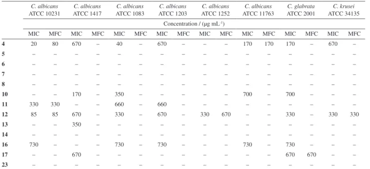
The synthetic compounds were tested against three Candida strains and five clinical isolates. The best antifungal activities were observed against C. albicans ATCC 10231 (Table 1). The compounds 4, 11 and 12 showed the greatest activity, with MICs ranging from 20 to 330 µg mL-1 and
MFCs ranging from 80 to 330 µg mL-1 for the C. albicans
strains. Compound 4 (MIC = 20 µg mL-1) showed the highest
inhibitory effect growth of C. albicans when compared to ketoconazole (MIC = 62.5 µg mL-1), the standard antifungal
agent utilized in this assay. The compounds 4, 11 and 12 showed fungicidal activity against C. albicans ATCC 10231, which was not observed for the antifungal ketoconazole. The MIC value of compound 4 obtained against this yeast was lower than the MFC value, which suggests that this compound is fungistatic at a lower concentration and fungicidal at a slightly higher concentration.
It is worth mentioning that C. glabrata ATCC 2001 displayed sensitivity towards the compounds 4 and 12 that showed fungistatic activity. In addition, C. krusei ATCC 34135 was susceptible to the compound 12 that showed MIC and MFC of 330 µg mL-1. All the yeasts tested were
resistant to compounds 5, 6, 7, 8, 14 and 23.
Regarding clinical strains, the compound 4 showed the best fungistatic activity against C. albicans clinical strains CA1, CA2, CA4 and CA5 with MICs ranging from 40-670 µg mL-1. The compound 10 showed fungistatic
activity against CA1 with an MIC of 170 µg mL-1. The
other compounds showed less activity.
In this study, the compounds 4, 11 and 12 had the greatest activity against C. albicans strains; therefore, their effect on membrane ergosterol was determined using
the exogenous ergosterol assay. Compound 4 showed
binding action on membrane ergosterol, since the MIC was enhanced 4 fold against C. albicans ATCC 10231, when performed in the presence of exogenous ergosterol (MIC = 80 µg mL-1) indicating their binding to ergosterol
(Table 2).
The cytotoxicity assay was performed with the compounds 4, 11 and 12, which showed best MIC values against C. albicans ATCC 10231. The CC50 obtained for
those compounds were 52.81, 52.58 and 85.94 µg mL-1,
respectively.
Molecular modeling studies
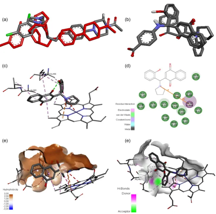
In order to initiate a lead optimization process, docking studies (Figure 1) were performed between the most active lawsone compounds and the molecular target of ketoconazole (3OZW).28 Initially, the docking methodology
was evaluated by redocking34 of ketoconazole, which
yielded a root mean square deviation (RMSD) of 2.29 Å (Figure 1a). Even though this value deviates 0.29 Å from the ideal value (2.0 Å), as can be seen in Figure 1a, it could keep the moieties in the similar order of crystallographic ligand. Next, the compounds 4, 11 and 12 were docked into the
binding site following a similar conformation (Figure 1b). The intermolecular interactions between the most active compound 4 and target are illustrated by Figures 1c and 1d. As can be observed, the pyrrolidine moiety is able to bind to the heme group by a cation-pi, whereas the naphthoquinone and phenyl moieties perform van der Waals interactions. Further, the binding site cavity was studied by hydrophobic
(Figure 1e) and hydrogen bond (Figure 1f) surfaces. In general, the binding site showed a high hydrophobic character (Figure 1e). However, close to the phenyl moiety, the binding site can recognize a hydrogen bond acceptor and donor. In other words, nonpolar moieties and hydrogen acceptor and donor moieties in the phenyl ring can improve the biological activity.
Scheme 5. Synthesis of compound 16.
Table 1. Antifungal activity (µg mL-1) of 2-hydroxy-1,4-naphthoquinone derivatives against Candida spp. strains C. albicans
ATCC 10231
C. albicans ATCC 1417
C. albicans ATCC 1083
C. albicans ATCC 1203
C. albicans ATCC 1252
C. albicans ATCC 11763
C. glabrata ATCC 2001
C. krusei ATCC 34135 Concentration / (µg mL-1)
MIC MFC MIC MFC MIC MFC MIC MFC MIC MFC MIC MFC MIC MFC MIC MFC
4 20 80 670 – 40 – 670 – – – 170 170 170 – 670 –
5 – – – – – – – – – – – – – – – –
6 – – – – – – – – – – – – – – – –
7 – – – – – – – – – – – – – – – –
8 – – – – – – – – – – – – – – – –
10 – – 170 – 350 – – – – – 700 – 700 – – –
11 330 330 – – 660 – 660 – – – – – – – – –
12 85 85 670 – 330 – 670 – 330 670 – – 330 – 330 330 13 – – 350 – – – – – – – – – – – – –
14 – – – – – – – – – – – – – – – –
16 730 – – – 730 – 730 – – – 730 – 730 – – –
17 – – 670 – – – – – – – – – 670 670 – –
23 – – – – – – – – – – – – – – – –
MIC: minimum inhibitory concentration; MFC: minimum fungicidal concentration; –: no activity.
Table 2. Mechanism of action on membrane ergosterol against three Candida spp. strains
C. albicans ATCC 10231
C. glabrata ATCC 2001
C. krusei ATCC 34135
MIC with ergosterol / (µg mL-1)
MIC without ergosterol / (µg mL-1)
MIC with ergosterol / (µg mL-1)
MIC without ergosterol / (µg mL-1)
MIC with ergosterol / (µg mL-1)
MIC without ergosterol / (µg mL-1)
4 80 20 170 170 N N
11 330 330 N N N N
12 85 85 330 330 330 330
Nystatin 31.25 3.9 31.25 3.9 31.25 3.9
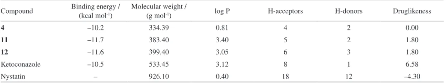
Finally, tests on the physicochemical properties were carried out to evaluate the most promising ligand35
among compounds 4, 11 and 12 to reach the market (Table 3). According to the rule of 5 (Ro5) proposed by Lipinski et al.,36 the lawsone derivatives 4, 11 and 12 do not
violate any rule. They showed molecular weights less than 500, the number of hydrogen bond donors and acceptors less than 5 and 10, respectively, and log P less than 5. In addition, the binding energy obtained from docking studies is similar to that of ketoconazole. On the other hand, ketoconazole and nystatin violated one and two Ro5 rules,
respectively. Moreover, druglikeness37 shows that these
compounds contain fragments present in commercial drugs, of which 80% have positive druglikeness values close to 3. These findings suggest that the compounds 4, 11 and 12 are more promising drugs than ketoconazole and nystatin.
Conclusions
In this study, we determined the MICs and MFCs of thirteen new naphthoquinones derivatives 4-8, 10-14, 16, 17 and 23 obtained by multicomponent Mannich reaction
containing 2-phenyl-2H-1,2,3-triazole or substituted and non-substituted phenyl groups against Candida strains and clinical isolates. Among them, compounds 4, 11 and 12 were the most active. These compounds showed fungistatic and fungicidal activity against strains of C. albicans ATCC 10231. This finding indicates that they are strong candidates for clinical use, since the complete elimination of the pathogen is the safest option.38 Moreover, the results indicate that these
compounds were more active against C. albicans ATCC 10231, which shows greater sensitivity than C. glabrata and C. krusei. It is noteworthy that C. albicans is the causative agent in 90% of fungal infections such as VVC.39 Besides,
C. albicans and C. tropicalis strains are responsible for a number of major diseases as well as providing recent cases of resistance to the current main antifungals.40
In addition, the effect on the membrane ergosterol was determined for compounds 4, 11 and 12. The evaluation of this mechanism of action is important due to the ergosterol molecule, or enzymes used in its biosynthesis, being important targets for the action of azole antifungals.41
Hence, the antifungal activity of the compound 4 against C. albicans ATCC 10231 can be attributed to its action on membrane ergosterol. The antifungal activity of the compounds 11 and 12 against C. albicans ATCC 10231 can be attributed probably to the reactive oxygen species (ROS) generation, because these compounds do not have the triazole group and because of the presence of naphthoquinone core with recognized pharmacological action via formation of ROS.1,42
The MICs for the compounds 4, 11 and 12 against yeasts indicate that these compounds are excellent choices for the development of novel drugs to treat fungal infections. However, our data highlighted that the compound 4 showed superior antifungal activity against C. albicans ATCC 10231 when compared to ketoconazole, the basis for most drugs available for treatment of fungal infections currently on the market. In our study, these compounds presented moderate to low cytotoxicity in BHK cells. These results also indicate that the compounds present low cytotoxicity on the cell strain tested.43
Table 3. Physicochemical properties for the bioactive lawsone derivatives
Compound Binding energy / (kcal mol-1)
Molecular weight /
(g mol-1) log P H-acceptors H-donors Druglikeness
4 –10.2 334.39 0.81 4 2 0.00
11 –11.7 383.40 3.40 5 2 1.80
12 –11.6 399.40 3.05 6 3 1.80
Ketoconazole –10.5 533.45 3.12 8 1 6.58
Nystatin – 926.10 0.40 18 12 –4.30
log P: partition coefficient.
The most active compounds showed similar binding energy with ketoconazole, suggesting their mechanism of action. In general, the cavity of the binding site has a hydrophobic character, addressing for lipophilic moieties in phenyl ring. Furthermore, the compounds 4, 11 and 12 are under Ro5 with a druglikeness score better than that for ketoconazole and nystatin. These findings suggest a therapeutic alternative against the increased resistance of Candida spp. to azole-based antifungals.40
Supplementary Information
Supplementary information (spectra for IR, 1H and 13C NMR and FT-ICR MS of the synthesized compounds)
is available free of charge at http://jbcs.sbq.org.br as PDF file.
Acknowledgments
The authors thank FAPES (process 54684722/2012), CNPq (process 449984/2014-1) and FAPEMIG (APQ-00557-14) for financial support.
References
1. Delarmelina, M.; Daltoé, R. D.; Cerri, M. F.; Madeira, K. P.; Rangel, L. B. A.; Lacerda Jr., V.; Romão, W.; Taranto, A. G.; Greco, S. J.; J. Braz. Chem. Soc. 2015, 26, 1804; Rahmoun, N. M.; Boucherit-Otmani, Z.; Boucherit, K.; Benabdallah, M.; Villemin, D.; Choukchou-Braham, N.; Med. Mal. Infect.2012, 42, 270; Silva, M. N.; Ferreira, V. F.; de Souza, M. C. B. V.;
Quim. Nova 2003, 26, 407; Liu, K. C.; Li, J.; Sakya, S.; Mini-Rev. Med. Chem. 2004, 4, 1105; Asche, C.; Mini-Rev. Med. Chem. 2005, 5, 449; Barbosa, T. P.; Camara, C. A.; Silva, T. M. S.; Martins, R. M.; Pinto, A. C.; Vargas, M. D.; Bioorg. Med. Chem. 2005, 13, 6464; Teixeira, M. J.; Almeida, Y. M.; Viana, J. R.; Holanda Filha, J. G.; Rodrigues, T. P.; Prata Jr., J. R. C.; Coelho, I. C. B.; Rao, V. S.; Pompeu, M. M. L.; Phytother. Res.
Wolfender, J. L.; Nianga, M.; Stoeckli-Evans, H.; Hostettmann, K.; Phytochemistry 1996, 42, 1315; de Moura, K. C. G.; Emery, F. S.; Neves-Pinto, C.; Pinto, M. C. F. R.; Dantas, A. P.; Salomão, K.; de Castro, S. L.; Pinto, A. V.; J. Braz. Chem. Soc. 2001,
12, 325; Zani, C. L.; Chiari, E.; Krettli, A. U.; Murta, S. M. F.; Cunningham, M. L.; Fairlamb, A. H.; Romanha, A. J.; Bioorg. Med. Chem. 1997, 5, 2185; Stagliano, K. W.; Emadi, A.; Lu, Z.; Malinakova, H. C.; Twenter, B.; Yu, M.; Holland, L. E.; Rom, A. M.; Harwood, J. S.; Amin, R.; Johnson, A.; Yves, P.; Bioorg. Med. Chem. 2006, 14, 5651; Bringmann, G.; Reichert, Y.; Kane, V. V.; Tetrahedron 2004, 60, 3539.
2. Kathiravan, M. K.; Salake, A. B.; Chothe, A. S.; Dudhe, P. B.; Watode, R. P.; Mukta, M. S.; Gadhwe, S.; Bioorg. Med. Chem.2012, 20, 5678; Gafner, S.; Wolfender, J. L.; Nianga, M.; Stoeckli-Evans, H.; Hostettman, K.; Phytochemistry1996, 42, 1315; Ibis, C.; Tuyun, A. F.; Ozsoy-Gunes, Z.; Bahar, H.; Stasevych, M. V.; Musyanovych, R. Y.; Komarovska-Porokhnyavets, O.; Novikov, V.; Eur. J. Med. Chem.2011, 46, 5861; Ibis, C.; Tuyun, A. F.; Bahar, H.; Ayla, S. S.; Stasevych, M. V.; Musyanovych, R. Y.; Komarovska-Porokhnyavets, O.; Novikov, V.; Med. Chem. Res. 2013,22, 2879; Arif, T.; Mandal, T. K.; Dabur, R.; Oppor., Challenge Scope Nat. Prod. Med. Chem.2011, 81, 283; Sacau, E. P.; Braun, A. E.; Ravelo, A. G.; Ferro, E. A.; Tokuda, H.; Mukainaka, T.; Nishino, H.; Bioorg. Med. Chem.2003, 11, 483; Ferreira, V. F.; Jorqueira, A.; Souza, A. M. T.; Silva, M. N.; de Souza, M. C. B. V.; Gouvêa, R. M.; Rodrigues, C. R.; Pinto, A. V.; Castro, H. C.; Santos, D. O.; Araújo, H. P.; Bourguignon, S. C.; Bioorg. Med. Chem.2006,14, 5459; Mickevičienė, K.; Baranauskaitė, R.; Kantminienė, K.; Stasevych, M.; Komarovska-Porokhnyavets, O.; Novikov, V.; Molecules 2015, 20, 3170; Tuyun, A. F.; Bayrak, N.; Yjldjrjm, H.; Onul, N.; Kara, E. M.; Celik, B. O.; J. Chem. 2015, 2015, 1; Tandon, V. K.; Maurya, H. K.; Mishra, N. N.; Shukla, P. K.; Eur. J. Med. Chem.2009,44, 3137; Sreelatha, T.; Kandhasamy, S.; Dinesh, R.; Shruthy, S.; Shweta, S.; Mukesh, D.; Karunagaran, D.; Balaji, R.; Mathivanan, N.; Perumal, P. T.; Bioorg. Med. Chem. Lett. 2014, 24, 3647.
3. Fiorot, R. G.; Allochio Filho, J. F.; Pereira, T. M. C.; Lacerda, V.; dos Santos, R. B.; Romão, W.; Greco, S. J.; Tetrahedron Lett.
2014, 55, 4373; Allochio Filho, J. F.; Fiorot, R. G.; Lacerda, V.; dos Santos, R. B.; Vanini, G.; Romão, W.; Greco, S. J.; Colloids Interface Sci. Commun.2015, 4, 14; Neves, A. P.; Barbosa, C. C.; Greco, S. J.; Vargas, M. D.; Visentin, L. C.; Pinheiro, C. B.; Mangrich, A. S.; Barbosa, J. P.; da Costa, G. L.; J. Braz. Chem. Soc.2009, 20, 712.
4. Dalgliesh, C. E.; J. Am. Chem. Soc.1949, 71, 1697; Silva Jr., E. N.; Melo, I. M. M.; Diogo, E. B. T.; Costa, V. A.; Souza Filho, J. D.; Valença, W. O.; Camara, C. A.; Oliveira, R. N.; Araújo, A. S.; Emery, F. S.; Santos, M. R.; Simone, C. A.; Menna-Barreto, R. F. S.; de Castro, S. L.; Eur. J. Med. Chem. 2012, 52, 304; Lagrota, M. H. C.; Wigg, M. D.; Santos, M. G. M.; Pinto, A.
V.; Pinto, M. C. F. R.; Rev. Microbiol.1988,19,338; Lima, N. M. F.; Correia, C. S.; Ferraz, P. A. L.; Pinto, A. V.; Pinto, M. C. R. F.; Santana, A. E. G.; Goulart, M. O. F.; J. Braz. Chem. Soc. 2002, 13, 822; Santos, A. F.; Ferraz, P. A. L.; Pinto, A. V.; Pinto, M. C. F. R.; Goulart, M. O. F.; Sant’Ana, A. E. G.; Int. J. Parasitol.2000, 30, 1199; Leffer, M. T.; Hathaway, R. J.; J. Am. Chem. Soc. 1948, 70, 3222.
5. Eggimann, P.; Garbino, J.; Pittet, D.; Lancet Infect. Dis.2003,
3, 685.
6. Ortega, M.; Marco, F.; Soriano, A.; Almela, M.; Martínez, J. A.; López, J.; Pitart, C.; Mensa, J.; J. Hosp. Infect.2011, 77, 157.
7. Sobel, J. D.; Zervos, M.; Reed, B. D.; Hooton, T.; Soper, D.; Nyirjesy, P.; Heine, M. W.; Willems, J.; Panzer, H.; Antimicrob. Agents Chemother.2003, 47, 34.
8. Sardi, J. C. O.; Scorzoni, L.; Bernardi, T.; Fusco-Almeida, A. M.; Mendes Giannini, M. J. S.; J. Med. Microbiol.2013, 62, 10.
9. Vale-Silva, L. A.; Coste, A. T.; Ischer, F.; Parker, J. E.; Kelly, S. L.; Pinto, E.; Sanglard, D.; Antimicrob. Agents Chemother.
2012, 56, 1960.
10. Keserü, G. M.; Makara, G. M.; Nat. Rev. Drug Discovery2009,
8, 203.
11. Elokely, K. M.; Doerksen, R. J.; J. Chem. Inf. Model.2013, 53, 1934.
12. de Oliveira, M. E.; Cenzi, G.; Nunes, R. R.; Andrighetti, C. R.; de Sousa Valadão, D. M.; dos Reis, C.; Simões, C. M. O.; Nunes, R. J.; Júnior, M. C.; Taranto, A. G.; Sanchez, B. A. M.; Viana, G. H. R.; de Pilla Varotti, F.; Molecules2013, 18, 15276. 13. Guimarães, D. S. M.; Fonseca, A. L.; Batista, R.; Comar Jr.,
M.; Oliveira, A. B.; Taranto, A. G.; Varotti, F. D. P.; Mem. Inst. Oswaldo Cruz2015, 110, 255.
14. Baramee, A.; Coppin, A.; Mortuaire, M.; Pelinski, L.; Tomavo, S.; Brocard, J.; Bioorg. Med. Chem.2006, 14, 1294.
15. Elavarasan, S.; Gopalakrishnan, M.; Spectrochim. Acta, Part A
2014, 133, 1.
16. Clayton, Y. M.; Proc. R. Soc. Med.1977, 70, 359.
17. National Committee for Clinical Laboratory Standards (NCCLS); Reference Method for Broth Dilution Antifungal Susceptibility Testing of Yeast: Approved Standard, CLSI document M27-A2; NCCLS: Wayne, PA, 2002.
18. Clinical and Laboratory Standards Institute (CLSI); Reference Method for Broth Dilution Antifungal Susceptibility Testing of
Yeasts, Document M27-A3; CLSI: Wayne, 2008.
19. Portillo, A.; Vila, R.; Freixa, B.; Ferro, E.; Parella, T.; Casanova, J.; Cañigueral, S.; J. Ethnopharmacol.2005, 97, 49.
20. Escalante, A.; Gattuso, M.; Pérez, P.; Zacchino, S.; J. Nat. Prod.
2008, 71, 1720.
21. Mosmann, T.; J. Immunol. Methods1983, 65, 55.
23. GraphPad; GraphPad QuickCalcs t test calculator, http:// graphpad.com/quickcalcs/ttest1/?Format=C accessed in March 2016.
24. Motulsky, H. J.; Christopoulos, A.; Fitting Models to Biological Data Using Linear and Nonlinear Regression. A Practical
Guide to Curve Fitting, 2nd ed.; GraphPad Software, Inc.: San Diego, 2003, pp. 351.
25. ten Brink, T.; Exner, T. E.; J. Comput.-Aided. Mol. Des.2010,
24, 935.
26. Stewart, J. J. P.; J. Mol. Model.2013, 19, 1.
27. Stewart, J. J. P.; MOPAC 2012; Stewart Computational Chemistry, Colorado Springs, 2012.
28. El Hammi, E.; Warkentin, E.; Demmer, U.; Limam, F.; Marzouki, N. M.; Ermler, U.; Baciou, L.; Biochemistry2011,
50, 1255.
29. Trott, O.; Olson, A. J.; J. Comput. Chem.2010, 31, 455. 30. Accelrys Software Inc.; Discovery Studio Modeling Environment,
Release 4.1; Accelrys Software Inc.: San Diego, 2014. 31. Sander, T.; Freyss, J.; von Korff, M.; Rufener, C.; J. Chem. Inf.
Model.2015, 55, 460.
32. Tornøe, C. W.; Christensen, C.; Meldal, M.; J. Org. Chem.2002, 67, 3057.
33. Jardim, G. A. M.; Reis, W. J.; Ribeiro, M. F.; Ottoni, F. M.; Alves, R. J.; Silva, T. L.; Goulart, M. O. F.; Braga, A. L.; Menna-Barreto, R. F. S.; Salomão, K.; de Castro, S. L.; da Silva Júnior, E. N.; RSC Adv. 2015, 5, 78047; Jardim, G. A. M.; Guimarães, T. T.; Pinto, M. C. F. R.; Cavalcanti, B. C.; de Farias, K. M.; Pessoa, C.; Gatto, C. C.; Nair, D. K.; Namboothiri, I. N. N.; da Silva Jr., E. N.; Med. Chem. Commun.2015, 6, 120; da Cruz, E. H. G.; Carvalho, P. H. P. R.; Corrêa, J. R.; Silva, D. A. C.; Diogo, E. B. T.; de Souza Filho, J. D.; Cavalcanti, B. C.; Pessoa, C.; de Oliveira, H. C. B.; Guido, B. C.; da Silva Filho, D. A.;
Neto, B. A. D.; da Silva Jr., E. N.; New J. Chem.2014, 38, 2569; Devi Bala, B.; Muthusaravanan, S.; Choon, T. S.; Ashraf Ali, M.; Perumal, S.; Eur. J. Med. Chem.2014, 85, 737.
34. Santos Jr., M. C.; de Assis, S. A.; Góes-Neto, A.; Duarte, A. A.; Alves, R. J.; Comar Jr., M.; Taranto, A. G.; Chem. Cent. J.
2013, 7, 48.
35. de Oliveira, M. E.; Cenzi, G.; Nunes, R. R.; Andrighetti, C. R.; de Sousa Valadão, D. M.; dos Reis, C.; Simões, C. M. O.; Nunes, R. J.; Júnior, M. C.; Taranto, A. G.; Sanchez, B. A. M.; Viana, G. H. R.; de Pilla Varotti, F.; Molecules2013, 18, 15276. 36. Lipinski, C. A.; Lombardo, F.; Dominy, B. W.; Feeney, P. J.;
Adv. Drug Delivery Rev.1997, 23, 3.
37. Verdonk, M. L.; Giangreco, I.; Hall, R. J.; Korb, O.; Mortenson, P. N.; Murray, C. W.; J. Med. Chem.2011, 54, 5422.
38. Wong, S. S. W.; Kao, R. Y. T.; Yuen, K. Y.; Wang, Y.; Yang, D.; Samaranayake, L. P.; Seneviratne, C. J.; PLoS One2014, 9, e85836.
39. Fisher, J. F.; Kavanagh, K.; Sobel, J. D.; Kauffman, C. A.; Newman, C. A.; Clin. Infect. Dis.2011, 52, 437.
40. Silva, S.; Negri, M.; Henriques, M.; Oliveira, R.; Williams, D. W.; Azeredo, J.; FEMS Microbiol. Rev.2012, 36, 288. 41. Onyewu, C.; Blankenship, J. R.; Del Poeta, M.; Heitman, J.;
Antimicrob. Agents Chemother.2003, 47, 956.
42. Amarantes-Mendes, G. P.; Green D. R.; Braz. J. Med. Biol. Res. 1999, 32, 1053; Avendaño, C.; Menéndez, J. C.; Medicinal Chemistry of Anticancer Drugs;Elsevier: Oxford, 2008, pp. 93. 43. Brandão, G. C.; Kroon, E. G.; Duarte, M. G. R.; Braga, F. C.;
de Souza Filho, J. D.; de Oliveira, A. B.; Phytomedicine2010,
17, 926.
Submitted: February 5, 2016