Tsetse Immune System Maturation Requires the
Presence of Obligate Symbionts in Larvae
Brian L. Weiss*, Jingwen Wang, Serap Aksoy
Department of Epidemiology and Public Health, Division of Epidemiology of Microbial Diseases, Yale University School of Medicine, New Haven, Connecticut, United States of America
Abstract
Beneficial microbial symbionts serve important functions within their hosts, including dietary supplementation and maintenance of immune system homeostasis. Little is known about the mechanisms that enable these bacteria to induce specific host phenotypes during development and into adulthood. Here we used the tsetse fly,Glossina morsitans, and its obligate mutualist, Wigglesworthia glossinidia, to investigate the co-evolutionary adaptations that influence the development of host physiological processes. Wigglesworthia is maternally transmitted to tsetse’s intrauterine larvae through milk gland secretions. We can produce flies that lackWigglesworthia(GmmWgm2) yet retain their other symbiotic microbes. Such offspring give rise to adults that exhibit a largely normal phenotype, with the exception being that they are reproductively sterile. Our results indicate that when reared under normal environmental conditionsGmmWgm2adults are also immuno-compromised and highly susceptible to hemocoelicE. coliinfections while age-matched wild-type individuals are refractory. Adults that lackWigglesworthiaduring larval development exhibit exceptionally compromised cellular and humoral immune responses following microbial challenge, including reduced expression of genes that encode antimicrobial peptides (cecropin and attacin), hemocyte-mediated processes (thioester-containing proteins 2 and 4 and
prophenoloxidase), and signal-mediating molecules (inducible nitric oxide synthase). Furthermore,GmmWgm2adults harbor a reduced population of sessile and circulating hemocytes, a phenomenon that likely results from a significant decrease in larval expression ofserpentandlozenge, both of which are associated with the process of early hemocyte differentiation. Our results demonstrate thatWigglesworthiamust be present during the development of immature progeny in order for the immune system to function properly in adult tsetse. This phenomenon provides evidence of yet another important physiological adaptation that further anchors the obligate symbiosis between tsetse andWigglesworthia.
Citation:Weiss BL, Wang J, Aksoy S (2011) Tsetse Immune System Maturation Requires the Presence of Obligate Symbionts in Larvae. PLoS Biol 9(5): e1000619. doi:10.1371/journal.pbio.1000619
Academic Editor:David S. Schneider, Stanford University, United States of America
ReceivedAugust 18, 2010;AcceptedApril 11, 2011;PublishedMay 31, 2011
Copyright:ß2011 Weiss et al. This is an open-access article distributed under the terms of the Creative Commons Attribution License, which permits unrestricted use, distribution, and reproduction in any medium, provided the original author and source are credited.
Funding:This work was generously funded by grants to S.A. from the NIAID AI051584, NIGMS 069449 and Ambrose Monell Foundation (http://www. monellvetlesen.org/). The funders had no role in study design, data collection and analysis, decision to publish, or preparation of the manuscript.
Competing Interests:Serap Aksoy, coauthor on this manuscript, is also Editor-in-Chief ofPLoS Neglected Tropical Diseases.
Abbreviations:AMP, antimicrobial protein; CFU, colony-forming units; dpi, days post-infection; GFP, green fluorescent protein;GmmWgm2, Wigglesworthia-free Glossina morsitans morsitans;GmmWT, wild-type Glossina morsitans morsitans; GmmWT/Wgm2, adult wild-type Glossina morsitans morsitans treated with tetracycline; hpi, hours post-infection; iNOS, inducible nitric oxide synthase; PPO, prophenoloxidase; qPCR, real-time quantitative PCR; rRNA, ribosomal RNA; TEP, thioester-containing protein.
* E-mail: [email protected]
Introduction
Bacteria comprise the most abundant and diverse life form on earth. The ubiquity of bacteria means they have colonized virtually every ecological niche, including habitation within more evolutionarily sophisticated multi-cellular animals. Co-evolution over millions of years has provided an opportunity for beneficial symbiotic associations to develop between phylogenetically distant taxa. Such affiliations are often mutualistic, meaning both partners benefit so that each can successfully inhabit diverse environments that neither could survive in on its own [1,2]. Deciphering the mutualistic relationships between prokaryotic bacteria and multi-cellular eukaryotic animals is a rapidly advancing field of research. Performing detailed investigations into the relationships be-tween symbiotic bacteria and higher eukaryotes often involves costly and complex procedures. However, insects have well-documented symbioses that are attractive to study because they have relatively short generation times and are easy and less costly
are the sole vector of pathogenic African trypanosomes, the causative agent of sleeping sickness in humans [7]. In laboratory experiments infection with immunogenic trypanosomes results in a decrease in tsetse fecundity [8]. Furthermore, parasite infection prevalence is higher in flies that lackWigglesworthiawhen compared to age-matched wild-type (WT) individuals [9]. Wigglesworthia is thought to influence tsetse’s vectorial competence by modulating its host’s humoral immune system [10]. Thus, by preventing energetically costly parasite infections, this obligate symbiont may indirectly benefit the reproductive fitness of tsetse.
Symbiotic bacteria are rapidly gaining recognition for their important contributions to host development and immunity. The most well-known example of this type of interaction involves the mammalian microbiome, which modulates gut development during early postnatal life and subsequently shapes our mucosal and systemic immune systems [11,12]. Symbionts also serve similar functions in invertebrate hosts. For example, light organ morphogenesis in juvenile bobtail squid (Euprymna scolopes) initiates only after symbiotic Vibrio fischeri cells have stably colonized this tissue [13]. The pea aphid,Acyrthosiphon pisum, harbors a secondary symbiont, Hamiltonella defensa, which can be infected with a lysogenic bacteriophage (A. pisum secondary endosymbiont; APSE). A. pisum that harbor both H. defensa and APSE are protected from being consumed by a parasitic wasp larvae through the action of phage-encoded toxins [14].
To the best of our knowledge no evidence exists that demonstrates insect symbionts can confer an impending protective phenotype in their adult host by directing immune system development during immature stages. In the present study we investigate the mechanism by whichWigglesworthiacontributes to the development and function of cellular and humoral immune responses in adult tsetse. Our results show that the maturation and normal function of adult cellular immune responses in tsetse are severely compromised whenWigglesworthiais absent during larval development. This study reveals an important new facet that further anchors the obligate relationship between tsetse and Wigglesworthiaand may serve as a useful model to understand the
highly integrated and dynamic relationship between hosts and their beneficial bacterial fauna.
Results
Tsetse’s Susceptibility toE. coliInfection Is Dependent Upon Age and Symbiotic Status
Insects are normally capable of mounting an immune response that combats infection with various groups of bacteria. Interest-ingly, in comparison to Drosophila, tsetse flies are uniquely susceptible to septic infection with 103 colony-forming units (CFU) of normally non-pathogenicEscherichia coli(E. coli) K12 [15]. In the present study we further investigated tsetse’s unique susceptibility toE. coliinfection by subjecting wild-type (GmmWT
) and adults from two age groups to hemocoelic infections with varying quantities of E. coli K12. Three-day-old GmmWT individuals (flies from this age group are hereafter referred to as ‘‘young’’) were highly susceptible to this treatment, as 103 CFU resulted in the death of all flies by 8 d post-infection (dpi; Figure 1A, top graph). In contrast, 77% and 55% of 8-d-old WT individuals (flies 8 d old and older are hereafter referred to as ‘‘mature’’) survived for 14 dpi with 103 and 106 CFU of E. coli K12, respectively (Figure 1A, middle graph).
We demonstrated previously that feeding pregnant female tsetse the antibiotic ampicillin results in the generation offspring that lack Wigglesworthia(GmmWgm2) but still harborSodalis [9] and presum-ablyWolbachia. In the present study we determined the survival outcome of matureGmmWgm2inoculated with 106CFU ofE. coli K12 (the dose required to kill ,50% of mature WT flies; Figure 1A, middle graph). In comparison to age-matched WT tsetse,GmmWgm2 flies were highly susceptible to this infection. In fact, at 14 dpi, only 1% of these individuals remained (Figure 1A, bottom graph). We reasoned that the dramatic susceptibility of matureGmmWgm2flies to infection with E. coli could result from one of two scenarios. In the first scenario Wigglesworthia may directly contribute to the adult immune response to a foreign micro-organism. In the second scenario, the absence of Wiggle-sworthiaduring larvagensis may give rise toGmmWgm2adults with compromised immune functions. To determine if the first scenario is correct we treated mature WT tsetse with the antibiotic tetracycline to eliminate all of their microbiota, including bacteriome-associatedWigglesworthia(Figure 1B), and subsequently challenged these adult flies (GmmWT/Wgm2) with 106 CFU of tetracycline resistantE. coli K12. We found that, similar to their WT counterparts (GmmWT; Figure 1A, middle panel), about 70% of GmmWT/Wgm2 individuals survived this infection (Figure 1C).
Conversely,GmmWgm2 adults were highly susceptible (Figure 1A, bottom graph). This result suggests that when Wigglesworthia is absent from tsetse during larval development subsequent adults are severely immuno-compromised.
We demonstrated that when larval tsetse lacks exposure to Wigglesworthiain utero their immune system is highly compromised during adulthood. This finding does not exclude the possibility that treatment of female flies with ampicillin to produceGmmWgm2 offspring induces perturbations in other constituents of tsetse’s microbiota. Because tsetse’s other two known endosymbionts, Sodalisand Wolbachia, are present at similar densities inGmmWT andGmmWgm2 adults (Figure 1D), we looked for the presence of uncharacterized endosymbionts or digestive tract-associated mi-crobes that are potentially passed on to developing intra-uterine larvae where they subsequently impact immunity. We generated a clone library containing 16s rRNA gene sequences PCR amplified from 3rdinstarGmmWgm2andGmmWTlarvae and then sequenced multiple clones from each tsetse line. Our results indicate that the Author Summary
Beneficial bacterial symbionts, which are ubiquitous in nature, are often characterized by the extent to which they interact with the host. In the case of mutualistic symbioses, both partners benefit so that each one can inhabit diverse ecological niches where neither could survive on its own. Unfortunately, little is known about the functional mechanisms that underlie mutualistic relationships. Insects represent a group of advanced multi-cellular organisms that harbor well-documented symbiotic associations. One such insect, the tsetse fly, harbors a maternally transmitted bacterial mutualist calledWigglesworthiathat provides its host with essential metabolites missing from its vertebrate blood-specific diet. In this study, we further examine the relationship between tsetse andWigglesworthiaby inves-tigating the interaction between this bacterium and its host’s immune system. We have found that when
proportion of Wigglesworthia, Sodalis, and Wolbachia in 3rd instar GmmWTlarvae is 19%, 75%, and 6%, respectively. As expected no Wigglesworthia 16s rRNA sequences were present in GmmWgm2 larvae, and the proportion of Sodalis and Wolbachia sequences present was 71% and 29%, respectively (Figure 1E). No uncharacterized endosymbionts or gut-associated environmental microbes were present in any sample. Taken together, these results suggest that the presence ofWigglesworthiaspecifically is responsible for enabling immune system maturation in WT tsetse.
Our host survival curves indicate that mature GmmWT and GmmWT/Wgm2 can survive infection with E. coli while young GmmWTand mature GmmWgm2perish. To determine a cause for the variation in survival we observed between the four groups
following infection with E. coli, we monitored bacterial growth dynamics over the course of the experiment in each group. Bacterial number within mature GmmWT
and GmmWT/Wgm2
peaked at 3.86104and 1.96104E. coli, respectively, over the
2-wk period, indicating that these groups appeared able to control their infections. In contrast, E. coli increased exponentially in young GmmWT and matureGmmWgm2 and reached a maximum
density at 6 dpi of 4.26106and 8.86106, respectively (Figure 1F).
These results implicate bacterial sepsis as the cause of high mortality observed in youngGmmWT
and matureGmmWgm2. All of
the above-mentioned results taken together indicate that mature GmmWTare considerably more resistant to infection with a foreign microbe than are their younger counterparts. Furthermore, tsetse’s
Figure 1. Host survival correlates with symbiont status following septic infection withE. coliK12.(A) The effects of age and symbiont status on the survival of tsetse following systemic infection withE. coliK12. Mature adultGmmWgm-flies were significantly more susceptible to
infection with 106CFU ofE. colithan were their wild-type counterparts (bottom and middle panels;p,0.001). (B)GmmWT/Wgm2 flies harbored
Wigglesworthiaduring immature development but lacked the bacteria as mature adults. (C)GmmWT/Wgm2adults were infected with tetracycline
resistantE. coli1 d after their last antibiotic-supplemented blood meal. Unlike their counterparts that lackedWigglesworthiathroughout immature development,GmmWT/Wgm2was able to survive infection withE. coli. No significant difference in survival was observed between mature adult
GmmWTversusGmmWT/Wgm2adults infected with 106CFU ofE. coli(Figure 1A middle panel and Figure 1C;p = 0.07). (D) RelativeSodalisand Wolbachiadensities in 40-d-oldGmmWTandGmmWgm2adults (n = 5 of each) were normalized against hostb-tubulincopy number. (E) Analysis of
bacterial 16s rRNA clone libraries indicates that GmmWTlarvae harboredWigglesworthia, Sodalis, andWolbachia, while their counterparts from
ampicillin treated females harbored onlySodalisandWolbachia. No other bacteria were identified from either fly line. (F) Average number (6SEM) of recE. colipILper tsetse strain over time (n= 3 individuals per strain per time point) following septic infection with 103CFU of bacteria. Values shown in
obligate mutualist, Wigglesworthia, must be present during the development of immature stages so that mature adults are able to overcome infection withE. coli.
Expression of Immunity-Related Genes in Adult Tsetse Varies Depending on Symbiont Status
To understand the basis of the compromised immunity we observed in mature GmmWgm2 flies, we evaluated the expression
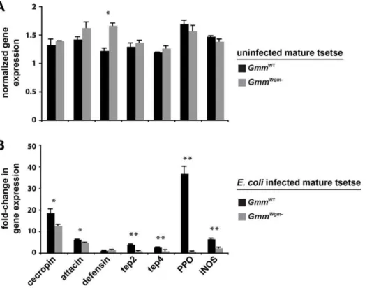
profile of a set of immunity-related genes from age-matched mature adult GmmWT and GmmWgm2 flies that were either uninfected or 3 dpi with E. coli K12. We included the antimicrobial peptides (AMPs)attacin, cecropin, anddefensin, which distinctly target gram-negative (attacin and cecropin) and gram-positive bacteria (defensin) [16]. Our analysis also included thioester-containing proteins (tep2 and tep4), prophenoloxidase (PPO), and inducible nitric oxide synthase (iNOS). In insects TEPs likely function as pathogen-specific opsonins that bind to bacteria or parasites and promote their phagocytosis/encapsulation [17], while PPO initiates a proteolytic cascade that results in melanin deposition [18]. iNOS catalyzes synthesis of the signaling molecule nitric oxide (NO), which plays a role in humoral and cellular immunity in Anopheline mosquitoes [19], reduvid bugs [20], and Drosophila [21,22] by inducing AMP expression and recruiting hemocytes to the site of infection.
Our expression results indicate that symbiont status plays little or no role in the expression of immunity-related genes in uninfected adults. In fact, with the exception of the AMPdefensin, no significant differences in immunity-related gene expression between mature uninfected GmmWT and GmmWgm2 adults
(Figure 2A). However, we observed a considerably different profile of immunity-related gene expression when these different fly strains were infected withE. coliK12. Under these circumstances, all of the genes evaluated (with the exception of defensin) were expressed at significantly higher levels in GmmWT
compared to GmmWgm2individuals (Figure 2B). Particular striking was the fact that the induction of pathways associated with cellular immunity, such as pathogen recognition (tep2 and tep4) and melanization (PPO), were significantly compromised in matureGmmWgm2adults. The absence of a robust cellular immune response is likely the cause of high mortality among these individuals followingE. coli infection. This analysis indicates that Wigglesworthia must be present during the development of immature tsetse in order for immune-related genes to subsequently be expressed in matureE. coli–infected adults.
Hemocytes Play an Integral Role in ControllingE. coli Infection Outcomes in Tsetse
Our analysis of immunity-related gene expression in tsetse suggests that cellular immune pathway functions in adult tsetse are particularly compromised when Wigglesworthia is absent during immature development. The most prominent cellular immune mechanisms include melanization and phagocytosis. These processes, which ultimately result in the removal of foreign invaders in Drosophila [23] and A. aegypti [24], both arise from distinct crystal cell and plasmatocyte hemocyte lineages, respectively [25].
We investigated the role hemocytes might play in determining the susceptible phenotype we observed in tsetse following
Figure 2. The effect of symbiont status on the expression of immunity-related genes in adult tsetse.(A) Target gene expression in uninfectedGmmWTandGmmWgm2adults is indicated relative to the constitutively expressed tsetseb-tubulin gene. (B) Fold-change in the expression of immunity-related genes inGmmWTandGmmWgm2tsetse 3 dpi withE. coliK12. All values for both tsetse strains are represented as a fraction of
average normalized gene expression levels in bacteria-infected flies relative to expression levels in PBS-injected controls. In (A) and (B), quantitative measurements were performed on three biological samples in duplicate. Values are represented as means (6SEM). *p,0.05, **p,0.005 (Student’s
t-test).
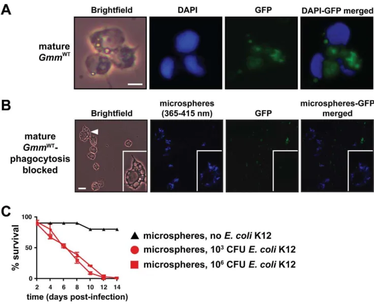
infection with E. coli. We infected mature GmmWT individuals with GFP-expressing E. coli and were able to observe that hemocytes had engulfed a large number of the introduced cells by 12 hpi (Figure 3A). We next inhibited phagocytosis by introducing blue fluorescent microspheres directly into tsetse’s hemocoel and 12 h later infected the bead-treated individuals with GFP-expressingE. coli. Microscopic inspection of hemocytes harvested 12 hpi withE. colirevealed the presence of internalized microspheres and the absence of engulfed E. coli. This observation indicated that we were successful in blocking hemocyte phagocytosis (Figure 3B). We subsequently maintained our microsphere-injected tsetse for 2 wk with the intention of determining the impact of impaired phagocytosis on host survival
outcome. MatureGmmWTflies exhibiting impaired phagocytosis were highly susceptible to infection with both 16103and 16106 E. coliK12. In fact, by day 12 post-infection, all of these flies had perished regardless of the initial dose used for infection (Figure 3C). This observation contrasts starkly with infection outcome in mature GmmWT that exhibit normally functioning hemocytes (Figure 1A, middle graph). Our results suggest that defects in phagocytosis severely compromise the ability of these tsetse flies to overcome bacterial infection.
A notable result of our immunity-related gene expression analysis was a 37-fold decrease inPPO levels inGmmWgm2 flies. This enzyme is an essential component of the melanization pathway, and its expression ultimately results in host wound
Figure 3. Hemocyte-mediated phagocytosis is a critical component of tsetse’s immune response.(A) 8-d-oldGmmWTwere subjected to septic infection with GFP-expressing E. coli K12. Twelve hours post-infection hemolymph was collected, fixed on glass slides using 2% paraformaldehyde, and microscopically examined for the presence of hemocyte-engulfed bacterial cells. Scale bar = 10mm. (B) The process of
hemocyte-mediated phagocytosis in tsetse was blocked by micro-injecting polystyrene beads into the hemocoel of 8-d-old WT individuals. In consecutive 12 h intervals following bead injection, flies were infected with GFP-expressingE. coliK12 and then hemolymph was collected and fixed as described above. Hemocytes appear to have engulfed the beads, thus prohibiting the subsequent uptake of bacterial cells. The inset in each panel shows a higher magnification image of one hemocyte, which is identified by a white triangle in the left-most panel. Scale bar = 20mm. (C) Tsetse flies that harbor hemocytes incapable of engulfingE. coliare susceptible to septic infection with this bacterium while their wild-type counterparts are not. The susceptible phenotype is exhibited regardless of whether phagocytosis-inhibited tsetse were inoculated with 103or 106CFU ofE. coli. Beads
alone had no effect on tsetse mortality. No significant difference existed in survival outcome between matureGmmWTphagocytosis inhibited flies infected with 103versus 106CFU ofE. coli(p= 0.47, log-rank analysis). Furthermore, no significant difference was present between matureGmmWgm2
healing and the melanization, encapsulation, and subsequent removal of foreign microorganisms [26–28]. In conjunction with the remarkable variation in PPO expression observed between GmmWTandGmmWgm2, we were also able to visually observe the absence of a melanization response to E. coli infection in flies lacking Wigglesworthia. In fact, 30 min post-injection with E. coli, hemolymph was still actively exuding from the inoculation wound ofGmmWgm2flies. Conversely, in WT individuals no hemolymph was detectable and melanin was deposited at the wound site (Figure 4). These results further suggest that hemocyte-mediated cellular immunity provides an imperative defense against the establishment of bacterial infections in tsetse’s hemocoel. Further-more, the absence of this response in GmmWgm2 individuals was likely responsible for the compromised host survival phenotype we observed following infection withE. coli.
Symbiotic Bacteria Impact the Maturation of Tsetse’s Cellular Immune Response During Development
We observed that young GmmWT were markedly more susceptible to infection withE. coliK12 than were their mature counterparts. Furthermore, symbiont status also altered infection outcome, as matureGmmWgm2perished followingE. coliinfection while age-matched WT individuals survived. These differential infection outcomes appeared to result from disparities in cellular immune system function between the different tsetse lines we examined. Based on these observations we hypothesized that the obligate mutualistWigglesworthiaplays a crucial role in regulating the development of cellular immunity in tsetse during immature stages. To test this hypothesis we quantified the number of circulating and sessile hemocytes present in young and mature adult GmmWT and mature adult GmmWgm2. Our results indicate
Figure 4. The effect of symbiont status on melanization in tsetse.MatureGmmWTandGmmWgm2tsetse (n = 10 of each strain) were
intra-thoracically inoculated with 16103E. coli K12. Thirty minutes post-inoculation the wound site on individuals from each strain was inspected microscopically for the presence of hemolymph clotting and melanin deposition. Thirty minutes post-inoculation, neither hemolymph clotting nor melanin were observed at the wound site ofGmmWgm2individuals (indicated by a red arrow). In contrast, within the same amount of time, hemolymph no longer exuded from the wound of WT flies and melanin was present surrounding the site (indicted by a white arrow).
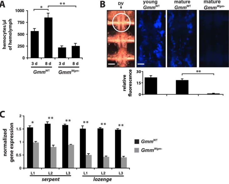
that a 1.4-fold increase in circulating hemocyte number occurs between day 3 and day 8 in WT tsetse, while no significant change in circulating hemocyte number was observed between young and matureGmmWgm2(Figure 5A). Interestingly, matureGmmWTadults harbored 3.46 more circulating hemocytes than did mature GmmWgm2adults (Figure 5A). We also looked at sessile hemocyte abundance as a further indicator ofWigglesworthia’simpact on the development of cellular immunity in tsetse. In Drosophila, this hemocyte subtype concentrates in large quantities around the anterior end of the fly’s dorsal vessel [29]. Thus, we indirectly quantified sessile hemocyte abundance immediately adjacent to the anterior-most chamber of tsetse’s dorsal vessel by measuring the fluorescent emission of microspheres that were found engulfed in this region. Young GmmWT adults engulfed 1.26 more
microspheres than their mature counterparts and 15.76more
than mature GmmWgm2 adults. Furthermore, mature WT adults engulfed 13.26 more microspheres than did age-matched GmmWgm2adults (Figure 5B).
Our results demonstrate that Wigglesworthia must be present during the development of immature tsetse in order for cellular immunity to develop and function properly in adults. Thus, we speculate that the absence of hemocytes in adult tsetse should reflect a lack of blood cell differentiation during the development of immature stages. In Drosophila the process of blood cell differentiation, or hematopoiesis, begins in the embryo and proceeds through all larval stages [30]. During Drosophila embryogenesis early hematopoiesis can be distinguished by the expression of the zinc finger transcription factor ‘‘Serpent.’’
Figure 5. The effect of age and symbiont status on the development of tsetse’s cellular immune response.(A) Number of hemocytes perml of hemolymph in young (3 d) and mature (8 d)GmmWTandGmmWgm-tsetse (n = 5 individuals from each tsetse strain at both time points).
(B) Quantitative analysis of sessile hemocyte abundance in young and matureGmmWTand matureGmmWgm
-tsetse (n = 4 individuals from each tsetse strain and age point). All tsetse strains tested were subjected to hemocoelic injection with blue fluorescent microspheres. Twelve hours post-injection, flies were dissected to reveal tsetse’s heart. The left-most panel is a Brightfield image of the three chambers that make up the dorsal vessel (DV; scale bar = 350mm). The anterior-most chamber is indicated within a white circle. The three remaining panels are close-ups of the anterior
chamber (scale bar for all 3 panels = 80mm), visualized by excitation with UV light (365/415 nm). Relative fluorescence per tsetse group was
determined using ImageJ software. (C) The presence ofWigglesworthia affects the expression of genes involved in hemocyte differentiation in immature larval tsetse. Target gene expression inGmmWTandGmmWgm2larval instars 1–3 is indicated relative to the constitutively expressed tsetse
b-tubulin gene. Quantitative measurements were performed on three biological samples in duplicate. All values in this figure are represented as means (6SEM). *p,0.05, **p,0.005 (Student’t-test).
Subsequently, another transcription factor, ‘‘Lozenge,’’ directs the differentiation of serpent-expressing precursor cells into a specific lineage of hemocytes called crystal cells. To address the relationship between the presence of Wigglesworthia and early hematopoiesis in tsetse, we used qPCR to evaluate the relative number ofserpentandlozengetranscripts present in 1st, 2nd, and 3rd instar larvae dissected from pregnant GmmWT and GmmWgm2 females. Larval instars L1, L2, and L3 from WT females expressed 1.7, 2.1, and 1.9 times moreserpenttranscripts, and 4, 4.4, and 3.9 times more lozenge transcripts, respectively, than did their counterparts from females that lacked Wigglesworthia(Figure 5C). This observed attenuated expression of bothserpentandlozengemay account for the depleted hemocyte population, and compromised cellular immune function, we observed in adult GmmWgm2 individuals.
Discussion
In the present study we demonstrate that Wigglesworthia is intimately involved in regulating the maturation and function of tsetse’s cellular immune system during immature larval develop-ment. We present a model that links the presence ofWigglesworthia in larval progeny with host immune system maturation during development and the subsequent ability of adult tsetse to overcome infection with foreign microbes (Figure 6). Obligate symbioses between intracellular bacteria and multi-cellular eukaryotes represent millions of years of co-evolution during which time both partners have adapted to increase each other’s overall fitness. The association between tsetse andWigglesworthiais an example of this reciprocal relationship in that neither organism can survive in the absence of the other.
We present several lines of evidence indicating that tsetse’s resistance toE. colipositively correlates with fly age and symbiont infection status during juvenile stages. Specifically, our results signify that matureGmmWT
adults are resistant toE. coliinfection, while young GmmWT adults are susceptible. In contrast, both young and old adult flies that lack Wigglesworthiathroughout all developmental stages are killed byE. coliinfections. Like their WT counterparts,GmmWgm2larvae acquire bothSodalisandWolbachia while in utero [31]. Although the intrauterine larval environment is otherwise aseptic, adultGmmWgm2can be exposed to a wide range of environmental microbes during adulthood. However, neither the presence of other symbiotic bacteria (SodalisandWolbachia) in larvae, nor environmental microbes acquired during adulthood, appear to be sufficient to induce immunity inGmmWgm2adults. Additionally, we show that mature GmmWT adults treated with antibiotics to eliminate Wigglesworthia(GmmWT/Wgm2) remain resistant toE. coli infections. The resistant phenotype ofGmmWT/Wgm2further signifies
that obligateWigglesworthiais not directly responsible for the ability of mature adult GmmWT to overcome septic bacterial infection. Instead, it appears that Wigglesworthia’s presence during the maturation of larval progeny stimulates the development of host immunity in adults.
Our quantitative analysis of gene expression indicates that GmmWTandGmmWgm2 adults exhibit no significant difference in the expression of genes that serve as hallmarks of humoral and cellular immunity in the absence of microbial challenge. However, following infection with E. coli, all pathways were significantly compromised in GmmWgm2 versus GmmWT
adults. The most notable discrepancy observed between the two tsetse lines involved the expression of prophenoloxidase. Following E. coli infection, the expression of this gene increased 37-fold in WT tsetse but remained virtually unchanged in individuals that lacked Wiggle-sworthia. In WT insects, PPO, which is an inactive zymogen, is
proteolytically cleaved to produce phenoloxidase upon mechanical injury or the presence of foreign pathogens. Phenoloxidase then facilitates the process of melanin deposition [18]. A functional melanotic pathway would likely increase tsetse’s resistance toE. coli infection in several ways. First, sequestration of E. coli in a melanotic capsule at the site of inoculation would likely help prevent their dissemination into adjacent host tissues. This phenomenon has been observed in both the hawk moth,Manduca sexta, and Drosophila following infection with pathogenic bacteria (Photorhabdus luminescens) and parasitic wasp eggs, respectively [32,33]. In both cases the lack of melanization at the wound site severely compromised the host’s ability to subsequently overcome the foreign invader. Second, the melanization cascade results in the production of reactive oxygen intermediates that are directly toxic to foreign pathogens. In the flesh flySarcophaga peregrinaand M. sexta, melanization intermediates such as DOPA exhibit direct antimicrobial activity [34,35]. Finally, melanin at the site of inoculation expedites wound healing that could prevent the spread of secondary infections [26].
TheE. coli–susceptible phenotype of matureGmmWgm2adults is likely reflective of their blood cell (hemocyte) deficit in comparison toE. coli–resistant WT flies. InDrosophila 90%–95% of the total hemocyte population is composed of sessile and circulating plasmatocytes, which are a distinct hemocyte lineage predomi-nantly responsible for engulfing and digesting foreign pathogens [36]. The inability of matureGmmWgm2tsetse to survive infection withE. colimay result from their significantly reduced population of phagocytic hemocytes available to engulf bacteria injected into the hemocoel. In fact, by injecting polystyrene beads as a means of blocking this physiological process, we demonstrate that phagocy-tosis is a critical component of tsetse’s ability to manage septic infection withE. coliin WT flies. Similarly, mutantDrosophilathat contain a depleted plasmatocyte population, or have been injected with beads to prohibit phagocytosis, exhibit a remarkable susceptibility to a variety of gram positive and negative bacteria [37,38]. Interestingly,Drosophilathat lack functional plasmatocytes also exhibit a reduced capacity to activate humoral immune responses, further inhibiting their ability to fight bacterial infection [29].
In Drosophila the crystal cell hemocyte lineage controls the humoral melanization cascade via the release of PPO stored in large cytoplasmic inclusion bodies [30,39]. In addition to the dramatic reduction in PPO expression in GmmWgm2 tsetse, two further lines of evidence indicate that this fly strain harbors a significantly reduced population of hemocytes that function in a homologous manner to Drosophila crystal cells. First, lozenge expression in all three larval instars from GmmWgm2 females is significantly lower than in theirGmmWTcounterparts. InDrosophila larval crystal cells fail to form in the absence oflozengeexpression, yet the differentiation of other hemocyte types proceeds normally [40]. Second, we observed that hemocyte-deficientGmmWgm2flies were unable to produce a viable clot at the site of bacterial inoculation. In mutantDrosophilastrains that lack crystal cells, PPO is absent from the hemolymph. Consequently, the melanization cascade fails to initiate and hard clots do not form at wound sites [41].
sessile hemocytes in young compared to mature GmmWT adults, although the number was not statistically significant between the two groups. We speculate that the increased abundance of circulating hemocytes we observed in mature compared to young GmmWTmay reflect a shift in the proportion of sessile to circulating cells instead of de novo production of new hemocytes. A comparable process occurs inDrosophilalarvae and adults, where the proportion of sessile to circulating hemocytes changes following immune stimulation [42,43]. Furthermore, after larval Drosophilareceive an epidermal wound, circulating hemocytes are rapidly recruited to the site of injury. These circulating cells are phagocytically active and likely function as a front-line surveillance system against tissue damage and microbial infection [44]. In the
mosquito malaria vectorAnopheles gambiae, exposure toPlasmodium parasites stimulated an increase in the number of granulocytes circulating in the hemolymph. These primed mosquitoes were subsequently more resistant to infection with pathogenic bacteria than their wild-type counterparts [45]. We propose that young GmmWTadults and matureGmmWig2adults may be susceptible to E. coli infection in part because they harbor significantly less circulating hemocytes than do older WT individuals that are resistant. Our previous studies indicated thatGmmWig2adults are highly susceptible to trypanosome infection [9,10] and that this phenotype may be modulated by tsetse’s humoral immune system [46–48]. Studies are ongoing to determine if cellular immunity also modulates tsetse’s trypanosome vectorial competence.
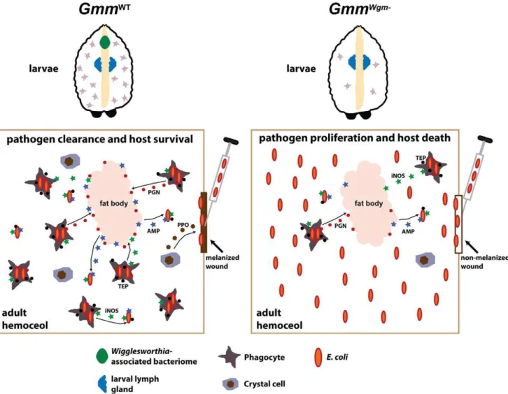
Figure 6.Wigglesworthiamodulates the development and function of tsetse’s immune system.Through a currently unknown mechanism, the presence ofWigglesworthiainGmmWTlarvae stimulates hemocyte differentiation in a specialized organ that is homologous toDrosophila’slymph
gland. Upon metamorphosis, specialized hemocyte subtypes are released from the lymph gland and carried over in a functional state to the adult. In the absence ofWigglesworthia,GmmWgm2larvae produce significantly less hemocytes than their WT counterparts. Several innate immunity pathways
are activated upon inoculation ofE. coliinto the hemocoel of mature adultGmmWT. Tsetse’s preliminary line of defense againstE. coliinfection likely involves melanization at the wound site. This process is initiated by localized crystal cells, which instigate the melanization cascade by secreting prophenoloxidase (PPO) into the hemolymph. Pathogens that circumvent the wound site then encounter phagocyte-mediated cellular and humoral immune responses. Soluble TEPs likely opsonize bacterial cells, thus tagging them for engulfment by phagocytes. Lysis of engulfed bacteria causes the release of bacterial peptidoglycan PGN that subsequently stimulates the production of AMPs by the fat body. AMPs, which are also generated by hemocytes, are then secreted into the hemolymph where they further abrogate microbial proliferation. Hemocytes also produce reactive oxygen intermediates, such as iNOS, that exhibit direct bacterial toxicity and further stimulate humoral immunity. In the case of GmmWgm2 adults,
incapacitated hematopoiesis during larval stages results in severely compromised immunity that renders these flies highly susceptible as adults to bacterial infection.
Neonatal humans (and presumably other mammals as well) acquire probiotic gut microbes through the act of breast feeding [49]. These bacteria subsequently stably colonize the naı¨ve intestine where they promote immune system maturation and enhance defense against infection with pathogenic microbes [50]. Most lower eukaryotes, including insects, hatch from an egg deposited into the environment and thus rely mainly on environmental microbes to stimulate their innate immune systems during the development of immature stages. For example, Anopheline mosquitoes and Drosophila are exposed to environmental microbes throughout all stages of their develop-ment. Under these natural conditions subsequent adults exhibit potent innate immunity [51,52]. However, when these insects are reared under germ-free conditions, they exhibit a severely compromised humoral immune response [51,53]. Tsetse flies are unique among other insects because they lead a relatively aseptic existence. Not only do they feed exclusively on sterile vertebrate blood, but they also exhibit a unique viviparous reproductive strategy. During this type of reproduction, all embryonic and larval stages develop within the female’s uterus. The protected in utero environment in which viviparous offspring develop limits their exposure to environmental microbes. However, throughout tsetse larvagenesis maternal milk gland secretions provide the developing offspring with nourishment as well asWigglesworthia,Sodalis, andWolbachia[31]. Interestingly, recently established Sodalis and Wolbachia appear unable to influence their host’s physiology to the same extent as mutualistWigglesworthia despite their presence throughout larval development. Future studies on this system will focus on determining the chemical and/or metabolic elements provided by Wigglesworthia that stimulate the maturation of its host’s immune system during larval development.
The results of this study provide evidence of a novel functional role for obligate insect symbionts–host immune system activation during immature developmental stages to ensure robust function during adulthood. We demonstrate that the obligateWigglesworthia provides tsetse offspring the stimuli necessary for immune system development, a process that exhibits functional parallels with the mammalian system following the transfer of beneficial microbes from mothers to their neonates during that act of breast feeding. This phenomenon represents an adaptation that further anchors the steadfast relationship shared between tsetse and its obligate mutualist. The essential nature of tsetse’s dependence on Wigglesworthia provides a potentially exploitable niche to experi-mentally modulate host immunity, with the intention of diminish-ing this insect’s capacity to vector deadly trypanosomes.
Materials and Methods
Tsetse and Septic Infections
Throughout the text, 3-d-old and 8-d-old tsetse are referred to as ‘‘young’’ and ‘‘mature,’’ respectively. Wild-typeG. m. morsitans (GmmWT) were maintained in Yale’s insectary at 24uC with 50%– 55% relative humidity. These flies received defibrinated bovine blood every 48 h through an artificial membrane feeding system [54]. TwoWigglesworthia-free tsetse lines were generated for use in our experiments. The first, GmmWgm2, was generated by supplementing the diet of pregnant females with 25mg of ampicillin per ml of blood.GmmWgm2 (offspring of ampicillin-fed females) adult are devoid of Wigglesworthia throughout all developmental stages [9]. PCR using Wigglesworthia thiamine C-specific primers (forward, 59 -TGAAAACATTTGCAAAATTTG-39; reverse, 59-GGTGTTACATAGCATAACAT-39) confirmed that this tsetse strain lacked Wigglesworthia. The second
Wiggle-sworthia-free tsetse line, GmmWT/Wgm2, was generated by feeding mature WT adults three blood meals supplemented with 40mg/ml tetracycline. GmmWT/Wgm2 individuals thus harbored Wiggle-sworthia,Sodalis, andWolbachiathroughout the development of all immature stages and early adulthood, but lacked these symbionts thereafter. The absence of Wigglesworthiafrom mature GmmWT/
Wgm2 adults was confirmed microscopically by comparing the
bacteriome (Wigglesworthia-harboring organ) contents of mature WT andGmmWT/Wgm2adults.
Septic infection of tsetse was achieved by anesthetizing flies with CO2and subsequently injecting individuals with live bacterial cells using glass needles and a Narashige IM300 micro-injector. The methods used to produce luciferase-expressingE. coliK12 (recE. colipIL), and the assay used to quantify luciferase expression in vivo, were described previously [15]. GFP-expressing and tetracycline-resistant E. coli K12 were produced via electroporation with pGFP-UV (Clontech, Mountain View, CA) and pBR322 (Pro-mega, Madison, WI) plasmid DNA, respectively. All flies treated with tetracycline were subsequently infected with tetracycline resistantE. coli K12. The number of bacterial cells injected and control group designations for all infection experiments are indicated in the corresponding figures and their legends. For all survival experiments, treatments were performed in triplicate, using 25 flies perE. coli treatment replicate. LB media controls were performed once using 25 flies.
Real-Time Quantitative PCR (qPCR)
For analysis of immunity-related gene expression, sample preparation and qPCR were performed as described previously [15]. Quantitative measurements were performed on three biological samples in duplicate and results were normalized relative to tsetse’s constitutively expressedb-tubulin gene (deter-mined from each corresponding sample). Fold-change data are represented as a fraction of average normalized gene expression levels in bacteria-infected flies relative to expression levels in corresponding uninfected controls.
For symbiont quantification, total RNA was prepared from 40-d-old adult GmmWT and GmmWgm2 flies. Symbiont genome numbers were quantified using single-copy Sodalis fliC and Wolbachia groEL. Relative symbiont densities were normalized to tsetseb-tubulin. All qPCR was performed with an icycler iQ real time PCR detection system (Bio-Rad). Values are represented as the mean (6SEM). qPCR primer sequences are shown in Table S1.
Bacterial 16s rRNA Clone Libraries
Hemolymph Collection and Hemocyte Quantification
Depending on the subsequent experiment, tsetse hemolymph was collected using one of two methods. For hemocyte quantification, undiluted hemolymph was collected by removing one front fly leg at the joint nearest the thorax and then applying gentle pressure to the distal tip of the abdomen. Hemolymph exuding from the wound was collected using a glass micro-pipette and placed into a microfuge tube on ice. Hemocytes were quantified microscopically using a Bright-Line hemocytometer, and hemocyte numbers are represented as cells per ml of hemolymph.
When hemocytes were required for microscopic visualization, hemolymph was collected by employing a modified version of the high injection/recovery method previously developed for use in mosquitoes [56]. In brief, tsetse flies were sedated on ice and injected with 25ml of chilled anticoagulant buffer [70% MM medium, 30% anticoagulant citrate buffer (98 mM NaOH, 186 mM NaCl, 1.7 mM EDTA, and 41 mM citric acid, buffer pH 4.5), vol/vol] between the last two abdominal schlerites using a glass needle and a Narashige IM300 micro-injector. Following a 30 min incubation on ice, a front leg was removed at the joint most proximal to the thorax. At this point internal pressure forced hemolymph diluted with anticoagulant buffer to be expelled from the wound site (more liquid could be recovered by applying gentle pressure to the distal end of the abdomen). Liquid was collected using a pipette and either placed into a chilled microfuge tube or directly into a 24-well cell culture plate. In the latter case, cells were allowed to adhere to the plate bottom, after which anticoagulant buffer was replaced with MM media.
Sessile hemocytes were observed by intra-thoracically injecting young and matureGmmWT, and matureGmmWgm2 (n= 5 of each strain), with 2ml of blue fluorescent (365/415 nm) 0.2mm carboxylate-modified beads (Invitrogen corp.). Prior to use, beads were washed once in PBS and resuspended in 100% of their original volume. Flies were dissected 12 h post-injection to reveal their dorsal vessel and surrounding tissue, which was gently washed 3 times with PBS to remove any potentially contaminating circulating (non-adherent) hemocytes. Engulfed microspheres were visualized using a Zeiss steriomicroscope (Discovery v8) equipped with a coaxial fluorescence module. Semi-quantitative comparison of sessile hemocyte number between young and mature GmmWT adults, and matureGmmWTandGmmWgm2adults, was performed by quantifying fluorescent signal intensity (n= 4 individuals from each group) using ImageJ software (http://rsbweb.nih.gov/ij/).
Phagocytosis and Melanization Assays
Phagocytosis by circulating tsetse hemocytes was observed by intra-thoracically infecting mature GmmWT flies (n = 10) with 16106 GFP-expressingE. coli K12. Twelve hours post-infection,
hemolymph was collected and hemocytes monitored to determine if they had engulfed the GFP-expressing bacterial cells.
Hemo-lymph samples were fixed on glass microscope slides via a 2 min incubation in 2% PFA. Prior to visualization using a Zeiss Axioscope microscope, slides were overlayed with VectaShield hard set mounting medium containing DAPI (Vector Laborato-ries, Burlingame, CA).
Phagocytosis by tsetse hemocytes was inhibited with blue fluorescent (365/415 nm) 0.2mm carboxylate-modified beads (Invitrogen Corp.). Prior to use, beads were washed once in PBS and resuspended in 100% of their original volume. Inhibition assays were performed by inoculating 8-d-oldGmmWTwith 2ml of beads via their thoracic compartment. Twelve hours later, these flies were similarly infected with 16103 and 16106
GFP-expressingE. coliK12 (experiment was performed in triplicate;n = 25 flies per replicate). Finally, 12 hours post-infection with E. coli, hemolymph was collected and processed as described above (these samples were overlayed with VectaShield hard set mounting medium that lacked DAPI).
Melanization assays were performed by intra-thoracically inoculating matureGmmWT
andGmmWgm2(n = 10 of each strain) with 16103E. coliK12. Subsequently, three individuals from each
group were monitored microscopically every 10 min for the presence of melanin at the wound site. The remaining seven flies from each group were maintained for 2 wk in order to observe infection outcome.
Statistics
Statistical significance of survival curves was determined by log-rank analysis using JMP (v8.02) software (www.jmp.com). Statistical analysis of qPCR data was performed by Student’s t test using JMP (v8.02) software. Statistical significance between various treatments, and treatments and controls, is indicated in corresponding figure legends.
Supporting Information
Table S1 Primers used for quantitative real-time PCR.
Found at: doi:10.1371/journal.pbio.1000619.s001 (0.02 MB XLS)
Acknowledgments
We are grateful to Dr. Yineng Wu for assistance with qPCR, Dr. Corey Brelsford for assistance with statistical analysis, and Dan Summers for his help processing bacterial 16s rRNA clone libraries. We also thank members of the Aksoy lab for critical review of the manuscript.
Author Contributions
The author(s) have made the following declarations about their contributions: Conceived and designed the experiments: BLW SA. Performed the experiments: BLW JW. Analyzed the data: BLW SA. Contributed reagents/materials/analysis tools: BLW SA. Wrote the paper: BLW SA.
References
1. Moran NA (2006) Symbiosis. Curr Biol 16: R866–R871.
2. Wernegreen JJ (2002) Genome evolution in bacterial endosymbionts of insects. Nat Rev Genet 3: 850–861.
3. Aksoy S (2000) Tsetse – a haven for microorganisms. Parasitol Today 16: 114–118.
4. Chen X, Li S, Aksoy S (1999) Concordant evolution of a symbiont with its host insect species: molecular phylogeny of genus Glossina and its bacteriome-associated endosymbiont,Wigglesworthia glossinidia. J Mol Evol 48: 49–58. 5. Akman L, Yamashita A, Watanabe H, Oshima K, Shiba T, et al. (2002)
Genome sequence of the endocellular obligate symbiont of tsetse flies,
Wigglseworthia glossinidia.Nat Genet 32: 402–407.
6. Nogge G (1976) Sterility in tsetse flies (Glossina morsitansWestwood) caused by loss of symbionts. Experientia 32: 995–996.
7. Aksoy S, Gibson WC, Lehane MJ (2003) Interactions between tsetse and trypanosomes with implications for the control of trypanosomiasis. Adv Parasitol 53: 1–83.
8. Hu C, Rio RV, Medlock J, Haines LR, Nayduch D, et al. (2008) Infections with immunogenic trypanosomes reduce tsetse reproductive fitness: potential impact of different parasite strains on vector population structure. PLoS Neg Trop Dis 2: 1–10. doi:10.1371/journal.pntd.0000192.
9. Pais R, Lohs C, Wu Y, Wang J, Aksoy S (2008) The obligate mutualist
Wigglesworthia glossinidia influences reproduction, digestion, and immunity processes of its host, the tsetse fly. Appl Environ Microbiol 74: 5965–5974. 10. Wang J, Wu Y, Yang G, Aksoy S (2009) Interactions between mutualist
11. Hooper LV (2004) Bacterial contributions to mammalian gut development. Trends Microbiol 12: 129–134.
12. Macpherson AJ, Harris NL (2004) Interactions between commensal intestinal bacteria and the immune system. Nat Rev Immunol 4: 478–485.
13. Koropatnick TA, Engle JT, Apicella MA, Stabb EV, Goldman WE, et al. (2004) Microbial factor-mediated development in a host-bacterial mutualism. Science 306: 1186–1188.
14. Oliver KM, Degnan PH, Hunter MS, Moran NA (2009) Bacteriophages encode factors required for protection in a symbiotic mutualism. Science 325: 992–994. 15. Weiss BL, Wu Y, Schwank JJ, Tolwinski NS, Aksoy S (2008) An insect symbiosis is influenced by bacterium-specific polymorphisms in outer membrane protein A. Proc Natl Acad Sci U S A 105: 15088–15093.
16. Hoffmann JA (2003) The immune response ofDrosophila. Nature 426: 33–38. 17. Blandin SA, Levashina EA (2007) Phagocytosis in mosquito immune responses.
Immunol Rev 219: 8–16.
18. Cerenius L, So¨derha¨ll K (2004) The prophenoloxidase-activating system in invertebrates. Immunol Rev 198: 116–126.
19. Dimopoulos G, Richman A, Muller HM, Kafatos FC (1997) Molecular immune responses of the mosquitoAnopheles gambiaeto bacteria and malaria parasites. Proc Natl Acad Sci U S A 94: 11508–11513.
20. Whitten M, Sun F, Tew I, Schaub G, Soukou C, et al. (2007) Differential modulation ofRhodnius prolixusnitric oxide activities following challenge with
Trypanosoma rangeli,T. cruziand bacterial cell wall components. Insect Biochem Mol Biol 37: 440–452.
21. Foley E, O’Farrell PH (2003) Nitric oxide contributes to induction of innate immune responses to Gram-negative bacteria inDrosophila. Genes Dev 17: 115–125.
22. Carton Y, Frey F, Nappi AJ (2009) Parasite-induced changes in nitric oxide levels inDrosophila paramelanica. J Parasitol 95: 1134–1141.
23. Matova N, Anderson KV (2006) Rel/NF-kB double mutants reveal that cellular immunity is central toDrosophilahost defense. Proc Natl Acad Sci U S A 103: 16424–16429.
24. Hillyer JF, Schmidt SL, Christensen BM (2003) Rapid phagocytosis and melanization of bacteria and Plasmodium sporozoites by hemocytes of the mosquitoAedes aegypti. J. Parasitol 89: 62–69.
25. Meister M, Lagueux M (2003)Drosophilablood cells. Cell Microbiol 5: 573–580. 26. Galko MJ, Krasnow MA (2004) Cellular and genetic analysis of wound healing inDrosophila larvae. PLoS Biol 2: 1114–1126. doi:10.1371/journal. pbio.0020239.
27. Christensen BM, Li J, Chen CC, Nappi AJ (2005) Melanization immune responses in mosquito vectors. Trends Parasitol 21: 192–199.
28. Ayres JS, Schneider DS (2008) A signaling protease required for melanization in
Drosophilaaffects resistance and tolerance of infections. PLoS Biol 6: 2764–2773. doi:10.1371/journal.pbio.0060305.
29. Elrod-Erickson M, Mishra S, Schneider DS (2000) Interactions between the cellular and humoral immune responses inDrosophila.Curr Biol 10: 781–784. 30. Evans CJ, Sinenko SA, Mandal L, Martinez-Agosto JA, Hartenstein V, et al.
(2008) Genetic dissection of hematopoiesis usingDrosophilaas a model system. In: Bodmer, R, eds. Advances in developmental biology, Volume 18 Elsevier. pp 259–298.
31. Attardo GM, Lohs C, Heddi A, Alam UH, Yildirim S, et al. (2008) Analysis of milk gland structure and function inGlossina morsitans: milk protein production, symbiont populations and fecundity. J Insect Physiol 54: 1236–1242. 32. Eleftherianos I, Boundy S, Joyce SA, Aslam S, Marshall JW, et al. (2007) An
antibiotic produced by an insect-pathogenic bacterium suppresses host defenses through phenoloxidase inhibition. Proc Natl Acad Sci U S A 104: 2419–2424. 33. Rizki TM, Rizki RM (1990) Encapsulation of parasitoid eggs in
phenoloxidase-deficient mutants ofDrosophila melanogaster. J Insect Physiol 36: 523–529. 34. Leem JY, Nishimura C, Kurata S, Shimada I, Kobayashi A, et al. (1996)
Purification and characterization of N-beta-alanyl-5-S-glutathionyl-3,4-dihy-droxyphenylalanine, a novel antibacterial substance ofSarcophaga peregrina(flesh fly). J Biol Chem 271: 13573–13577.
35. Zhao P, Li J, Wang Y, Jiang H (2007) Broad-spectrum antimicrobial activity of the reactive compounds generatedin vitrobyManduca sextaphenoloxidase. Insect Biochem Mol Biol 37: 952–959.
36. Lebestky T, Chang T, Hartenstein V, Banerjee U (2000) Specification of
Drosophilahematopoietic lineage by conserved transcription factors. Science 288: 146–149.
37. Charroux B, Royet J (2009) Elimination of plasmatocytes by targeted apoptosis reveals their role in multiple aspects of theDrosophilaimmune response. Proc Natl Acad Sci U S A 106: 9797–9802.
38. Pham LN, Dionne MS, Shirasu-Hiza M, Schneider DS (2007) A specific primed immune response inDrosophilais dependent on phagocytes. PLoS Pathog 3: 1–8. doi:10.1371/journal.ppat.0030026.
39. Bidla G, Dushay MS, Theopold U (2007) Crystal cell rupture after injury in
Drosophilarequires the JNK pathway, small GTPases and the TNF homolog Eiger. J Cell Sci 120: 1209–1215.
40. Lanot R, Zachary D, Holder F, Meister M (2001) Postembryonic hematopoiesis inDrosophila. Dev Biol 230: 243–257.
41. Bidla G, Lindgren M, Theopold U, Dushay MS (2005) Hemolymph coagulation and phenoloxidase inDrosophilalarvae. Dev Comp Immunol 29: 669–679. 42. Zettervall C, Anderl I, Williams MJ, Palmer R, Kurucz E, et al. (2004) A direct
screen for genes involved inDrosophilablood cell activation. Proc Natl Acad Sci U S A 101: 14192–14197.
43. Brandt SM, Schneider DS (2007) Bacterial infection of fly ovaries reduces egg production and induces local hemocyte activation. Dev Comp Immunol 31: 1121–1130.
44. Babcock DT, Brock AR, Fish GS, Wang Y, Perrin L, et al. (2008) Circulating blood cells function as a surveillance system for damaged tissue inDrosophila
larvae. Proc Natl Acad Sci U S A 105: 10017–10022.
45. Rodrigues J, Brayner FA, Alves LC, Dixit R, Barillas-Mury C (2010) Hemocyte differentiation mediates innate immune memory inAnopheles gambiaemosquitoes. Science 329: 1353–1355.
46. Hao Z, Kasumba I, Lehane MJ, Gibson WC, Kwon J, et al. (2001) Tsetse immune responses and trypanosome transmission: implications for the development of tsetse-based strategies to reduce trypanosomiasis. Proc Natl Acad Sci U S A 98: 12648–12653.
47. Hu Y, Aksoy S (2005) An antimicrobial peptide with trypanocidal activity characterized fromGlossina morsitans morsitans. Insect Biochem Mol Biol 35: 105–115.
48. Hu C, Aksoy S (2006) Innate immune responses regulate trypanosome parasite infection of the tsetse fly Glossina morsitans morsitans. Mol Microbiol 60: 1194–1204.
49. Martin R, Langa S, Reviriego C, Jimenez E, Marin ML, et al. (2003) Human milk is a source of lactic acid bacteria for the infant gut. J Pediatr 143: 754–758. 50. Lara-Villoslada F, Olivares M, Sierra S, Rodriguez JM, Boza J, et al. (2007) Beneficial effects of probiotic bacteria isolated from breast milk. Br J Nutr 98: S96–S100.
51. Dong Y, Manfredini F, Dimopolous G (2009) Implications of the mosquito midgut microbiota in the defense against malaria parasites. PLoS Pathog 5: 1–10. doi:10.1371/journal.ppat.1000423.
52. Charroux B, Royet J (2010) Drosophila immune response: from systemic antimicrobial peptide production in fat body cells to local defense in the intestinal tract. Fly 4: 40–47.
53. Ryu J, Kim S, Lee H, Bai J, Nam Y, et al. (2008) Innate immune homeostasis by the homeobox gene caudal and commensal-gut mutualism inDrosophila. Science 319: 777–782.
54. Moloo SK (1971) An artificial feeding technique forGlossina. Parasitol 68: 507–512.
55. Dale C, Beeton M, Harbison C, Jones T, Pontes M (2006) Isolation, pure culture, and characterization of ‘‘Candidatus arsenophonus arthropodicus,’’ and intracellular secondary endosymbiont from the hippoboscid louse fly Pseudolyn-chia canariensis. App Environ Microbiol 72: 2997–3004.