Patrícia Costinha Marques
Degree in Biochemistry
The role of PGN hydrolases in the
ability of
Staphylococcus aureus
to
evade the host innate immune system
Dissertation to obtain the degree of Master in Biochemistry for
Health
Orientador: Sérgio R. Filipe, Professor Auxiliar, FCT/UNL
iii
Patrícia Costinha Marques
Degree in Biochemistry
The role of PGN hydrolases in the
ability of
Staphylococcus aureus
to
evade the host innate immune system
Dissertation to obtain the degree of Master in Biochemistry for
Health
Orientador: Sérgio R. Filipe, Professor Auxiliar, FCT-UNL
Juri:
Presidente: Prof. Doutora Maria Teresa Nunes Mangas Catarino
Arguente: Prof. Doutor Luís Jaime Gomes Ferreira da Silva Mota
Vogal: Prof. Doutor Sergio Raposo Filipe
Faculdade de Ciências e Tecnologias – Universidade Nova de Lisboa
v
The role of PGN hydrolases in the ability of Staphylococcus aureus to evade the host innate immune system
Copyright Patrícia Costinha Marques, FCT/UNL, UNL
vii
Acknowledgements
Ao Professor Sérgio Filipe, deixo um grande obrigado por todo apoio e disponibilidade ao longo deste ultimo ano; pela oportunidade de trabalhar no seu laboratório, pelos concelhos, desafios e orientação essenciais para o desenvolvimento deste projeto.
Ao Gonçalo Covas, agradeço por toda a orientação e paciência, por apesar da distância ou do trabalho inesgotável, fazer todos os possíveis para nos ajudar.
A Ana Rita Narciso e a Joana Figueiredo agradeço toda a ajuda e orientação no laboratório. Ao pessoal dos laboratórios MGPI e MGPII, no ITQB, agradeço pelo acolhimento caloroso nos laboratórios e por todo o conhecimento transmitido.
Ao pessoal do laboratório 333, deixo um grande obrigado pela eterna amabilidade com que nos receberam e por todo o tempo disponibilizado, que tanto nos ajudou quando mais precisávamos. Ao pessoal do laboratório 349, agradeço por todo o apoio e auxílio.
Aos meus colegas que me acompanharam durante todo o ano: António Lopez, Liliane Raposo, Rúben Silva, Lígia Martins, Bruno Guerreiro e Diana Silva. Pelas cantigas e noites ao luar, pela amizade inegável, pelo humor e companhia; deixo-vos um grande obrigado pelas memórias e palavras que nunca vou esquecer.
Devo um enorme obrigado (e chocolate) a Jéssica Gomes, a minha parceira do crime, porque esta viagem nunca teria sido a mesma sem a tua presença e humor. Por todo o conhecimento e ajuda que me deste, pela eterna paciência para me explicar todas as minhas dúvidas, por mais elementares que fossem; pela confiança e pelo respeito; e claro, por toda a amizade que me deste.
Girl, you know what to do: Go Forth and Conquer!
Aos meus amigos: começo pela Joana Coelho, sempre amável e de braços abertos, pelas conversas no carro, pelos concelhos, por me ouvir e principalmente, pelo sorriso e carinho, devo-te um edevo-terno obrigado e desejo-devo-te o mundo de oportunidades.
Ao pessoal do “D&D”, Marco Galvão, Reinaldo Muralha e Diogo Filipe, devo-vos um grandessíssimo obrigado, pelas sessões de sábado à tarde, pelas noites de jogo, pelos copos no café, e por toda a amizade e carinho que me entregaram sem rodeios.
viii
To Bart Selie, I can only thank you with my heart, for all the care and endless support you offered me. For all your devotion, friendship and love that you delivered so selflessly, I will be forever grateful.
À minha família, quero deixar um enorme obrigado, pelo apoio constante, pelos jantares caóticos e espalhafatosos que tanto aquecem o coração, pelas palavras de coragem e de ânimo que oferecem de sorriso rasgado.
E finalmente, aos meus pais, a quem devo o mundo. Pelo amor incondicional e por nunca me deixarem cair; pela confiança, ajuda e sabedoria. Agradeço-vos o mundo que me entregaram, e por nunca desistirem de mim.
ix
Resumo
Staphylococcus aureus é um microorganismo importante que pode ser encontrado na pele
e membranas mucosas do corpo humano. Apesar da habilidade de colonizar o hospedeiro humano, a invasão deste patogénico para os tecidos pode originar severas condições de saúde. À medida que antibióticos actuais perdem eficiência contra este patogénico, torna-se urgente o desenvolvimento de novas estratégias de tratamento.
Peptidoglicano está presente em bactérias gram-negativas e gram-positivas, como uma molécula fundamental para a estrutura celular. Trata-se também de uma molécula que denuncia a presença da bactéria quando reconhecida por recetores inatos ao hospedeiro como proteínas PGRP.
De modo a evitar deteção, S.aureus desenvolveu estratégias para evadir o reconhecimento
de PGN na superfície. Foi demonstrado que Atl (Atilano et al.), uma autolisina principal de
S.aureus, é capaz de aparar os fragmentos mais externos de peptidoglicano de modo a impedir a
ligação de PGRPs.
Este projecto foca-se no estudo da proteína SAOUHSC_00671 da estirpe S.aureus NCTC
8325-4, semelhante à Sle1, uma hidrolase capaz de aparar os fragmentos de PGN na divisão do septo da bactéria. Neste projeto focou-se à purificação da proteína SAOUHSC_00671 e à construção de mutantes knock-out de S.aureus, que carecem da sequencia codificante para
SAOUHSC_00671.
xi
Abstract
Staphylococcus aureus is an important microorganism that can be found on the skin and
mucous membranes of the human body. Despite its ability to colonize the human host, invasion of this pathogen into the host tissue can lead to severe health conditions. As current antibiotics become less effective in the combat to this pathogen, it becomes urgent to develop new strategies of treatment.
Peptidoglycan is present in gram-negative and gram-positive bacteria alike, as a fundamental molecule for cell structure. It is a telltale molecule that betrays the presence of the bacteria when recognized by host innate immune receptors such as the PGRP proteins.
In order to avoid detection, S. aureus uses different strategies to circumvent recognition of the PGN at its surface. It has been shown that Atl, a major autolysin of S. aureus, is able to trim the outermost fragments of peptidoglycan in order to prevent the binding of PGRPs.
This project focused in studying the protein SAOUHSC_00671 from S. aureus NCTC 8324-5 strain, which belongs to the same protein family as SleI, a PGN hydrolase capable of trimming PGN fragments at the staphylococcal division septum. This project aimed purify the SAOUHSC_00671 protein and construct knock-out S. aureus mutants, which lack the coding sequence for SAOUHSC_00671.
xiii
Index
Acknowledgements ... vii
Resumo ... ix
Abstract ... xi
Index ... xiii
Index of Figures ... xv
Index of Tables... xvii
Abbreviations ...xix
1 Introduction ...1
1.1 Staphylococcus aureus ...1
1.2 Peptidoglycan and PGRPs ...2
1.3 PGN Hydrolases ...3
1.4 S.aureus NCTC 8325-4 SAOUHSC_00671 ...4
2 Materials and Equipment ...5
3 Procedures and Protocols ...7
3.1 E.coli competent cells ...7
3.2 S.aureus genomic DNA extraction ...8
3.3 Amplification of gene 671 ...8
3.4 Construction of pET21a vector for protein expression ...9
3.5 E.coli transformation ... 12
3.6 Colony Screening PCR ... 12
3.7 E.coli BL21(DE3) induction test ... 13
3.8 SDS-PAGE Gel Electrophoresis ... 14
3.9 Sanger Sequencing ... 15
3.10 S.aureus competent cells... 15
3.11 Purification of Up and Down regions of gene 671... 16
3.12 Construction of pMAD vector for gene excision ... 18
3.13 S.aureus electroporation ... 20
3.14 Transduction protocol ... 20
3.15 Gene knock-out protocol ... 22
3.16 S.aureus genomic DNA extraction for PCR screening. ... 23
3.17 PCR screening for gene knock-out confirmation ... 23
4 Results and Discussion ... 27
4.1 Construction of pET21a-671 I ... 27
4.2 Acquiring pET21a-671 I positive colonies. ... 29
xiv
4.4 Sequencing of pET21a-671 I ... 33
4.5 Construction of pET21a-671 II ... 34
4.6 Acquiring pET21a-671 II positive colonies ... 34
4.7 Induction test of pET21a-671 II A and B ... 36
4.8 Sequencing of samples pET21a-671 II A and B ... 39
4.9 Additional inductions of pET21a-671 II A ... 39
4.10 Acquiring positives of pMAD del 671... 42
4.11 Sequencing of pMAD del 671 ... 44
4.12 Integration of pMAD del 671 in NCTC 8325-4 genome ... 44
4.13 Excision of pMAD del 671 ... 45
5 Conclusion ... 47
6 References ... 48
xv
Index of Figures
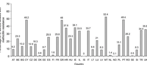
Figure 1.1Prevalence of methicillin-resistant Staphylococcus aureus among patients with
bacteremia in Europe in 2007, as reported by the European Antimicrobial Resistance
Surveillance System(5). AT, Austria; BE, Belgium; BG, Bulgaria; CY, Cyprus; CZ, Czech
Republic; DE, Germany; DK, Denmark; EE, Estonia; ES, Spain; FI, Finland; FR, France; GR, Greece; HR, Croatia; HU, Hungary; IE, Ireland; IL, Israel; IS, Iceland; IT, Italy; LT, Lithuania; LU, Luxembourg; LV, Latvia; MT, Malta; NL, The Netherlands; NO, Norway; PL, Poland; PT, Portugal; RO, Romania; SE, Sweden; SI, Slovenia; TR, Turkey; UK, United Kingdom. ...1
Figure 1.2: The structure of S. aureus peptidoglycan (11). The arrows indicate the cleavage
sites: 1 N-acetyl muramidase, 2 N-acetyl-β-D-glucosaminidase, 3 Nacetylmuramoyl-L-alanine amidase, 4 L-alanoyl-D-glutamate endopeptídases and 5 interpeptide bridge endopeptidases (e.g., lysostaphin). Abbreviations: GlcNAc N-acetylglucosamine, MurNAc N-acetylmuramic acid ...3 Figure 4.1: Amplification of gene 671. Expected length of amplified fragment is
approximately 850bp. 1 to 8- Amplified 671 insert. 9- control. ... 27 Figure 4.2: Plasmid pET21a digested with restriction enzymes XbaI, EagI and NheI. 1- Plasmid pET21a. 2- pET21a digested with XbaI. 3- pET21a digested with EagI. 4- pET21a digested with NheI. 5- pET21a digested with XbaI and EagI. ... 28 Figure 4.3: Cloning PCR of pET21a-671 I plasmid. Expected length of the amplified 671 insert is of 850bp, which verifies a successful insertion. 1- PCR product of pET21a-671 I. 2-PCR product of auto-ligated pET21a. 3- 2-PCR product of pET21a digested with XbaI and EagI. 4- pET21a digested with XbaI and EagI. 5- pET21a digested with Xbal. 6- PCR product of pET21a. 7- pET21a. 8- Control. ... 29 Figure 4.4: Screening PCR for pET21a-671 I positives. 1 to 11- Amplification from
transformed Dh5α with pET21a-671 I. 12- Amplification from transformed Dh5α with auto-ligated pET21a. 13- DNA ladder 14- Amplification from pET21a-671 I (positive control). 15- Amplification from auto-ligated pET21a (negative control). 16- Amplification from pET21a digested with XbaI and EagI. 17- Amplification from pET21a digested with XbaI. 18-
Amplification from pET21a. ... 29 Figure 4.5: Restriction sites of NaeI in plasmid pET21a-671 I. ... 30 Figure 4.6: Digestive profile of pET21a-671 I by NaeI. Digestion with NaeI enzyme will result in three fragments for both pET21a-671 I and pET21a. The size difference in the smallest fragment attests for a positive insertion of gene 671. 1- pET21a non-digested. 2- Digested pET21a. 3- pET21a-671 I non-digested. 4- Digested pET21a-671 I. ... 31 Figure 4.7: Optical density measurements along time of induction. 671+: culture induced with 1mM IPTG. 671-: culture not induced (control) ... 32 Figure 4.8:SDS/PAGE gel of pET21a-671 I protein induction test. T0: time 0 of induction. T3+: time 3-hour induced. T3-: time 3-hour non-induced. TON+: time overnight induced. TON-: time overnight non-induced. ... 33 Figure 4.9:Sequencing of pET21a-671 I. A- Segment of sequencing traces assembled by SeqMan software from Lasergene’s DNAStar and provided by STABvida laboratories. B: Segment of in-silico sequence depicting the deleted nucleotides. ... 33 Figure 4.10: Cloning PCR of pET21a-671 II plasmid. Expected size of amplified fragment is approximately 850 bp, which verifies a proper insertion of gene 671. 1- PCR product of
pET21a-671 II 10μL. 2- PCR product of pET21a-671 II 7μL. 3- PCR product of pET21a-671 II 5μL. 4- PCR product of auto-ligated pET21a. 5- PCR product of pET21a digested with NheI and EagI. 6- PCR product of pET21a. 7- Control. ... 34 Figure 4.11: Colony Screening PCR of pET21a-671 II. 1 to 23- Amplification from
auto-xvi
ligated pET21a. 25- DNA Ladder. 26- Amplification from pET21a-671 II 10μL (positive control). 27- Amplification from pET21a-671 II 7μL (positive control) 28- Amplification from auto-ligated pET21a (negative control). 29- Amplification from pET21a. ... 35 Figure 4.12 Digestive profile of pET21a-671 II by NaeI. 1- pET21a non-digested. 2- Digested pET21a. 3- pET21a-671 II A non-digested. 4- Digested pET21a-671 II A. 5- pET21a-671 II B non-digested. 6- Digested pET21a-671 II B. ... 36 Figure 4.13: SDS/PAGE gel of pET21a-671 II A protein induction test. 1- Time 0:00. 2- Time 0:30 non-induced. 3- Time 0:30 induced. 4- Time 1:00 non-induced. 5- Time 1:00 induced. 6- Time 1:30 induced. 7- Time 1:30 non-induced. 8- Time 2:00 non-induced. 9- Time 2:00 induced. 10- Time 2:30 non-induced. 11- Time 2:30 induced. 12- Time 3:00 non-induced. 13- Time 3:00 induced. 14- Time ON non-induced. 15- Time ON induced. ... 37 Figure 4.14 Optical density measurements along time of induction. 671+: culture induced with 1mM IPTG. 671-: culture not induced (control) ... 37 Figure 4.15: SDS/PAGE gel of pET21a-671 II B protein induction test. 1- T 0:00. 2- T 0:30 induced. 3- T 0:30 induced. 4- T 1:00 induced. 5- T 1:00 induced. 6- T 1:30 non-induced. 7- T 1:30 non-induced. 8- T 2:00 non-non-induced. 9- T 2:00 non-induced. 10- T 2:30 non-non-induced. 11- T 2:30 induced. 12- T 3:00 non-induced. 13- T 3:00 induced. 14- T ON non-induced. 15- T ON induced. ... 38 Figure 4.16: Optical density measurements along time of induction. 671+: culture induced with 1mM IPTG. 671-: culture not induced (control). ... 38 Figure 4.17: SDS/PAGE gel of protein 671 induction. 1- Time 0 pET21a-671 II A. 2- Time 0 pET21a-671 II B. 3- Time 0 pET21a-671 I. 4- Time 0 pET21a. 5- Time 3 hours pET21a-671 II A non-induced. 6- Time 3 hours pET21a-671 II A induced. 7- Time 3 hours pET21a-671 II B induced. 8- Time 3 hours pET21a-671 II B induced. 9- Time 3 hours pET21a-671 I non-induced. 10- Time 3 hours pET21a-671 I non-induced. 11- Time 3 hours pET21a non-non-induced. 12- Time 3 hours pET21a induced. ... 39 Figure 4.18: Colony Screening PCR of pET21a-671 II A. 1 to 8- Amplification from
transformed BL21(DE3) with pET21a-671 II A. 9- Amplification from pET21a-671 II 10μL (positive control). 10- Control ... 40 Figure 4.19: SDS/PAGE gel of pET21a-671 II A induction. 1- Time 0 hour Colony 1. 2- Time 3 hours induced Colony 1. 3- Time 3 hours non-induced Colony 1. 4- Time 0 hour Colony 2. 5- Time 3 hours induced Colony 2. 6- Time 3 hours non-induced Colony 2. 7- Time 0 hours Colony 3. 8- Time 3 hours induced Colony 3. 9- Time 3 hours non-induced Colony 3. ... 41 Figure 4.20: Colony Screening PCR of pMAD del 671. 1 to 13- Amplification from
transformed DC10b with pMAD del 671. 14 DNA Ladder 15- Amplification from pMAD del 671 (positive control). 16- Amplification from auto-ligated pMAD. 17- Amplification from pMAD digested with BamHI. 18- Amplification from pMAD digested with EcoRI. 19-
Amplification from pMAD del Sle1. 20- Amplification from pMAD. 21- Control... 42 Figure 4.21: Digestive Profile of pMAD del 671 by AvaI. 1- pMAD non-digested. 2- Digested pMAD. 3- pMAD del 671 digested. 4- Digested pMAD del 671. 5- pMAD del 671 B non-digested. 6- Digested pMAD del 671 B... 43 Figure 4.22: Integration Screening of pMAD del 671. 1- amplification pair negative
integration. 2- amplification pair positive for Up-region integration. 3- amplification pair with mixed colonies. 4- amplification pair positive for Down-region integration. ... 45
Figure 4.23: PCR screening of NCTC Δ671. 1- amplification from screened sample A. 2-
amplification from screened sample B. 3- amplification from screened sample C. 4- amplification from pMAD del 671. 5- amplification from NCTC 8325-4 wildtype. 6-
xvii
Index of Tables
Table 3.1: Constitution of solutions RF1 and RF2………...………....7 Table 3.2: Content of Master Mix solution for 671 amplification……….8 Table 3.3: PCR conditions for 671 amplification. Number of cycles (a cycle comprising the steps of Denaturation, Annealing and Extension) was of 30……….9 Table 3.4: Content of Master Mix solution for pET21a-671 I verification……….….10 Table 3.5: PCR conditions for pET21a-671 I verification. Number of cycles (a cycle comprising the steps of Denaturation, Annealing and Extension) was of 30……….….10 Table 3.6: Content of Master Mix solution for pET21a-671 II verification………....11 Table 3.7: PCR conditions for pET21a-671 II verification. Number of cycles (a cycle
comprising the steps of Denaturation, Annealing and Extension) was of 30………...12 Table 3.8: Contents of Master Mix solution for Colony Screening PCR of E.coli colonies……13 Table 3.9: PCR conditions for Colony Screening PCR for E.coli colonies. Number of cycles (a cycle comprising the steps of Denaturation, Annealing and Extension) was of 30……….13 Table 3.10: Preparation of the Resolution Gel with 12,5% of Acrylamide……….14 Table 3.11: Preparation of the Stacking Gel with 4% Acrylamide………..14 Table 3.12: Preparation of SDS/Page Protein Loading Buffer 5x. Sample stored at -20ºC…….15 Table 3.13: Forward and reverse primers for amplification of fragments Up and Down …….16 Table 3.14: Contents of Master Mix solution for Up and Down fragments amplification……..16 Table 3.15: PCR conditions for Up and Down cloning PCR. Number of cycles (a cycle
comprising the steps of Denaturation, Annealing and Extension) was of 30………...17 Table 3.16: Contents of Master Mix solution for overlapping PCR of Up and Down
fragments………...17 Table 3.17: PCR conditions for Overlapping PCR of Up and Down regions. Number of cycles (a cycle comprising the steps of Denaturation, Annealing and Extension) was of 30………….18 Table 3.18: Contents of Master Mix solution for pMAD del 671 verification………19 Table 3.19: PCR conditions for pMAD del 671 verification. Number of cycles (a cycle
comprising the steps of Denaturation, Annealing and Extension) was of 30………...19 Table 3.20: Preparation of 0,3GL Top and Bottom Agar. Medium was autoclaved at 120ºC for
40 minutes………20
Table 3.21: Preparation of Phage Top and Bottom Agar. Medium was autoclaved at 120ºC for
20 minutes………....20
xviii
Table 3.25: PCR conditions for integration screening PCR. Number of cycles (a cycle
comprising the steps of Denaturation, Annealing and Extension) was of 30………...……24 Table 3.26: Contents of Master Mix solution excision verification PCR………..…..25 Table 3.27: PCR conditions for excision verification PCR. Number of cycles (a cycle
comprising the steps of Denaturation, Annealing and Extension) was of 30………...25
xix
Abbreviations
o APS – Ammonium persulfate o BSI – Bloodstream Infections o CFU – Colony Forming Units o ddH2O – Bi-distilled water o DNA – Deoxyribonucleic Acid o dNTPs – Deoxyribonucleotides
o EDTA – Ethylenediamine tetra-acetic acid o Fw - Foward
o g – Grams
o IE – Infective Endocarditis o L - Liters
o LA – Luria-Bertani Agar o LB – Luria-Bertani Broth o M – Molar
o MCS – Multi Cloning Site o mg – Milligrams
o mL – Milliliters
o MRSA – Methicillin-resistant Staphylococcus aureus o nm – Nanometers
o OD – Over day
o OD600– Optical Density at 600nm wavelength o ON – Overnight
o PBP – Penicillin binding proteins o PCR – Polymerase Chain Reaction o PGRP – Peptidoglycan receptor proteins o RPM – Rotations per minute
o RT – Room Temperature o Rv - Reverse
o SA – Staphylococcus aureus o SDS – Sodium Dodecyl Sulfate
o SDS/PAGE LB – Protein Loading Buffer o ST – Sterile
o TAE – Tris- Acetate - EDTA o TE – Tris-EDTA
xx
o w/v – weight per volume o WT – Wild type
o X-gal – 5-bromo-4-3-indolyl-β-D-galactopyranoside o μg – Micrograms
1
1
Introduction
1.1 Staphylococcus aureus
Staphylococcus bacteria are gram-positive cocci that are commonly found in the form
irregular grape-like clusters. These organisms are widespread in nature, more specifically in the skin and mucous membranes, such as the mouth, mammary glands and intestinal, genitourinary, and upper respiratory tracts of mammals and birds (1). The relationship established between the bacteria and the host is generally symbiotic, however, infiltration onto the host tissue can lead to severe health conditions such as skin infections, Bloodstream Infections (BSI) (2) and infective endocarditis (3).
The major incidence of Staphylococcus aureus related infections is in medical facilities
(4). Yilmaz et al. estimated that 42.1% of a series of 255 diagnosed cases of Staphylococcus
aureus Bacteremia (SAB), were originated from vascular catheters and other surgical site
infections. Healthcare associated SAB was defined in 55.7%.
Figure 1.1Prevalence of methicillin-resistant Staphylococcus aureus among patients with bacteremia in Europe in 2007, as reported by the European Antimicrobial Resistance Surveillance System(5). AT, Austria; BE, Belgium; BG, Bulgaria; CY, Cyprus; CZ, Czech Republic; DE, Germany; DK, Denmark; EE, Estonia; ES, Spain; FI, Finland; FR, France; GR, Greece; HR, Croatia; HU, Hungary; IE, Ireland; IL, Israel; IS, Iceland; IT, Italy; LT, Lithuania; LU, Luxembourg; LV, Latvia; MT, Malta; NL, The Netherlands; NO, Norway; PL, Poland; PT, Portugal; RO, Romania; SE, Sweden; SI, Slovenia; TR, Turkey; UK, United Kingdom.
2
increase of the annual rate of found Methicilin Resistant S.aureus (MRSA) isolates, in European
countries during 1999-2006 (8).
S.aureus are majorly treated with administration of β-lactam antibiotics. These molecules
bind to the native penicillin-binding proteins (PBPs) present in the cell wall interrupting the synthesis of peptidoglycan layer (9). In methicillin-resistant strains, the gene mecA codifies for an additional, foreign, penicillin-binding protein (PBP2a) that prevents the binding of β-lactams and ensures the peptidoglycan synthesis is not disrupted (10). Furthermore, MRSA tend to accumulate and develop resistances to other antibiotics (9,10).
1.2 Peptidoglycan and PGRPs
Peptidoglycan is the principal component of bacterial cell wall and is composed of long glycan chain made up of alternating amino sugars N-acetylglucosamine and N-acetylmuramic acid, and cross-linked by short peptides of variable length and composition (11).
In gram-positive bacteria, this peptidoglycan is covered by a series of different molecules, such as surface proteins, capsular polysaccharides (12) and wall-teichoic acids (13) (phosphate-rich glycopolymers involved in resistance to environments stress and regulation of bacterial division)
This peptidoglycan, however, can be recognized by peptidoglycan recognition proteins (PGRP), innate immunity molecules, as a pathogen-associated molecular pattern, or PAMP, and trigger inflammatory response against the bacterial pathogen (14).
Insects possess two response pathways able to discern between positive and gram-negative bacteria (toll pathway for gram-positive bacteria, and Immune Deficiency pathway for gram-negative bacteria). When stimulation of these two pathways occurs, there are activated a series of responses, such as transcriptional regulators, antimicrobial peptides and signaling cascades, specific to each pathway (15).
In mammals, PGRPs can act as antibacterial agents due to their bactericidal and/or bacteriostatic activity, that is mediated by PGN hydrolytic activity (16), or by the binding of PGRPs to targets on the bacterial cell surface, which causes the activation of specific bacterial two-component systems (17).
3
1.3 PGN Hydrolases
Bacteria organisms possess a variety of enzymes capable of cleaving bonds in polymeric peptidoglycan. These are known as hydrolases. They play vital role in regulating cell wall growth as well as other lysis phenomena. It has been documented the participation of PGN hydrolases in the release of turnover products from E.coli peptidoglycan during cell growth (19).
PGN hydrolases have different cleavage sites. N.Acetylmuramyl-L-alanine amidases disrupt the bond between MurNAc and N-terminal L-Alanine, thus separating the glycan strand from the peptide (20). Carboxy- and endopeptidases hydrolyse amide bonds between amino acids. N-Acetyl-β-D-muramidades (or N-acetylmuramidases) cleave the bond β1,4-glycosidic between residues MurNAc and GlcNAc of peptidoglycan (20). This can occur in two different ways: hydrolysis of the glycosidic bond by lysozymes results in terminal reducing MurNAc residue, whereas lytic transglycosylases enacts a transglycosylation reaction that forms a 1,6-anhydro ring at the MurNAc residue (20).
Figure 1.2: The structure of S. aureus peptidoglycan (11). The arrows indicate the cleavage sites: 1 N-acetyl muramidase, 2 N-N-acetyl-β-D-glucosaminidase, 3 Nacetylmuramoyl-L-alanine amidase, 4 L-alanoyl-D-glutamate endopeptídases and 5 interpeptide bridge endopeptidases (e.g., lysostaphin). Abbreviations: GlcNAc N-acetylglucosamine, MurNAc N-acetylmuramic acid
The major autolysin of S.aureus is AtlA, composed of an amidase domain and a
glucosaminidase domain that, upon proteolytic cleavage, yield two active proteins: amidase (AM) and glucosaminidase (GL) (21). The role of this protein is essentially in cell division regulation but it has been documented the participation of AtlA in biofilm formation.
4
inaccessible to host PGN receptors. As such, targeting bacterial autolysins to prevent their activity may constitute a new strategy against these pathogens.
The hydrolase Sle1 (also known as Aaa) has also been characterized (23,24). Kajimura et al. (23) demonstrated this protein as a N-acetylmuramyl-L-alanine amidase and assessed its crucial role in the process of cell separation. Parallel to this study, Heilmann et al. (24) deduced the sequence of Sle1 from homologous DNA sequences from S.aureus strain Col, which revealed three
identical domains highly homologous to the lysin motif (LysM) followed by a Chap domain in the C-terminal of the protein. Lysin motifs and Chap domains are often found in cell wall enzymes. (25,26,27)
1.3.1
S.aureus
NCTC 8325-4 SAOUHSC_00671
Screening of the genome of S.aureus strain NCTC 8325-4 for similar proteins to Sle1,
revealed two putative autolytic proteins: SAOUHSC_00671 and SAOUHSC_00773. The former revealed in its structure two LysM domains followed by a Chap domain, while the latter presented one LysM domain and a Chap domain.
5
2
Materials and Equipment
All S.aureus and E.coli strains were provided from the research laboratory that hosted the
student.
Cloning vector pET21a was chosen for the construction of the protein expression mutant for E.coli BL21(DE3). Gene 671 was cloned to the Multi Cloning Site (MCS) with primers
NCTC671ClonFwXbaI (forward) and NCTC671ClonRvEagI (reverse), built in-silico with
Lasergene’s DNAStar software, SeqBuilder. Vector plasmid pET21a was provided from Novagen.
Shuttle vector pMAD (Arnaud et al., 2004) selected for the construction of the Δ671 mutants for S.aureus NCTC 8325-4. Regions 1000bp Up and 1000bp Down of gene 671 were
cloned into the MCS.
All mutants and vectors in-silicosequences were built with Lasergene’s DNAStar
software, SeqBuilder, as well as the necessary primers. DNA template sequence of S.aureus
NCTC 8325-4 was downloaded from National Center for Biotechnology Information (NCBI). All restriction enzymes and their respective buffers were provided by New England Biolabs.
o 1kb plus DNA ladder (Thermo Scientific)
o 1kb GeneRuler DNA Ladder (Thermo Scientific, catalog number: SM0312 o 1 μl Loops (Sarstedt AG, catalog number: 86.1567.010 )
o 25 ml Glass (or plastic disposable) Pipettes (Normax, catalog number: 4.5434334 ) o 30% Acrylamide/Bis Solution (Bio-Rad Laboratories, catalog number: 161-0158 ) o 50 ml Falcon tubes (Sarstedt AG, catalog number: 62.548.004 )
o Ammonium persulfate (APS) [(NH4)2S2O8] (Sigma-Aldrich, catalog number:
A3678 )
o Bromophenol Blue sodium salt (Sigma-Aldrich, catalog number: B8026 )
o Calcium chloride dihydrate (CaCl2.2H2O) (Sigma-Aldrich, catalog number: C3306
)
o Clean Up Kit: Wizard SV Gel and PCR Clean-Up System (Promega Corporation,
catalog number: A9285)
o DreamTaq Hot Start DNA Polymerase (Thermo Scientific, catalog number:
EP1701)
6
o Hydrochloric acid (Merck Millipore Corporation, catalog number: 1.01834.2500 ) o Luria Agar (Miller`s LB agar) (LA) (VWR, catalog number: 84684.0500 )
o Luria Broth (Miller`s LB broth) (LB) (Sigma-Aldrich, catalog number: L3022 ) o Lysostaphin (Sigma-Aldrich, catalog number: L7386)
o Magnesium chloride hexahydrate (Sigma-Aldrich, catalog number: M9272 ) o Methylene Blue hydrate (Sigma-Aldrich, catalog number: 66720 )
o Miniprep Kit: Wizard Plus SV Minipreps DNA Purification Systems (Promega
Corporation, catalog number: A1465)
o Mini-PROTEAN® Comb, 10-well, 0.75 mm (Bio-Rad Laboratories, catalog
number: 165-3354 )
o N, N, N′, N′-Tetramethylethylenediamine (TEMED) (Sigma-Aldrich, catalog
number: T9281 )
o Nuclei Lysis Solution (Promega, catalog number: A7941) o Petri dishes (Sarstedt AG, catalog number: 82.1473 )
o Phusion High-Fidelity PCR Master Mix with HF buffer (Thermo Scientific, catalog
number: F531S)
o Potassium hydroxide (KOH) (Sigma-Aldrich ,catalog number: P5958 )
o Precision Plus ProteinTM Dual Color Standards (PPPS) (Bio-Rad Laboratories,
catalog number: 1610374 )
o Protein Precipitation Solution (Promega, catalog number: A7953) o Rubidium Chloride, RbCl (Sigma-Aldrich, catalog number: 7791-11-9)
o Sodium chloride (Merck Millipore Corporation, catalog number: 1.06444.1000 ) o Sodium Dodecyl Sulfate (Sigma-Aldrich, catalog number: L5750 )
o Spacer Plates with 0.75 mm Integrated Spacers (Bio-Rad Laboratories, catalog
number: 165-3310 )
o Tris (Trizma® base) (Sigma-Aldrich, catalog number: T1503 ) o Triton X-100 (Sigma-Aldrich, catalog number: T8787 )
7
3
Procedures and Protocols
3.1 E.coli competent cells
For the preparation of E.coli Dh5α, DC10b and BL21(DE3) competent cells was prepared
the following solutions.
Table 3.1: Constitution of solutions RF1 and RF2.
RF1 Total Volume
200mL RF2
Total Volume
60mL
RbCl 2,4 g RbCl 0,072g
MnCl2. 4 H2O 1,98g CaCl2 . 2 H2O 0,66g
CaCl2 . 2 H2O 0,30g Glicerol 9g
Glycerol 30g MOPS 0,5M pH
6,8 1,2 mL
KAc 1M pH 7,5 6 mL
The pH of solutions RF1 and RF2 was corrected to 5,8 and 6,8, respectively and sterilized with a 0,22 μm filter. Solutions were stored at 4ºC until used.
E.coli strains Dh5α, DC10b and BL21(DE3) were inoculated in 5mL of LB medium and
grown overnight at 37ºC with shaking.
The following day, 100μL of each overnight culture was retrieved and added to 100mL of LB medium. Cultures were incubated at 37ºC with shaking and grown until OD600= 0,35 – 0,45. Upon reaching recommended OD, cultures were put on ice for 15 minutes. Samples were then transferred to falcon tubes of 50 mL and centrifuged at 4 ºC for 15 minutes at 3500 rpm.
Supernatant of centrifuged samples was discarded and each pellet was re-suspended in 15mL of RF1 solution. Falcons were placed on ice for 15 minutes and then centrifuged at 4ºC for 15 minutes at 3500 rpm. Supernatants were discarded and each pellet was re-suspended in 4mL of RF2 solution. Samples were placed on ice for 15 minutes.
8 3.2 S.aureus genomic DNA extraction
S. aureus strain NCTC 8325-4 was plated in plates of TSA and grown overnight at 37 ºC.
Confluent growth was scrapped from overnight plates with the help of 10μL loops, and re-suspended in 50μL of EDTA pH 8. It was then added 1μL of Lysostaphin (10mg/mL) and 1μL of RNase (10mg/mL). Mixture was incubated at 37ºC for 30 minutes.
After incubation, it was added 200μL of 50mM EDTA followed by 250μL of Nuclei Lysis Solution. Mixture was incubated at 80ºC for 5 minutes and then cooled at room temperature.
It was then added 100μL of Protein Precipitation Solution to the solutions and vortex for 20 seconds. Samples were put on ice for 10 minutes and then centrifuged at top speed, for 20 minutes, at room temperature.
Resulting supernatant was transferred to a clean Eppendorf and added 420μL of Isopropanol. Samples were centrifuged again at top speed for 30 minutes.
Carefully, the supernatant was poured off and the tube cleaned with absorbent paper. Mixtures were added 250μL of 70% ethanol to wash the DNA pellet. It followed a new centrifugation at top speed for 3 minutes.
Ethanol was aspirated using a clean absorbent paper and air-dried for 5 minutes. DNA pellets re-suspended in 50μL of TE.
3.3 Amplification of gene 671
Gene 671 was amplified with the primers NCTC671ClonFwXbaI, GCGTCTAGACATCACCATCACCATCACTCTACACAACAT, and NCTC671ClonRvEagI, CGCGGCCGTTAATGGATGAATGCATAGC. Polymerase chain reaction was performed with the following Master Mix and reaction conditions:
Table 3.2: Content of Master Mix solution for 671 amplification.
Master Mix 50μL
HF Phusion Buffer 5x 10μL
dNTP (10mM) 1μL
9
Primer Rv (10mM) 1μL
Phusion enzyme 0,5μL
MilliQ H2O 31,5μL
DNA template 1μL
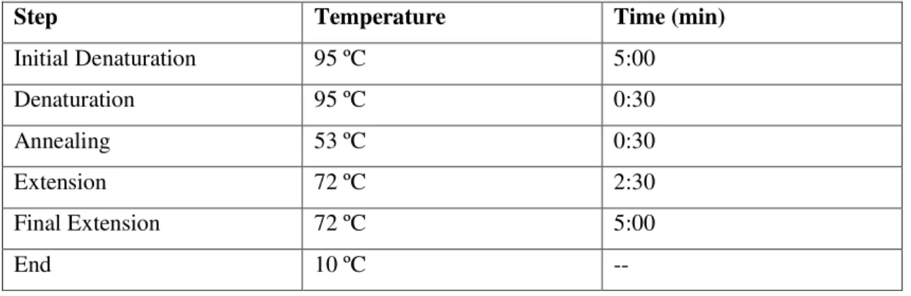
Table 3.3: PCR conditions for 671 amplification. Number of cycles (a cycle comprising the steps of Denaturation, Annealing and Extension) was of 30.
Step Temperature Time (min)
Initial Denaturation 98ºC 1:00
Denaturation 98ºC 0:10
Annealing 58ºC 0:30
Extension 72ºC 2:00
Final Extension 72 ºC 10:00
End 10 ºC --
The resulting amplified product was loaded into a 0,8% agarose gel and separated at 120mV for 40 minutes. Sequentially, sample was cleaned with a Clean Up kit.
It followed the digestion of the purified insert, 35μL of DNA was digested with 1μL of restriction enzyme XbaI, 5μL of 10x NEBuffer CutSmart and water MiliQ to a total volume of 50μL. Mixture was kept overnight at a temperature of 37 ºC, after which, sample was cleaned with Clean Up kit and re-suspended on 50μL water MilliQ. From the cleaned mixture was retrieved a volume of 35μL and added 1μL of restriction enzyme EagI, 5μL of 10x NEBuffer 3.1 and water MilliQ to a total volume of 50μL. Digestion proceeded overnight at a temperature of 37 ºC, after which, mixture was purified with Clean Up kit and stored at a -20 ºC.
3.4 Construction of pET21a vector for protein expression
It was constructed two pET21a – 671 mutants intended for protein expression.
10
temperature of 37ºC, after which, sample was purified with Clean Up kit, and re-suspended in 50μL water MilliQ. From the purified sample was retrieved a volume of 35μL and added 1,5μL of restriction enzyme EagI, 5μL of 10x NEBuffer 3.1 and water MilliQ to a total volume of 50μL. Digestion was kept overnight at a temperature of 37 ºC, and purified with Clean Up kit afterwards. Re-suspended in 50μL of water MilliQ.
Ligase reaction was prepared with 1μL of T4 Ligase enzyme, 2μL of 10x Ligase buffer, 10μL of digested plasmid pET21a and 7μL of digested insert 671. Auto-ligated sample was prepared with the same volumes with the exception of digested insert 671, which was replaced with 7μL MilliQ water. Mixture was kept overnight at room temperature and stored at -20 ºC. In order to confirm a successful ligation, it was performed a PCR with the commercial primers T7 promoter and T7 terminator, and the enzyme DreamTaq. Master Mix solution were as follows:
Table 3.4: Content of Master Mix solution for pET21a-671 I verification.
Master Mix 20μL
DreamTaq Green Buffer 10x 2μL
dNTP (10mM) 0,4μL
Primer T7 promoter (20mM) 0,2μL
Primer T7 terminator (20mM) 0,2μL
DreamTaq enzyme 0,1μL
MilliQ H2O 14,1μL
DNA template 3μL
PCR conditions were as follows:
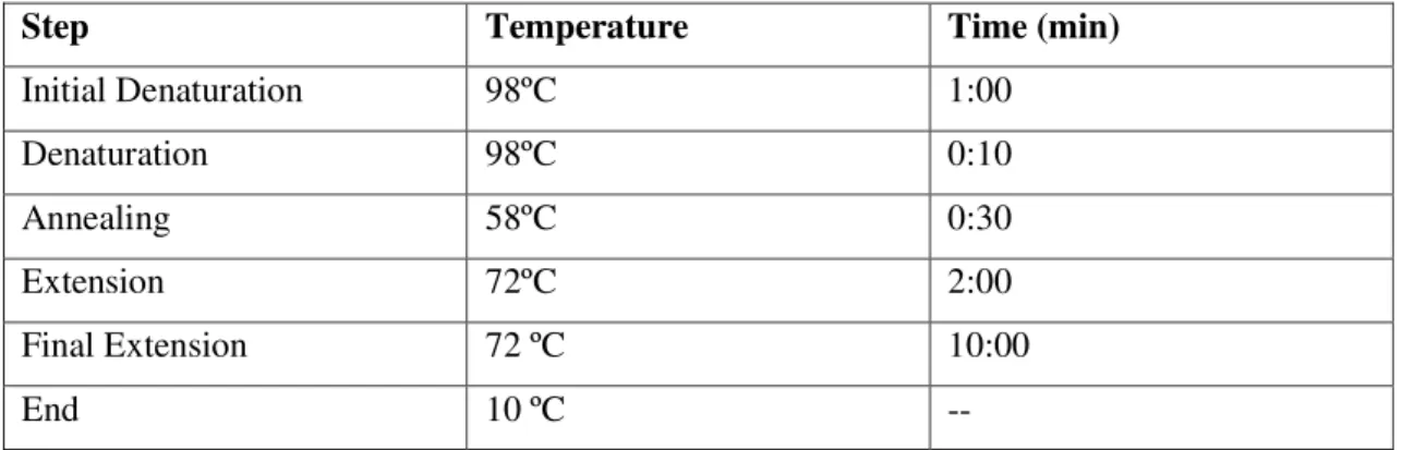
Table 3.5: PCR conditions for pET21a-671 I verification. Number of cycles (a cycle comprising the steps
of Denaturation, Annealing and Extension) was of 30.
Step Temperature Time (min)
Initial Denaturation 95 ºC 3:00
Denaturation 95 ºC 0:30
Annealing 53 ºC 0:30
11
Final Extension 72 ºC 5:00
End 10 ºC --
Following the PCR reaction, the product was loaded on a 0,8% agarose gel with 5μL of RedSafe and separated at 120mV for 40 minutes.
The second plasmid was denominated pET21a – 671 II and was constructed by digesting 20μL (2,960μg) of purified plasmid DNA with 1,5μL of restriction enzyme NheI, 3μL of 10x NEBuffer 2.1 and water MilliQ to a total volume of 30 μL. Digestion mixture was kept overnight at a temperature of 37 ºC, after which, sample was purified with Clean Up kit, and re-suspended in 50μL water MilliQ. From the purified sample was retrieved a volume of 20μL (521,2 ng) and added 1μL of restriction enzyme EagI, 5μL of 10x NEBuffer 2.1 and water MilliQ to a total volume of 30μL. Digestion was kept overnight at a temperature of 37 ºC, and neutralized by heat inactivation at 65 ºC for 30 minutes.
Ligase reaction was prepared with 1μL of T4 Ligase enzyme, 2μL of 10x Ligase buffer, 3μL (3,909μg) of digested plasmid pET21a and the following volumes of digested insert 671: 10μL, 7μL, 5μL and 0μL (auto-ligated plasmid). Mixture was kept overnight at room temperature and stored at -20 ºC.
In order to confirm a successful ligation, it was performed a PCR with the commercial primers T7 promoter and T7 terminator, and the enzyme DreamTaq. Master Mix solution were as follows:
Table 3.6: Content of Master Mix solution for pET21a-671 II verification.
Master Mix 20μL
DreamTaq Green Buffer 10x 2μL
dNTP (10mM) 0,4μL
Primer T7 promoter (20mM) 0,2μL
Primer T7 terminator (20mM) 0,2μL
DreamTaq enzyme 0,1μL
12
DNA template 3μL
PCR conditions were as follows:
Table 3.7: PCR conditions for pET21a-671 II verification. Number of cycles (a cycle comprising the steps
of Denaturation, Annealing and Extension) was of 30.
Step Temperature Time (min)
Initial Denaturation 95 ºC 5:00
Denaturation 95 ºC 0:30
Annealing 53 ºC 0:30
Extension 72 ºC 2:30
Final Extension 72 ºC 5:00
End 10 ºC --
PCR product was loaded on 0,8% agarose gel with 5μL RedSafe and separated at 120mV for 40 minutes.
3.5 E.coli transformation
Competent cells of the strain Dh5α were prepared according to the protocol previously described (Section 3.1). Cells were retrieved from –80ºC storage and thawed in ice. Once defrosted it was added 5μL of ligated DNA to the cell aliquots and placed on ice for 10 minutes.
Sequentially, samples were incubated at 42 ºC for 1 minute and then immediately placed on ice for 5 minutes. After ice incubation, it was added 1mL LB to cell samples and incubated under agitation at 37 ºC for 1 hour.
Samples were plated in LA plates supplemented with 100μg/mL of Ampicillin and incubated overnight at 37 ºC.
3.6 Colony Screening PCR
Transformed colonies of E.coli were confirmed via PCR. From ON plates was streaked
13
PCR reaction was performed with the commercial primers T7 promoter and T7 terminator, and the enzyme DreamTaq. Master Mix solution were as follows:
Table 3.8: Contents of Master Mix solution for Colony Screening PCR of E.coli colonies.
Master Mix 20μL
DreamTaq Green Buffer 10x 2μL
dNTP (10mM) 0,4μL
Primer T7 promoter (20mM) 0,2μL
Primer T7 terminator (20mM) 0,2μL
DreamTaq enzyme 0,1μL
MilliQ H2O 17,1μL
PCR conditions were as follows:
Table 3.9: PCR conditions for Colony Screening PCR for E.coli colonies. Number of cycles (a cycle
comprising the steps of Denaturation, Annealing and Extension) was of 30.
Step Temperature Time (min)
Initial Denaturation 95 ºC 10:00
Denaturation 95 ºC 0:30
Annealing 53 ºC 0:30
Extension 72 ºC 2:30
Final Extension 72 ºC 5:00
End 10 ºC --
PCR product was run on 0,8% agarose gel with 5μL of RedSafe and separated at 120mV for 40 minutes.
3.7 E.coli BL21(DE3) induction test
E.coli strain BL21(DE3) was transformed with pET21a plasmid according to stated
14
Grown ON culture was diluted to 0,05 OD600 in 20mL of LB medium, supplemented with 100μg/mL, incubated at a temperature of 37 ºC and agitation of 150 rpm until it reached 0,5 OD600.
When culture reached recommended OD600, it was divided in equal volume to another Erlenmeyer and added 1mM of IPTG. Nothing was added to the initial Erlenmeyer (negative control).
Both cultures proceeded with incubation at 37 ºC under agitation of 150 rpm. Aliquots of each cell culture were retrieved and stored at -20 ºC.
3.8 SDS-PAGE Gel Electrophoresis
Acrylamide gel matrix for SDS-PAGE gel electrophoresis was prepared following:
Table 3.10: Preparation of the Resolution Gel with 12,5% of Acrylamide.
Resolution Gel 12,5% Acrylamide Total Volume 10mL
MilliQ H2O 3,4mL
Tris-HCl 1,5M pH 8,8 2,48ml
Acrylamide 4,1mL
SDS 10% (w/v) 100μL
APS 10% (w/v) 50μL
TEMED 5μL
Table 3.11: Preparation of the Stacking Gel with 4% Acrylamide.
Stacking Gel 4% Acrylamide Total Volume 3mL
MilliQ H2O 1,8mL
Tris-HCl 0,5M pH 8,8 750μL
Acrylamide 400μL
SDS 10% (w/v) 30μL
APS 10% (w/v) 15μL
TEMED 3μL
15
Table 3.12: Preparation of SDS/Page Protein Loading Buffer 5x. Sample stored at -20ºC.
Protein Loading Buffer 5x Total Volume 5mL
SDS 10% (w/v) 1mL
Glicerol 0,5mL
Tris 1M pH 6,8 0,8mL
Bromophenol Blue 0,2% (w/v) 0,1mL
β-Mercaptoethanol 0,5mL
MilliQ H2O 2,1mL
Band separation was performed at 80mV in 1x Tris-Glycine SDS Running Buffer for approximately two hours. Gel was stained in Blue Safe overnight.
3.9 Sanger Sequencing
Sanger sequencing was provided by STABvida Laboratories, in FCT UNL campus. (http://www.stabvida.com)
3.10 S.aureus competent cells
For the preparation of S.aureus competent cells was prepared a solution of Sucrose 0,5M,
filtered and stored at 4ºC until used.
S.aureus strain RN4220 was inoculated in 5mL of TSB medium and grown overnight at 37ºC.
The following day, overnight culture was diluted 1:200 ratio into 100mL of TSB medium and incubated at 37ºC with shaking until OD600= 0,4-0,6. Upon reaching recommended OD, cultures were transferred into four pre-cooled 50mL Falcon tubes and centrifuged at top speed, 4ºC for 15 minutes.
Supernatant of centrifuged samples was discarded and pellet re-suspended in 50mL of sucrose 0,5M. Samples were centrifuged again at top speed, 4ºC for 15 minutes. Supernatant was discarded and pellet re-suspended 25mL of sucrose 0,5M.
16
Pellets were re-suspended in 300μL of sucrose 0,5M and all the content was combined into one single Falcon tube. The resulting mixture was distributed into aliquots of 50μL and stored at -80ºC.
3.11 Purification of Up and Down regions of gene 671
Regions 1kb Upstream and 1kb Downstream of gene 671 were amplified from genomic DNA extracted from S.aureus NCTC 8325-4 with the following primers:
Table 3.11: Forward and reverse primers for amplification of fragments Up and Down
Up
NCTC671UpFwEcoRI (P1) CGC GCG AAT TCT GGG TAT TAT TAC
GTT GGC ATT G
NCTC671UpRegOver (P2) GGA GTG CCT TTG TAA TTA AAT AAA
TTG TAC TG Down
NCTC671DwRegOver (P3) CAA TTT ATT TAA TTA CAA AGG GAC
TCC TCC
NCTC671DwRevBamHI (P4) CGC GGA TCC TTC TGA ACA TAT TAG
CAA GGT AAA G
Master Mix constitution and reaction conditions for both PCRs are as follows:
Table 3.12: Contents of Master Mix solution for Up and Down fragments amplification.
Master Mix 50μL
HF Phusion Buffer 5x 10μL
dNTP (10mM) 1μL
Primer Fw (P1 or P3) (10mM) 1μL
Primer Rv (P2 or P4) (10mM) 1μL
Phusion enzyme 051μL
17
DNA template 1μL
Table 3.13: PCR conditions for Up and Down cloning PCR. Number of cycles (a cycle comprising the
steps of Denaturation, Annealing and Extension) was of 30.
Step Temperature Time (min)
Initial Denaturation 98ºC 1:00
Denaturation 98ºC 0:10
Annealing 58ºC 0:30
Extension 72ºC 2:00
Final Extension 72 ºC 10:00
End 10 ºC --
After PCR, all samples were loaded into a 0,8% agarose gel with 5μL RedSafe and separated at 120mV for 40 minutes. Afterwards, the amplified product was cleaned using a Clean Up kit.
Once inserts Up and Down were purified, it followed an Overlapping PCR with primers NCTC671UpFwEcoRI (P1) as forward primer and NCTC671DwRevBamHI (P4) as reverse primer. The resulting amplification will consist of inserts Up and Down combined into a single, longer DNA fragment (UP-DOWN). PCR conditions and Master Mix constitution were as follows:
Table 3.14: Contents of Master Mix solution for overlapping PCR of Up and Down fragments.
Master Mix 50μL
HF Phusion Buffer 5x 10μL
dNTP (10mM) 1μL
Primer Fw P1 (10mM) 1μL
Primer Rv P4 (10mM) 1μL
Phusion enzyme 0,5μL
18
DNA template Up 1μL
DNA template Down 1μL
Table 3.15: PCR conditions for Overlapping PCR of Up and Down regions. Number of cycles (a cycle comprising the steps of Denaturation, Annealing and Extension) was of 30.
Step Temperature Time (min)
Initial Denaturation 98ºC 1:00
Denaturation 98ºC 0:10
Annealing 58ºC 0:30
Extension 72ºC 4:00
Final Extension 72 ºC 10:00
End 10 ºC --
After PCR, all samples were loaded into a 0,8% agarose gel with 5μL RedSafe and separated at 120mV for 40 minutes. Amplified band was cut from the gel and cleaned with a Clean Up kit.
It followed the sequential digestion of the purified UP-DOWN insert. For the first reaction 6,35μL (800ng) of insert was digested with 1μL of restriction enzyme EcoRI, 3μL of 10x NEBuffer 3.1 and water MiliQ to a total volume of 30μL. Mixture was kept overnight at a temperature of 37 ºC, after which, sample was cleaned with Clean Up kit and re-suspended on 50μL water MilliQ, From the cleaned mixture was retrieved a volume of 20μL and added 1μL of restriction enzyme BamHI, 5μL of 10x NEBuffer 3.1 and water MilliQ to a total volume of 50μL. Digestion proceeded overnight at a temperature of 37 ºC, after which, mixture was purified with Clean Up kit and stored at a -20 ºC.
3.12 Construction of pMAD vector for gene excision
19
Digestion was kept overnight at a temperature of 37 ºC, and cleaned with Clean Up kit afterwards. Re-suspended in 50μL of water MilliQ.
Ligase reaction was prepared with 1μL of T4 Ligase enzyme, 2μL of 10x Ligase buffer, 10μL (67ng) of digested plasmid pMAD and the following volumes of digested insert UP -DOWN: 10μL, 7μL, 5μL and 0μL (auto-ligated plasmid). Mixture was kept overnight at room temperature and stored at -20 ºC.
To confirm a successful ligation, it was performed a PCR with the primers pMAD1 (forward) and pMAD2 (reverse). PCR conditions and Master Mix constitution are as follows:
Table 3.16: Contents of Master Mix solution for pMAD del 671 verification.
Master Mix 20μL
DreamTaq Green Buffer 10x 2μL
dNTP (10mM) 0,4μL
Primer pMAD1 (10mM) 0,4μL
Primer pMAD2 (10mM) 0,4μL
DreamTaq enzyme 0,1μL
MilliQ H2O 16,7μL
DNA template 3μL
Table 3.17: PCR conditions for pMAD del 671 verification. Number of cycles (a cycle comprising the steps of Denaturation, Annealing and Extension) was of 30.
Step Temperature Time (min)
Initial Denaturation 95 ºC 3:00
Denaturation 95 ºC 0:30
Annealing 53 ºC 0:30
Extension 72 ºC 3:00
Final Extension 72 ºC 5:00
20
PCR product was loaded on a 0,8% agarose gel with 5μL of RedSafe and separated at 120mV for 40 minutes.
Once verified a successful ligation of pMAD del 671, the plasmid was transformed to
E.coli strain of DC10b according to the E.coli transformation protocol (section 3.5). Transformed
colonies were screened via Colony Screening PCR (section 3.6) with the primers pMAD1 and pMAD2. Plasmid DNA was extracted with a Miniprep kit from positive colonies and stored at -20ºC.
3.13 S.aureus electroporation
S.aureuscompetent cells were thawed in ice and 5μL of purified DNA plasmid was added
to the aliquot. Mixture was then transferred to a 0,2 cm BioRad Gene Pulser cuvette and incubated on ice for 5 minutes.
Samples were then retrieved and pulsated once at 2500V, 25μF, 100Ω for 2,5 msec. Cells were immediately rescued with 1mL of TSB medium and placed on ice for 15 minutes. Samples were then incubated at 30 ºC, under agitation for 2 hours.
3.14 Transduction protocol
The following solutions were prepared:
Table 3.20: Preparation of 0,3GL Top and Bottom Agar. Medium was autoclaved at 120ºC for 40
minutes.
0,3GL Top Agar (Bottom Agar) pH 7,8 Concentration
Casamino Acids 3 g/L
Yeast Extract 3 g/L
NaCl 5,9 g/L
Sodium Lactate, 60% syrup/DL-Lactate Acid 3,3 mL/L
50% Glicerol 2 mL/L
Tri-Sodium Citrate 0,5 g/L
Agar for Top agar (for Bottom agar) 7,5 g/L (15 g/L)
Table 3.21: Preparation of Phage Top and Bottom Agar. Medium was autoclaved at 120ºC for 20
minutes.
Phage Top Agar (Bottom Agar) pH 7,8 Concentration
21
Yeast Extract 3 g/L
NaCl 5,9 g/L
Agar for Top agar (for Bottom agar) 5 g/L (15 g/L)
Table 3.8: Preparation of Phage Buffer. Solution as sterilized with 0,22 μm filter.
Phage Buffer Concentration
0,1M MgSO4 10 mL/L (1mM)
0,4M CaCl2 10 mL/L (4mM)
2,5M Tris pH 7,8 20 mL/L (50mM)
NaCl 5,9 g/L
Gelatin 1 g/L
All media and buffers were stored at 4ºC.
In order to make the phage lysate, a single colony of transformed S.aureus strain RN4220
was plated and incubated at 30ºC overnight.
It was retrieved two 10μL loops of confluent growth and re-suspended in 1mL of TSB medium where it was added 5mM of CaCl2.
Several dilutions of Phage 80α lysate were prepared with Phage Buffer and kept at 4ºC. Additionally, it was prepared 3mL of Phage Top Agar supplemented with 5mM CaCl2, 10μL of cell suspension and 10μL of diluted phage lysate. The mixture was poured onto 20mL Bottom Phage Agar plates sustained with 5mM of CaCl2. Plates were incubated overnight at 30ºC.
The following day, 4mL of Phage Buffer was added to plates showing confluent lysis and place at 4ºC for one hour. Sequentially, the Phage Top Agar layer was collected into a Falcon tube, vortexed and incubated at 4ºC for one hour. Mixture was then centrifuged at top speed for 15 minutes at 4ºC.
Supernatant was recovered and filtered with a 0,45μm filter and stored at 4ºC. As a sterility control, 50μL of recovered phage lysate was plated on a TSA plate and incubated overnight at 37ºC.
For the transduction procedure it was plated a single colony of the receiving strain of
S.aureus NCTC 8325-4 and incubated overnight at 30ºC.
22
It was poured into petri dishes 10mL of 0,3GL Bottom Agar containing 3x 10μg/mL Erythromycin. After agar is set, it was poured a new layer of 20mL 0,3GL Bottom Agar without antibiotic.
The following transduction mixtures were prepared:
Table 3.23: Transduction mixtures prepared.
Sample Culture (μL) Phage Lysate (μL) Phage Buffer μL
1 100 0,1 100
2 100 1 100
3 100 10 100
4 100 100 100
Control 100 - 100
Mixtures were incubated under agitation for 20 minutes at 37ºC.
Each transduction mixture was added to 3mL of Top Agar in a test tube, shaken and immediately poured onto a prepared 0,3GL Bottom Agar plate. After solidified, plates were incubated at 30ºC for 48 hours.
3.15 Gene knock-out protocol
For the integration step of gene knock-out procedure, inoculates of transduced strain of
S.aureus NCTC 8325-4 + pMAD del 671 were prepared in TSB medium supplemented with
10μg/ml of Erythromycin and incubated overnight at 30ºC. The following day, cultures were diluted 1:1000 in fresh medium of TSB with 10μg/mL Erythromycin and incubated at 30ºC for a period of 8 hours. After incubation over day, cultures were once again diluted at 1:1000 ratio in TSB medium with 10μg/mL Erythromycin and incubated at 43ºC overnight.
The following day, grown cultures were plated in fresh plates of TSA medium supplemented with 10μg/mL Erythromycin and 100μg/mL X-Gal with the following dilutions: 10-4, 10-5 and 10-6. Plates were then incubated overnight at 43ºC. The following day, isolated blue colonies were stricken to fresh plates of TSA + 10μg/mL Erythromycin + 100μg/mL X-Gal and incubated at 43ºC overnight.
23
Positive colonies with integrated pMAD del 671 plasmid were selected for the excision step of gene knock-out procedure. Samples were inoculated in TSB medium with no antibiotic and incubated at 30ºC overnight. The following day, cultures were diluted 1:500 in fresh TSB medium with no antibiotic and incubated over day at 30ºC for a period of 8 hours. Afterwards, cultures were diluted 10-4, 10-5 and 10-6 and plated in fresh plates of TSA + 100μg/mL X-Gal. Plates were incubated at 43ºC overnight.
The following day, the same isolated white colonies were stricken in fresh plates TSA + 10μg/mL Erythromycin + 100μg/mL X-Gal as well as plates TSA + 100μg/mL X-Gal. Both set of plates were incubated overnight at 37ºC.
After incubation, white colonies that did not grow in TSA medium supplemented with Erythromycin were stricken into fresh plates of TSA + 100μg/mL X-Gal and incubated overnight at 37ºC. The following day, genomic DNA of grown white colonies was extracted (section 3.16) and a PCR (section 3.17) was performed to confirm the successful excision of the gene 671.
3.16 S.aureus genomic DNA extraction for PCR screening.
The following solutions were prepared:
• Alkaline Wash Solution
o 0,05M Sodium Citrate o 0,5M NaOH
• 0,5M Tris-HCl pH 8,0
Cells were scraped from plates and re-suspended in 500μL of Alkaline Wash Solution and followed by room temperature incubation for 20 minutes. Sequentially, samples were centrifuged at top speed for 1 minute and the resulting pellet was washed with 500μL of 0,5M Tris-HCl pH 8,0. A new centrifugation was performed and pelleted cells were re-suspended in 100μL of MilliQ water.
Samples were boiled for 10 minutes followed by centrifugation at top speed for 5 minutes. Supernatant was then transferred to a new, clean Eppendorf and stored at -20ºC.
3.17 PCR screening for gene knock-out confirmation
24
“NCTC671UpFwEcoRI” (P1) and reverse pMAD2. The resulting amplification will verify if the crossing over took place on the Up region of the 671 gene. The second set of primers consisted of the forward primer pMAD1 and the reverse primer “NCTC671DwRevBamHI” (P4). This set of primers will amplify for successful crossing over on the Down region of the gene 671.
The Master Mix constitution and PCR conditions for integration screening are as follows:
Table 3.24: Contents of Master Mix solution for integration screening PCR.
Master Mix 20μL
DreamTaq Green Buffer 10x 2μL
dNTP (10mM) 0,4μL
Primer P1/pMAD1 (10mM) 0,4μL
Primer pMAD2/P4 (10mM) 0,4μL
DreamTaq enzyme 0,1μL
MilliQ H2O 14,1μL
Genomic DNA 3μL
Table 3.25:PCR conditions for integration screening PCR. Number of cycles (a cycle comprising the steps of Denaturation, Annealing and Extension) was of 30.
Step Temperature Time (min)
Initial Denaturation 95 ºC 10:00
Denaturation 95 ºC 0:30
Annealing 53 ºC 0:30
Extension 72 ºC 3:00
Final Extension 72 ºC 10:00
End 10 ºC --
After PCR, all samples were loaded into a 0,8% agarose gel with 5μL RedSafe and separated at 120mV for 40 minutes.
25
“NCTC671DwRevBamHI” (P4) as reverse primer. The constitution of the reaction Master Mix and the PCR conditions are as follows:
Table 3.26: Contents of Master Mix solution excision verification PCR.
Master Mix 20μL
DreamTaq Green Buffer 10x 2μL
dNTP (10mM) 0,4μL
Primer P1 (10mM) 0,4μL
Primer P4 (10mM) 0,4μL
DreamTaq enzyme 0,1μL
MilliQ H2O 14,1μL
Genomic DNA 3μL
Table 3.27: PCR conditions for excision verification PCR. Number of cycles (a cycle comprising the steps
of Denaturation, Annealing and Extension) was of 30.
Step Temperature Time (min)
Initial Denaturation 95 ºC 10:00
Denaturation 95 ºC 0:30
Annealing 53 ºC 0:30
Extension 72 ºC 2:00
Final Extension 72 ºC 10:00
End 10 ºC --
After PCR, all samples were loaded into a 0,8% agarose gel with 5μL RedSafe and separated at 120mV for 40 minutes.
27
4
Results and Discussion
4.1 Construction of pET21a-671 I
As it was described in section 3.3, gene 671 was amplified from the extracted DNA of
S.aureus strain NCTC 8325-4. PCR reaction amplified a DNA fragment of approximately 800bp
which is in conformance with the in-silico sequence. As figure 4.1 demonstrates, the amplified
band is inferior to the 850bp ladder band and shows no other amplification products. Control sample was performed with MilliQ water (last lane before ladder) and it displays no amplification.
Figure 4.1: Amplification of gene 671.Expected length ofamplified fragment is approximately 850bp. 1 to 8- Amplified 671 insert. 9- control.
Cloning vector pET21a was extracted using a Miniprep kit from E.coli strain Dh5α and
digested with restriction enzymes XbaI, EagI and NheI.
In order to assess the integrity of the extracted and digested DNA, it was performed an agarose gel (Figure 4.2) where pET21a non-digested was loaded next to the digested samples. All lanes display two bands with no visible smears: the first band at 5000bp is in accordance to the expected size of pET21a (which has approximately 5500bp) whereas the second band over the 12000bp ladder mark corresponds to super-coiled form of DNA.
All used enzymes have a unique restriction site that will linearize the circular plasmid, and therefore display a band of the same weight as the circular plasmid. The existence of the second band from the super-coiled form confirms an incomplete digestion.
850 bp
28
Figure 4.2: Plasmid pET21a digested with restriction enzymes XbaI, EagI and NheI. 1- Plasmid pET21a. 2- pET21a digested with XbaI. 3- pET21a digested with EagI. 4- pET21a digested with NheI. 5- pET21a digested with XbaI and EagI.
Digested plasmid and insert 671 were ligated using a T4 Ligase enzyme. Considering the gene 671 was inserted in the multiple cloning site of pET21a it is possible to verify a successful ligation with the primers T7 promoter and T7 terminator, located upstream and downstream of
the MCS. PCR conditions are described in section 3.4 and the agarose gel is shown in figure 4.3. The amplification product of a successful ligation will consist of a DNA fragment with the approximate size of 850bp, as is shown in lane 1 of figure 4.3. The auto-ligated plasmid (lane 2) will amplify a DNA fragment of 300bp.
A plasmid digested with either XbaI, EagI or both enzymes will become linear and will not allow any amplification. The existence of a 300bp band in lane 3 indicates the digestion of pET21a with either XbaI or EagI was incomplete and the plasmid returned to its original circular form.
In lane 6 is displayed the amplification of 300bp fragment that is expected of pET21a plasmid.
1 2 3 4 5 6
29
Figure 4.3: Cloning PCR of pET21a-671 I plasmid. Expected length of the amplified 671 insert is of 850bp, which verifies a successful insertion. 1- PCR product of pET21a-671 I. 2-PCR product of auto-ligated pET21a. 3- PCR product of pET21a digested with XbaI and EagI. 4- pET21a digested with XbaI and EagI. 5- pET21a digested with Xbal. 6- PCR product of pET21a. 7- pET21a. 8- Control.
4.2 Positive colonies of pET21a-671 I.
The confirmed ligated pET21a-671 I was transformed into E.coli strain Dh5α according
to the protocol described in section 3.5. The auto-ligated pET21a was also transformed. Several of the resulting colonies were stricken into fresh plates and screened via Colony Screening PCR (section 3.6)
Figure 4.4: Screening PCR for pET21a-671 I positives. 1 to 11- Amplification from transformed Dh5α with pET21a-671 I. 12- Amplification from transformed Dh5α with auto-ligated pET21a. 13- DNA ladder 14- Amplification from pET21a-671 I (positive control). 15- Amplification from auto-ligated pET21a (negative control). 16- Amplification from pET21a digested with XbaI and EagI. 17- Amplification from pET21a digested with XbaI. 18- Amplification from pET21a.
1 2 3 4 5 6 7 8
5 000 bp
850 bp
300 bp
1 2 3 4 5 6 7 8 9 10 11 12 13 14 15 16 17 18
30
PCR conditions and Master Mix constitution were as it is described in section 3.6. From a selection of 11 transformed Dh5α colonies with plasmid pET21a-671 I, only one showed the amplification of the 800bp band respective to gene 671 (figure 4.4).
Positive colony of pET21a-671 I was stricken into fresh plate and plasmid DNA was extracted with a Miniprep kit. It followed a digestion with restriction enzyme NaeI in order to assess a Digestive Profile. Enzyme NaeI possesses three restriction sites as it is demonstrated in figure 4.5.
Figure 4.5: Restriction sites of NaeI in plasmid pET21a-671 I.
It was digested 1μg of plasmid pET21a-671 I and plasmid pET21a that served as negative control.
31
Table 4.1: Size comparison of DNA fragments produced by NaeI digestion.
pET21a pET21a-671 I
3336 3336
1588 1588
456 1197
Figure 4.6: Digestive profile of pET21a-671 I by NaeI. Digestion with NaeI enzyme will result in three
fragments for both pET21a-671 I and pET21a. The size difference in the smallest fragment attests for a positive insertion of gene 671. 1- pET21a digested. 2- Digested pET21a. 3- pET21a-671 I non-digested. 4- Digested pET21a-671 I.
The agarose gel in figure 4.6 depicts the digestion of both plasmids by NaeI. As it was expected, the two plasmids display two bands of equal size: a first with 3000bp followed by the second close to the 1650bp ladder band.
The last band of the sample pET21a is barely visible in the agarose gel but is clearly below the 1000bp ladder mark. The positive sample pET21a-671 I, however, exhibits the third band above the 1000bp which confirms the insertion of gene 671.
1 2 3 4
3 000 bp 1 650 bp 1 000 bp
32
4.3 Induction test of pET21a-671 I
Positive plasmid pET21a-671 was transformed into E.coli strain BL21(DE3) as it was
described in section 3.5. One transformed colony was selected and inoculated in LB medium supplemented with 100μg/ml Ampycilin, incubated overnight at 37ºC, 150rpm.
Protein induction test was performed according to section 3.7, with 1mM IPTG and temperature 37ºC. Aliquots were retrieved at time 0 of induction, at 3-hour induction and overnight induction. Optical density was also measured at the mentioned points.
Figure 4.7: Optical density measurements along time of induction. 671+: culture induced with 1mM
IPTG. 671-: culture not induced (control)
Retrieved samples were loaded into SDS/PAGE gel as is described in section 3.8. Samples were run at 80 mV for approximately 2 hours (figure 4.8). Expected protein is of 26,6 kDa, according to SeqBuilder software from Lasergene’s DNAStar.
As it is depicted in figure 4.8, it is possible to discern an accentuated band in the induced samples of pET21a-671 I. Considering the weight of said band, which stands above the 25 kDa ladder mark, it can be correlated to the successful expression of protein 671.
0 0,1 0,2 0,3 0,4 0,5 0,6
0 5 10 15 20
OD
Time (h)
pET21a-671 I induction
-33
Figure 4.8:SDS/PAGE gel of pET21a-671 I protein induction test.T0: time 0 of induction. T3+: time
3-hour induced. T3-: time 3-3-hour induced. TON+: time overnight induced. TON-: time overnight non-induced.
4.4 Sequencing of pET21a-671 I
Sanger sequencing of plasmid pET21a-671 I was also performed in order to verify coherence with the in-silico sequence (section 3.9).
As it is demonstrated in figure 4.9, sanger sequencing exposed an interruption of approximately 40bp in plasmid pET21a-671 I. This interruption implied the deletion of the conserved RBS of pET21a as well as the start codon, which could potentially hinder the expression of the protein.
T0
T3
+
T3
-
TON
+
TON
-25 kDa
A
B
Figure 4.9:Sequencing of pET21a-671 I.A- Segment of sequencing traces assembled by SeqMan