FACULDADE DE CIÊNCIAS
DEPARTAMENTO DE BIOLOGIA VEGETAL
Towards improvement of Haematococcus pluvialis cultures by
cell sorting and UV mutagenesis
Filipa Faria Rosa
Mestrado em Microbiologia Aplicada
Dissertação orientada por:
Doutor Luís Tiago Guerra
Professora Doutora Ana Tenreiro
Towards improvement of Haematococcus pluvialis cultures by cell
sorting and UV mutagenesis
Filipa Faria Rosa
2017
This thesis was fully performed at A4F – Algae for Future and Bugworkers Laboratory
| M&B
– BioISI | Teclabs under the direct supervision of Doutor Luís Tiago Guerra.
Professora Doutora Ana Tenreiro was the internal supervisor in the scope of the Master
in Applied Microbiology of the Faculty of Sciences of the University of Lisbon.
4 – RESULTS & DISCUSSION
I
ACKNOWLEDGMENTS
First of all, I would like to show my gratitude to the administration of A4F – Alga for Future. Not only they gave me the opportunity of carrying out my master thesis but also I was able to work in two laboratories, A4F and Bugworkers Laboratory | M&B – BioISI | Teclabs, thanks to their partnership. It was an exceptional and exclusive experience which I will never forget.
I want to thank Dr. Luis Tiago Guerra, my supervisor at A4F, and Professora Doutora Ana Tenreiro, my supervisor at Bugworkers Laboratory | M&B – BioISI | Teclabs. I am especially grateful for all the knowledge and guidance given along the entire thesis, as well as the availability they showed to help whenever I needed. Not least, thank for the advices, patience, trust and other contributions that allowed the improvement of this work.
I am thankful to all my A4F’s colleagues, especially the ones from the laboratory, who gave me all their support and most important, provided me great and unforgettable moments in the laboratory. Not forgetting, my professors and my colleagues from the Bugworkers Laboratory, for answering my questions and clear up my doubts always with the greatest positivity.
To all my friends and family, a huge thanks for being so supportive and always around when needed the most. In particular, a very special thanks to my parents and brother who are the major pillars of my being and who never gave up on my success. Thanks for your support, encouragement, strength and patience.
ABSTRACT
Nowadays, there is an increased interest in biological active compounds derived from natural sources, especially compounds with medical applications, nutrient rich food and feed, and health promoting compounds. Microalgae are a potential valuable resource for a biotech purposes, as new sources of biomolecules such as pigments, lipids, carbohydrates and proteins. Natural pigments have pharmacological properties and have increased marketability of products advantages over synthetic products. Commercial production of natural carotenoids from microalgae is an eco-friendlier and safer approach than synthetic manufacture by chemical procedures. Of several naturally occurring carotenoids, astaxanthin is considered one of the best, being able to protect cells, lipids and membrane lipoproteins against oxidative damage. Haematococcus pluvialis is the richest source of natural astaxanthin and is produced at large scale.
The work herein described had the objective to improve H. pluvialis strains with an enhanced accumulation of astaxanthin content. For the strain improvement three approaches have been used: (i) screening of different strains to select those with a superior performance; (ii) flow cytometry assisted cell sorting of cells subpopulations with increased astaxanthin production and (iii) random mutagenesis by UV-C irradiation and mutants selection. For this purpose, seven H. pluvialis strains were used and a characterization was conducted throughout the 3-steps cultivation experimental strategy: green vegetative growth, induction of astaxanthin accumulation and additional salinity stress stages. This integrative characterization focused on a number of microalgae physiological properties, intending to thoroughly assess the evolution and heterogeneity of their vitality and astaxanthin accumulation capacity.
HP_02 and HP_03 were the most promising selected strains, reaching respectively 3.8 % and 4.4 % (of their DW) of astaxanthin content, and presenting high growth rates of 0.58 day-1 and 0.52 day-1. The two strains were submitted to fluorescence activated cell sorting for enrichment of astaxanthin over-producers. Both strains showed similar performances and no specific cell properties and/or stresses responses could be point as particularly relevant. The HP_03 strain was further subjected to random mutagenesis by exposure to UV-C radiation in order to promote heterogeneity in the cell population. The isolation of cells sub-populations of interest, with high recovery and high degree of purity, was not achieved, due to the homogeneity of the populations of the analyzed strains, and due to technical impossibility of the sorting device. However, it is noteworthy that, flow cytometry allowed the monitoring of astaxanthin content during the induction phase of the culture, and showed that the autofluorescence of this pigment can be a good indicator of its intracellular accumulation and can therefore be monitored in real time by flow cytometry. The results of the preliminary random mutagenesis assay, by exposure to UV-C radiation seem to indicate that this strategy is promising to increase the subpopulation of astaxanthin producing cells. Further similar studies with different strains and additional parameters should be performed to better clarify this subject.
4 – RESULTS & DISCUSSION
III
RESUMO
As microalgas estão a emergir como uma das mais promissoras fontes sustentáveis de biomassa, combustível, alimentação, rações e outros produtos. Estes microrganismos têm um potencial enorme na área da biotecnologia por serem uma fonte valiosa de metabolitos como pigmentos (ex. astaxantina, β-caroteno), proteínas (ex. ficocianina), lípidos (ex. ómega-3, DHA, EPA) e hidratos de carbono. Embora os seus compostos ativos apresentem vantagens relativamente aos produtos sintéticos ou outros produtos de fontes naturais, têm contudo, desvantagens a nível de custos (Borowitzka, 2013; Demirbas, 2011; Milledge, 2010). O maior desafio na aplicação das microalgas para fins comerciais tem sido minimizar os custos de produção e extração dos compostos, devido à complexidade da fase de cultivo e dos processos a jusante (ex. extração dos compostos de valor acrescentado).
Entre os metabolitos de elevado valor acrescentado, a astaxantina (3,3’-dihidroxi-β,β’-caroteno-4,4’diona), carotenoide secundário, é considerada um composto valioso com um elevado número de aplicações, desde sectores alimentares, cosméticos a farmacêuticos. Este pigmento tem como fonte natural microalgas como Chlorella zofingiensis, Chlorococcum, Haematococcus pluvialis, leveduras vermelhas, Phaffia rhodozyma e bactérias, Paracoccus carotinifaciens. Haematococcus pluvialis, é considerada a maior fonte natural de astaxantina, podendo acumular até 6 % do seu peso seco e é a melhor fonte natural de astaxanthina para consumo humano (Ambati et al., 2014; Lorenz, 1999; Olaizola, 2003; Yuan et al., 2010).
A acumulação de carotenoides secundários, como a astaxantina, é uma característica de resposta ao
stress de certas microalgas como é o caso de H. pluvialis. Quando há privação de nutrientes, aumento
da intensidade luminosa, salinidade, etanol e hormonas entre outros fatores desfavoráveis ao crescimento da microalga, esta começa a induzir (Sarada et al., 2002a; Su et al., 2014; Zhang et al., 2014). A fase de indução, também conhecida como fase vermelha, corresponde à produção e acumulação da astaxantina, essencial para H. pluvialis sobreviver a condições adversas (Hagen et al., 2002; Wayama et al., 2013). Devido às flutuações das condições de stress que podem ser aplicadas para iniciar o processo de indução, todas as estratégias devem ser otimizadas para que a acumulação de astaxantina seja o mais reprodutível possível. O desenvolvimento de novas tecnologias para produção de microalgas, em sistemas abertos (ex. raceways) e sistemas fechados (ex. fotobioreactores), assim como a otimização das condições de cultivo (um ou duas fases de produção) têm maximizado o crescimento e a produção de biomassa, teor de pigmentos e outros produtos, reduzindo os custos de produção à escala industrial (Der Rio et al., 2007; Fábregas et al., 2001; Shah et al., 2016).
Contudo, existem ainda muitos desafios e problemas na produção a grande escala de H. pluvialis. A otimização não tem de ser restrita ao melhoramento das condições de cultivo e aos sistemas de produção. O melhoramento do desempenho das estirpes de H. pluvialis também tem sido alvo de estudos recentes. Para aumentar o conteúdo de astaxantina acumulado, as células podem ser submetidas a agentes mutagénicos químicos, como etil-metano-sulfanato (EMS) ou N-metil-N-nitro-N-nitrosoguanidina (NTG), ou agentes físicos, como radiação ultravioleta ou raio-X (Chen et al., 2003; Kamath et al., 2008; Tjahjono et al.,1994; Tripathi et al., 2001). Mutações mais direcionadas também foram aplicadas a estas microalgas, como modificações genéticas (Forján et al., 2015; Sharon-Gojman et al., 2015).
O trabalho desenvolvido no âmbito desta tese teve como principal objetivo o melhoramento de estirpes de H. pluvialis, pertencentes à coleção de microalgas da empresa A4F, para aumentar o teor intracelular
de astaxantina. Para o melhoramento das estirpes foram utilizadas três abordagens: (i) caracterização de diferentes estirpes para posterior seleção daquelas que apresentaram desempenho superior; (ii) separação física (cell sorting) de subpopulações de células com aumento da produção de astaxantina, por citometria de fluxo e (iii) mutagénese aleatória por irradiação com UV-C e seleção de mutantes. Neste trabalho foram inicialmente utilizadas sete estirpes de H. pluvialis (HP_01 to HP_07) cultivadas numa estratégia experimental em três fases: na primeira fase de crescimento vegetativo ou fase verde, as diferentes estirpes foram crescidas em condições ótimas durante 7 dias, avaliando-se e comparando as taxas de crescimento e produtividade da biomassa; na segunda fase de indução ou fase vermelha, aplicaram-se dois fatores de stress, privação de nutrientes e aumento da intensidade luminosa (150 µmol.m-2.s-1), durante 17 dias, analisando-se a produtividade e a acumulação máxima de astaxatina nas diferentes estirpes; na terceira fase aplicou-se um stress salino adicional, analisando-se o seu impacto na biomassa e no conteúdo máximo de astaxantina das culturas já induzidas. Esta caracterização integrativa enfocou um número de propriedades fisiológicas das microalgas, com a intenção de avaliar completamente a evolução e heterogeneidade de sua vitalidade e capacidade de acumulação de astaxantina. Os parâmetros avaliados foram: contagem de células, peso seco, produtividade e viabilidade de biomassa e análise de pigmentos.
Os resultados obtidos no decurso deste trabalho permitiram caracterizar as diferentes estirpes de H.
pluvialis e selecionar as que apresentaram as melhores características relativamente à taxa de
crescimento e produtividade de biomassa na fase de crescimento, e com especial interesse à sua produtividade e teor em astaxantina na fase de indução. Das sete, destacaram-se as estirpes HP_02 e HP_03, que durante a fase de crescimento apresentaram elevadas taxas de crescimento (0,52 e 0,58 dia -1, respetivamente) e produtividades similares de biomassa comparativamente às outras estirpes (0,27 e 0,22 g.L-1.dia-1, respetivamente). HP_02 e HP_03, obtiveram o maior conteúdo de astaxantina acumulado (3,8 % e 4,4 % respetivamente), e registaram produtividades de astaxanina elevadas, (2,31 e 2,58 mg.g-1.dia-1 respetivamente), superiores às outras estirpes estudadas. As duas estirpes foram submetidas a cell sorting para enriquecimento de sobre-produtores de astaxantina. Ambas as estirpes apresentaram desempenhos semelhantes e não foram consideradas particularmente relevantes propriedades específicas das células. A estirpe HP_03 foi ainda sujeita a mutagénese aleatória por exposição à radiação UV-C de modo a promover a heterogeneidade na população celular.
Os pigmentos fotossintéticos presentes nas microalgas são clorofilas e carotenóides, e a sua concentração intracelular depende das condições de cultivo. Esta concentração está linearmente correlacionada com a fluorescência dos pigmentos, o principal componente da fluorescência endógena (autofluorescência) (Hyka et al., 2013). Assim, a intensidade de autofluorescência foi utilizada para a identificação e quantificação de pigmentos de microalgas por citometria de fluxo. Esta metodologia permitiu ainda detetar alterações no estado fisiológico das células; a morfologia, incluindo tamanho e complexidade celular dependem das condições de cultivo e estão correlacionadas com os dois sinais de dispersão da luz, medidos por citometria, nomeadamente dispersão direta (FSC) e ortogonal (SSC). A acumulação de astaxantina das 7 estirpes, foi monitorizada por citometria de fluxo, no início e ao longo da fase de indução, assim como foi seguida a evolução do estado fisiológico das células. É de salientar que os resultados da quantificação de astaxantina por citometria de fluxo (valores médios das intensidades de fluorescência) foram consistentes com os determinados pelo método de extração de pigmentos totais, confirmados pela forte correlação linear encontrada (r = 0,98) entre o conjunto completo de valores dos dois parâmetros. Demonstrou-se assim que, a autofluorescência da astaxantina é um bom indicador da sua acumulação intracelular, podendo ser monitorizada em tempo real por citometria de fluxo.
4 – RESULTS & DISCUSSION
V
Para concluir a fase de caracterização das estirpes, os resultados obtidos após adição de NaCl (10 g/L) mostraram que o aumento do conteúdo de astaxantina não é estatisticamente significativo, à exceção de HP_01 que obteve um aumento de 2,5 % para 3,4 % (p < 0,001). A aplicação do stress salino adicional, ainda que apenas durante um curto período de tempo, 1 a 2 dias, determinou um aumento do teor de astaxantina, começando as culturas a perder biomassa após o terceiro dia, e como tal teve efeitos negativos no conteúdo de astaxantina acumulado. A maior produtividade global de astaxantina foi obtida pelas estirpes HP_02 e HP_03, atingindo 1,63 e 1,88 mg.g-1.dia-1, respetivamente. Este valor foi baseado no tempo essencial para as estirpes alcançarem o valor máximo de astaxantina, incluindo as duas fases de cultivo durante 24 dias.
A partir das culturas selecionadas, HP_02 e HP_03, o isolamento de subpopulações de células de interesse, com alta recuperação e alto grau de pureza, não foi alcançado, devido à homogeneidade das populações das estirpes analisadas, e devido à impossibilidade técnica do dispositivo de sorting. Como terceira abordagem para o melhoramento das estirpes, a HP_03 foi selecionada, realizou-se mutagénese aleatória por exposição à radiação ultravioleta (UV-C), criando condições propícias a que as células sofressem alterações genéticas de modo a potenciar a produção de astaxantina. O efeito letal da exposição à radiação foi estudado analisando a viabilidade celular por citometria de fluxo, usando como fluoróforo um oxonol [DiBAC4(3)], que responde ao potencial de membrana. Os resultados obtidos permitiram seguir a evolução da viabilidade celular com o aumento do tempo de exposição à radiação, observando-se uma despolarização progressiva da membrana, que terminou ao fim de 100 s de exposição por uma dissipação generalizada do potencial. No entanto, nos resultados obtidos pela análise de citometria de fluxo, não foi notável um aumento da heterogeneidade populacional, mantendo-se a dificuldade da realização do sorting das células com maior conteúdo de astaxantina. Este ensaio preliminar de mutágenese aleatória apresentou resultados promissores, na medida em que um dos mutantes isolados, Mut 2, atingiu valores superiores de astaxantina, 4,1 % por peso seco, relativamente ao controlo que obteve 3,1 % de astaxantina, para o mesmo período de tempo.
Uma vez que as estirpes selecionadas apresentaram um desempenho análogo e uma homogeneidade populacional, torna-se difícil identificar propriedades celulares ou respostas ao stress que possam ser particularmente relevantes para o melhoramento das estirpes com vista a uma maior acumulação de astaxantina. Deverão realizar-se novos estudos com recurso a estirpes com maior heterogeneidade, que possibilitem o isolamento das células com as propriedades desejadas, e as configurações do citómetro de fluxo e do dispositivo de sorting devem ser otimizadas de modo a permitir separação física (cell
sorting) de subpopulações de células com aumento da produção de astaxantina, com alta recuperação e
alto grau de pureza.
Palavras-chave: astaxantina; Haematococcus pluvialis; citometria de fluxo, cell sorting; mutagénese
INDEX
ACKNOWLEDGMENTS... i
ABSTRACT ... ii
RESUMO ... iii
INDEX ... vi
LIST OF FIGURES ... viii
LIST OF TABLES ... x
LIST OF EQUATIONS ... xi
1 – INTRODUCTION ... 1
1.1. MICROALGAE OVERVIEW ... 1
1.1.1. Microalgae playing an important role ... 1
1.1.2. Microalgae products & economic interests ... 1
1.2. ASTAXANTHIN PIGMENT ... 3
1.2.1. Biochemistry and biological activity of astaxanthin ... 5
1.2.2. Natural vs synthetic source of high value molecule: Astaxanthin ... 5
1.3. HAEMATOCOCCUS PLUVIALIS ... 6
1.3.1. Taxonomy, Morphology & Life cycle ... 6
1.3.2. Biochemical composition of H. pluvialis ... 9
1.3.3. Astaxanthin biosynthesis in H. pluvialis ... 9
1.3.4. H. pluvialis growth and astaxanthin accumulation requirements ... 10
1.3.5. Large scale production of H. pluvialis ... 12
1.3.6. Challenges for the improvement of H. pluvialis ... 13
2 – OBJECTIVES ... 14
3 – MATERIALS & METHODS ... 15
3.1. MICROORGANISM... 15
3.2. CULTIVATION CONDITIONS... 15
3.3. ANALYTICAL METHODS ... 16
3.3.1. Microscopy observation ... 16
3.3.2. Cell Counting ... 16
3.3.3. Cell Counting & viability analysis ... 16
3.3.4. Dry weight (DW) ... 17
3.3.5. Pigments analysis ... 17
3.3.7. Nitrate determination ... 18
3.4. FLOW CYTOMETRY AND CELL SORTING ... 19
4 – RESULTS & DISCUSSION
VII
3.4.2. The Instrument ... 19
3.4.3. Cell Sorting... 20
3.4.4. Defined settings to perform cell sorting ... 21
3.5. MUTAGENESIS BY ULTRAVIOLET LIGHT ... 21
3.5.1. Dose-response curve determination ... 21
3.5.2. Mutants generation ... 21
4 – RESULTS & DISCUSSION ... 22
4.1. THREE-STAGE CULTURES FOR ASTAXANTHIN PRODUCTION ... 22
4.1.1. Characterization of Vegetative Growth Stage ... 23
4.1.2. Characterization of Induction Stage ... 26
4.1.3. Additional stress phase ... 28
4.2. MONITORING INDUCTION STAGE THROUGH FLOW CYTOMETRY ... 30
4.3. SELECTION AND SORTING CELLS OF INCREASED ASTAXANTHIN CONTENT . 36 4.4. RE-CULTIVATION THE CULTURES SUBMITTED TO CELL SORT ... 38
4.5. MUTAGENESIS ... 38
4.5.1. Response of H. pluvialis to UV mutagenesis ... 38
4.5.2. Dose-response curve determination ... 40
4.5.3. Two-stage cultures for the production of astaxanthin ... 40
4.5.3.1. Characterization of Vegetative Growth Stage ... 41
4.5.3.2. Characterization of Induction Stage ... 42
4.5.3.2.1. Monitoring induction stage through flow cytometry ... 42
5 – CONCLUSION & FUTURE PERSPECTIVES ... 45
LIST OF FIGURES
Figure 1.1 – Global carotenoids market value by product type. Figure 1.2 – Molecular structure of different carotenoids.
Figure 1.3 – Transverse cell membrane orientation of 3S,3S’ astaxanthin. Figure 1.4 – Life cycle of H. pluvialis.
Figure 1.5 – Light (DIC) and fluorescent microscopy images of H. pluvialis life cycle. Figure 1.6 – Pathway of astaxanthin biosynthesis in H. pluvialis.
Figure 1.7 – Schematic diagram showing impact of environmental and nutrient factors on lipid and
carotenoid production.
Figure 1.8 – Two examples of cultivation systems used at industrial scale. Figure 3.1 – Flow cytometry CyFlow Space – Partec.
Figure 4.1 – Macroscopic evolution of the cultures during the assay. Figure 4.2 – Cultures evolution during the assay.
Figure 4.3 – Growth rate and biomass productivity in the vegetative stage. Figure 4.4 – Astaxanthin and chlorophyll contents evolution.
Figure 4.5 – Maximum astaxanthin accumulation.
Figure 4.6 – Astaxanthin content through the salinity assay.
Figure 4.7 – Example of flow cytometric acquisition of HP_03 strain data, on day 0 of induction stage. Figure 4.8 – Flow cytometry comparison of the seven strains on day 0 of induction stage.
Figure 4.9 – Multiparametric analysis of the evolution of HP_01 cells physiological state, throughout
the induction stage.
Figure 4.10 – Multiparametric analysis of the evolution of HP_03 cells physiological state, throughout
the induction stage.
Figure 4.11 – Multiparametric analysis of the evolution of HP_05 cells physiological state, throughout
the induction stage.
Figure 4.12 – Multiparametric analysis of the evolution of HP_07 cells physiological state, throughout
the induction stage.
Figure 4.13 – Correlation between astaxanthin content quantified by total pigments extraction, and
autofluorescence intensity (arbitrary units) determined by flow cytometry.
Figure 4.14 – Defined gate to perform cell sorting. Figure 4.15 – Sample from HP_03 sorted cells.
Figure 4.16 – Evaluation of H. pluvialis viability during the exposition to 40 and 100 s of UV radiation.
Figure 4.17 – Correlation between the viability determined by Muse® cell analyzer with PI and from
4 – RESULTS & DISCUSSION
IX
Figure 4.18 – Dose-response curve of H. pluvialis to UV light exposure. Figure 4.19 – Cultures evolution during the assay.
Figure 4.20 – Multiparametric analysis of the evolution of HP_03 cells physiological state, throughout
the induction stage.
Figure 4.21 – Multiparametric analysis of the evolution of Mut2 cells physiological state, throughout
LIST OF TABLES
Table 1.1 – Examples of microalgae products and applications. Table 1.2 – Taxonomic classification of H. pluvialis Flotow.
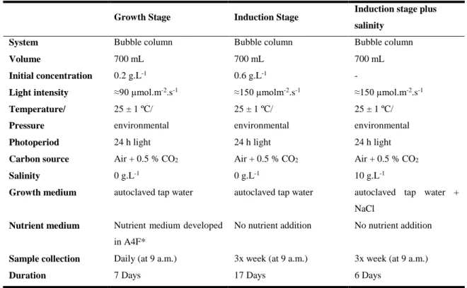
Table 1.3 – Composition of H. pluvialis biomass in green and red cultivation stages. Table 3.1 – Cultivation conditions for the 3 phases of the assay.
Table 3.2 – Emission and excitation of the filter, respectively. Table 3.3 – Basic components of Flow Cytometry.
Table 3.4 – Defined parameters to perform cell sorting. Table 4.1 – Average of weight per cell.
Table 4.2 – Total of chlorophylls and carotenoids in the vegetative stage in the first day of growth stage. Table 4.3 – Astaxanthin productivity in mg.g-1 DW.day-1 and pg.cell-1.day-1 after 17 days under stress conditions.
Table 4.4 – Average of weight per cell.
Table 4.5 – Astaxanthin global productivity in mg.g-1 DW.day-1, presented during 24 days.
Table 4.6 – Original culture vs culture from cell sort.
Table 4.7 – Growth rate and biomass productivity in the vegetative growth stage.
Table 4.8 – Total of chlorophylls and carotenoids in the vegetative growth stage in the first day. Table 4.9 – Maximum astaxanthin content, astaxanthin productivity and global productivity.
4 – RESULTS & DISCUSSION
XI
LIST OF EQUATIONS
Equation 3.1 – Determination of dry weight. Equation 3.2 – Beer-Lambert law.
Equation 3.3 – Determination of growth rate.
Equation 3.4 – Determination of volumetric biomass productivity. Equation 3.5 – Determination of astaxanthin productivity.
1 – INTRODUCTION
1
1 – INTRODUCTION
1.1. MICROALGAE OVERVIEW
1.1.1. Microalgae playing an important role
Microalgae are unicellular, colonial or filamentous, aquatic organisms that convert sunlight, nutrients and carbon dioxide into biomass via photosynthesis. Microalgae are one of the most primitive plants, consisting of the base of food chain of all aquatic ecosystems and the primary producers on earth. It can be grown almost anywhere, fresh water, salt-water and even on sewage and hypersaline waters. Around 200,000 species of microalgae are estimated to exist, but only a limited number, about 30,000, have been studied and analyzed (Mata et al., 2010; Sing and Saxena, 2015). Microalgae consist of a large and heterogeneous group of microorganisms, distinguished according the basic cellular structure, life cycle and pigment composition. The most important classes or categories of microalgae in terms of their abundance are: diatoms (Bacillariophyceae); green (Chlorophyceae); blue-green algae or cyanobacteria (Cyanophyceae); golden (Chrysophyceae); and red algae (Rhodophyceae) (Bharathiraja et al., 2015; Sing and Saxena, 2015; Vassilev and Vassilev, 2016).
Microalgae photosynthetic mechanism is similar to that of terrestrial plants, however, it has specific advantages over; it presents higher photosynthetic efficiency, since microalgae, during cellular metabolism, can convert more solar energy (4 - 7.5 %) than land plants (0.5 %). Moreover, microalgae present higher biomass production and faster growth, with growth rates of less than 24h. Microalgae also depend upon fewer resources than land plants, do not require fertile land or food crops, and processing consumes less energy than the land plants need (Raheem et al., 2015; Sing and Saxena, 2015). Although, its cultivation is very challenging, once variations in light, temperature, pH, salinity, qualitative and quantitative nutrient profiles, dissolved oxygen, among others, will affect the growth and the quality of microalgae. These conditions can be modified to accomplish high yields and reduce production costs (Tran et al., 2015).
To benefit the most from microalgae, since its isolation up to its large-scale production, several stages have to be studied, and microalgae potential is acquired step by step. The increasing concern for a better life quality, by consuming from natural sources and usage of renewable resources, is leading to a high investment in microalgae business and an increasing research for its biotechnological applications (Bharathiraja et al., 2015; Markou and Nerantzis, 2013; Vassilev and Vassilev, 2016).
1.1.2. Microalgae products & economic interests
Microalgae are a potential resource for biotechnological purposes as new sources of biomolecules such as pigments, lipids, carbohydrates and proteins. Microalgae in their natural environment, have adapted in order to inhabit a wide range of environmental conditions and habitats. Therefore, due to their variety of metabolic pathways, these microorganisms can produce an enormous diversity of compounds. Microalgae biomass from different strains can be processed and their active form from its compounds, such as pigments (e.g. β-carotene), antioxidants (e.g. astaxanthin), proteins (e.g. phycocyanin), and polyunsaturated fatty acids (e.g. omega-3, DHA, EPA) can be extracted to commercialize (Borowitzka, 2013; Demirbas, 2011).
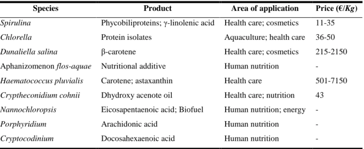
Although the current key commercial applications appear to be food additives and fuel, microalgae and its compounds have a tremendous range of applications. These may have advantages over synthetic products or products obtained from natural sources, but may have a cost disadvantage. These characteristics make them promising microorganisms with possible impact, on the chemical, pharmaceutical, cosmetic, energetic and on nutritional sectors (Table 1.1). The challenge in the application of microalgae for commercial purposes is to focus on these products with large market and profit, for which the use of microalgae is a clear competitive advantage (Milledge, 2010).
Table 1.1 – Examples of microalgae products and applications. (Adapted from Bharathiraja et al., 2015 and Enzing et al., 2014).
Species Product Area of application Price (€/Kg)
Spirulina Phycobiliproteins; γ-linolenic acid Health care; cosmetics 11-35
Chlorella Protein isolates Aquaculture; health care 36-50
Dunaliella salina β-carotene Health care; cosmetics 215-2150
Aphanizomenon flos-aquae Nutritional additive Human nutrition - Haematococcus pluvialis Carotene; astaxanthin Health care 501-7150 Cryptheconidium cohnii Dhydroxy acenote oil Health care; nutrition 43 Nannochloropsis Eicosapentaenoic acid; Biofuel Human nutrition; energy -
Porphyridium Arachidonic acid Human nutrition -
Cryptocodinium Docosahexaenoic acid Human nutrition -
The first report of human consumption of microalgae was in the 16th century with the harvest of
Spirulina (Arthrospira) from Lake Texcoco by the Aztec people, and latter of Lake Chad by Kanembu
population. During the natural Spirulina bloom, the populations collected and dried microalgae for later consumption as dried cakes. Nutritional properties of Spirulina showed an exceptionally high protein content, of the order of 60–70 % of its dry weight (Abdulqader et al., 2000; Ahsan et al., 2008). However, the industrial scale production of microalgae only began in the 1960s, in Japan with Chlorella production for human consumption. Chlorella vulgaris presents a total protein content up to 60% dry weight. It is considered to have a high protein nutritional quality according to the standard amino acid profile for human nutrition proposed by the World Health Organization (WHO) and the Food and Agricultural Organization (FAO) (Safi et al., 2014). This was followed in the 1970s by the commercialization of Spirulina, which is an excellent source of C-phycocyanin, followed in the 1980s by Dunaliella salina, source of β-carotene and later source of glycerol (Ben-Amotz and Avron, 1982; Spolaore et al., 2006) and astaxanthin from Haematococcus pluvialis in the 1990s (Lorenz and Cysewski, 2000). Thus, microalgae biotechnology industry has been growing and diversifying significantly.
However, the microalgae products currently on the market are still limited. The main limiting factor for the development of the markets is the production costs. The actual costs are related to the complexity of the cultivation phase and the downstream processes (extraction of the high-value compounds). The technical innovation and the market demand will result in further major advances and in an expansion of the commercially available products. Besides, efforts in improving the efficiency of systems and production operation are in progress to allow the cultivation of a larger diversity of microalgae. Nowadays, nutrition education programs could improve the microalgae products consumption. The main commercial product appears to be ‘‘health care’’ or “nutrition” that may produce health benefits, but may be subject to fashion and the current tendency. Also, to increase the microalgae products should be done a revision of the Novel Food Regulation. The complexity of the regulation on novel foods makes
1 – INTRODUCTION
3
it difficult the authorization of microalgae based products on the market (Milledge, 2010; Podola et al., 2016; Spolaore et al., 2006; Vigani et al., 2015).
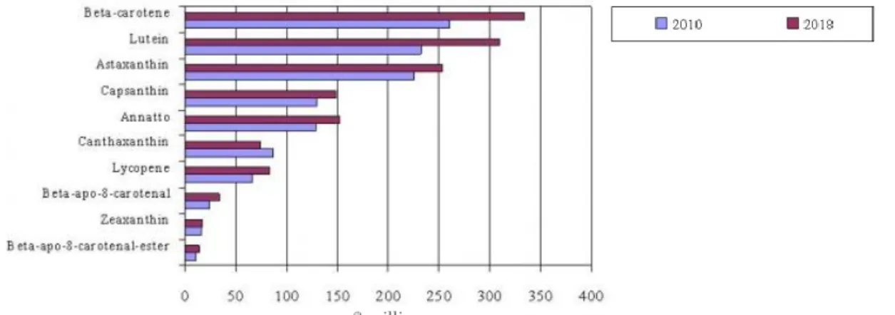
Alternative sources, as chemical synthesis products, are the major competitors of several of the microalgae products, especially carotenoids. According to BCC research press release (BCC research), the global carotenoids market generated about $1.2 billion in 2010, with the bulk of the carotenoids being produced by chemical synthesis (Borowitzka, 2013). In 2018, that value is projected to surpass $1.4 billion, increasing at an eight-year Compound Annual Growth Rate (CAGR) of 2.3 %. The global market for carotenoids comprise principally ten products: β-carotene, lutein, astaxanthin, capsanthin, annatto, canthaxanthin, lycopene, β-apo-8-carotenal, zeaxanthin, and β-apo-8-carotenal-ester (Figure 1.1).
The higher quality of microalgae compounds compared to the corresponding synthetic sources, is mainly due to their chemical conformation which is much more efficient than the synthetic variants. The products of high added value obtained from microalgae are subject to a range of rules and regulations affecting the production process. The concerns of production process are incremented when it is intended for human or animal nutrition. The alternative sources present a challenge to producers of microalgae-derived products, which have either to compete on price, or differentiate themselves from the synthetic source in the market place, in order to be able to be sold at a higher price. The manufacturing processes required to produce natural carotenoids are sophisticate and suffered a high development over the past years. Introduction of new manufacturing technologies is leading to a price reduction across most products allowing the preference for natural sources instead of synthetic. From this, biotechnology will play an important role in the near future, especially in production systems, as it can help to increase the productivity and reduce the production costs of micro-algae products (Borowitzka, 2013; Enzing et al., 2014; Guedes et al., 2011).
Figure 1.1 – Global carotenoids market value by product type. Estimative of evolution of carotenoid market from 2010 to 2018 ($ millions). Extracted from BCC Research.
1.2. ASTAXANTHIN PIGMENT
Nowadays there is an increased interest in biological active compounds derived from natural sources, especially the ones that can act on molecular targets, which are involved in some diseases. Astaxanthin (3,3’-dihydroxy-β,β’-carotene-4,4’dione) a compound of highly interest, has unique chemical properties based on its molecular structure, derived from lycopene. It is a hydrocarbon that contains two terminal ring systems joined by a chain of conjugated double bonds or poliene system (Figure 1.2) (Guerin et al., 2003). Its structure explains its unique chemical features, such as the ability to be esterified, a higher anti-oxidant activity and a more polar configuration than other carotenoids. Astaxanthin, a secondary carotenoid that results from secondary metabolism, belongs to the family of xanthophyll and it has a
stronger antioxidant activity when compared to β-carotene or α-tocopherol. It is proposed to be a super vitamin E and it can easily cross blood brain barrier in mammals, having proprieties that are believed to have a key role in the medicinal, pharmaceutical and food industries. (Goswami et al., 2010; Miki, 1991).
Of several naturally occurring carotenoids, astaxanthin is considered one of the best being able to protect cells, lipids and membrane lipoproteins against oxidative damage (Ambati et al., 2014). Numerous studies (Ambati et al., 2014; Guerin et al., 2003; Kidd, 2011; Yamashita, 2013; Yuan et al., 2011) have shown that astaxanthin has potential health-promoting effects in the prevention and treatment of various diseases, such as cancers, chronic inflammatory diseases, metabolic syndrome, diabetes, cardiovascular diseases, gastrointestinal diseases, liver diseases, neurodegenerative diseases, eye diseases, skin diseases, exercise-induced fatigue, male infertility, and renal failure.
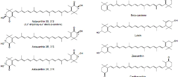
Figure 1.2 – Molecular structure of different carotenoids: the three stereoisomers of astaxanthin, β-carotene, lutein, zeaxanthin and canthaxanthin. Extracted from Guerin et al., 2003.
Astaxanthin shares many of the metabolic and physiological functions attributed to carotenoids, since it is closely related to other carotenoids, such as β-carotene, zeaxanthin and lutein (Figure 1.2) (Goswami
et al., 2010; Guerin et al., 2003). There are three stereoisomers for astaxanthin: two enantiomers (3R,
3’R and 3S, 3’S) and a meso form (3R, 3’S). Of all isomers, the 3S, 3’S is the most abundant in nature and different organisms produce astaxanthin in different stereoisomeric ratios. Esterified astaxanthin may increase biological activities especially since it can be easily absorbed into the metabolism, when compared to its free form. The stereoisomer 3S, 3’S in the esterified form (mono and di-esters) is predominantly found in Haematococcus pluvialis, while the 3R, 3’R stereoisomer in the unesterified form is found in Phaffia rhodozyma. Synthetic astaxanthin is produced as the unesterified xanthophyll and as a 1:2:1 mixture of the three stereoisomers: 3S, 3’S, 3R, 3’S and 3R, 3’R (Ambati et al., 2014; Higuera-Ciapara et al., 2006).
Astaxanthin is found in microalgae, microorganisms and aquatic animals, i.e. many types of seafood, including salmon, trout, red sea bream, shrimp and lobster, as well as in birds such as the flamingo and the quail. There are diverse natural sources of astaxanthin, such as microalgae Haematococcus pluvialis,
Chlorococcum, Chlorella zofingiensis, red yeast, Phaffia rhodozyma and bacteria, Paracoccus carotinifaciens. Haematococcus pluvialis is considered the richest source of natural astaxanthin (up to
1 – INTRODUCTION
5
6 % dry weight) as well as the best sources of astaxanthin for human consumption (Ambati et al., 2014; Lorenz, 1999; Olaizola, 2003; Yuan et al., 2010).
1.2.1. Biochemistry and biological activity of astaxanthin
An unavoidable consequence of aerobic metabolism is the production of reactive oxygen (ROS) and nitrogen (RNS) species. In microalgae, ROS are always formed by the leakage of electrons onto O2 from the electron transport activities of chloroplasts, mitochondria and plasma membranes or as a byproduct of various metabolic pathways. All ROS are extremely harmful to organisms at high concentrations and its enhanced production during environmental stresses can cause peroxidation of lipids, oxidation of proteins, damage of nucleic acids and enzyme inhibition, ultimately leading to cells death (Ambati et
al., 2014).
Due to the polyene chain, astaxanthin has an antioxidant activity by quenching single oxygen and scavenging radicals to terminate chain reactions. Specific physicochemical interactions of antioxidant compounds with membranes are responsible for their antioxidant properties and their biologic benefits, such as its transmembrane orientation which facilitates electron shuttling. The transmembraneous alignment of polar carotenoids provides exposure of the polar (hydrophilic) ends of the molecule to the internal cytoplasm and to the aqueous environment external to the cell (or the mitochondrial matrix and the intermembrane space of mitochondria), potentially facilitating electron transfer via the double bonds of the carbon scaffold of the compound (Figure 1.3) (Ambati et al., 2014; Kidd, 2011; Pashkow et al., 2008; Yuan et al., 2011).
1.2.2. Natural vs synthetic source of high value molecule: Astaxanthin
Natural pigments have pharmacological properties and have increased marketability advantages over synthetic products. The commercial production of natural carotenoids from microalgae is an eco-friendlier and safer approach than synthetic manufacturing by chemical processes (Aberoumand, 2011; Tuli et al., 2014). Currently, astaxanthin accounted for $226 million in 2010 and will be worth $253 million in 2018, a CAGR of 1.4 % (BCC Research). However, 95 % of this market consumes synthetic Figure 1.3 – Transverse cell membrane orientation of 3S,3S’ astaxanthin. The polar end groups overlap the polar boundary zones of the membrane, while the nonpolar middle fits the membrane’s nonpolar interior. The dashed red line speculatively indicates the conduction of electrons along the astaxanthin molecule, possibly to vitamin C or other antioxidants located outside the membrane. Extracted from Pashkow et al., 2008.
astaxanthin. Natural products make the synthetic pigments less desirables, since are derived from petrochemical sources raising issues related to food safety, pollution, and sustainability. Therefore, the chemical astaxanthin is only allowed to be used in aquaculture, not in human consumption or animal feed (Lemoine and Schoefs, 2010; Li et al., 2011; Lorenz and Cysewski, 2000). Due to consumers’ ability to differentiate between the benefits of natural pigments and hazardous effects of synthetic pigments have been greatly boosted the application of microbial pigments as food additive (Nigan and Luke, 2016).
There are three key areas where further improvements are required for a better implementation of microalgae products: (i) minimization of capital and operational costs; (ii) enhancement of cultivation efficiency; and (iii) astaxanthin isolation and purification. This will lead algae companies to successful commercial implementation. Since US Food and Drug Administration (FDA) granted "Generally Recognized As Safe" status (GRAS) astaxanthin from H. pluvialis, biotechnological innovation and continuous research are walking side by side to enhance microalgae production technology and strain improvement (e.g. genetic engineering), into a sustainable source of food/feed commodities, with enhanced yields of the desired products. (Bhosale, 2004; Pulz and Gross, 2004; Shah et al., 2016; Vigani
et al., 2015).
1.3. HAEMATOCOCCUS PLUVIALIS
Haematococcus pluvialis (hereafter referred to as H. pluvialis) is the microorganism used for the work
of this thesis.
1.3.1. Taxonomy, Morphology & Life cycle
H. pluvialis is a freshwater, unicellular, biflagellate green microalgae. Its scientific classification,
originally described by Flotow (1844), is presented in Table 1.2 (Algabase). Table 1.2 – Taxonomic classification of H. pluvialis Flotow. From Algabase.
Domain Eukaryota Kingdom Plantae Subkingdom Viridiplantae Phylum Chlorophyta Class Chlorophyceae Order Chlamydomonadales Family Haematococcaceae Genus Haematococcus
Species Haematococcus pluvialis
H. pluvialis is adapted to a diverse range of environmental and climate conditions, being distributed in
many fresh water habitats worldwide. It is capable of surviving in adverse conditions due to its ability to encyst, such as high light intensity, salt concentration, temperature, water availability and other adverse conditions (Proctor, 1957). This microalgae is frequently found in temperate regions around the world, like Europe, America and Africa (Pringsheim, 1966). However, it had been found to withstand adverse conditions revealing its presence at: low temperatures (4 - 10 °C), in Blomstrandhalvøya Island (Svalbard) (Klochkova et al., 2013); at high salinities (up to 25 ‰) on coastal rocks on Kost’yan Island, White Sea (Chekanov et al., 2014) and at a dried fountain near Rozhen village Blagoevgrad in Bulgaria (Gacheva et al., 2015).
1 – INTRODUCTION
7
Several ultrastructural changes occur during H. pluvialis life cycle which may be divided in four stages: (i) vegetative cell growth; (ii) encystment (vegetative to immature cyst cells); (iii) maturation (immature to mature cyst cells) and (iv) germination (mature cyst to vegetative cells) (Kobayashi et al., 1997a). In the life cycle four types of cells are produced (Figure 1.4): macrozooids (large, flagellated); microzooids (small, slender and flagellated); palmella forms (non-motile) and aplanospores (large, red hematocysts with a resistant cellulose wall). Cellular structure and chemical changes allow H. pluvialis, under nutritional and environmental factors, to transform from green flagellated cells into red cell, aplonospores, and vice versa (Hagen et al., 2002).
Figure 1.4 – Life cycle of H. pluvialis. a) Adult palmelloid cell; b) Aplanospore; c) cell division, microzoids released; d) Young palmelloid cell; e) Adult macrozoid; f) Young macrozoid; g) Palmelloid cell; h) cell division, microzoids and palmella cells released (Adapted from Elliot, 1934).
When a culture is performed with fresh medium, macrozooids (zoospores) predominate in the vegetative growth stage. Cells are spherical, ellipsoidal, or pear-shaped with two flagella of equal length emerging from anterior end, and a cup-shaped chloroplast with numerous, scattered pyrenoids (Kobayashi et al., 1997a). Macrozooid cells are between 8 and 20 μm long with a distinct gelatinous extracellular matrix with a median tripartite crystalline layer (Figure 1.5A) (Hagen et al., 2002). These cells might divide asexually, by mitosis, into 2 to 32 daughter cells (Figure 1.5B) (Wayama et al., 2013). As soon as environmental or culture conditions change inducting stress, macrozooids develop into a non-motile palmella form by losing their flagella while expanding the cell size. The transformation into palmella cells is characterized by the formation of a new two-layered amorphous, primary wall and simultaneously, the tripartite crystalline layer decomposition (Figure 1.5A) (Hagen et al., 2002). Through the continued environmental or cultural stress (e.g. nutrient starvation) the encystment process will continue. H. pluvialis turned into greenish-orange cells (Figure 1.5C), which can be referred as intermediate stage cells. In the aplanospore or cyst stage (Figure 1.5D) astaxanthin accumulates and cells form cysts. At this stage, further morphogenesis occurs. There is a formation of a voluminous multilayered cell wall, which enhances their tolerance against environmental impact (Damiani et al., 2006; Hagen et al., 2002). Along the transition to aplanospore cells, a large amount of astaxanthin is synthesized in lipid vesicles in the cytoplasm, in a way to storage carbon, energy and prevention from oxidative stress. The maturation of cysts is accompanied by the degradation of chloroplasts, remaining a low percentage that will play a role in the recovery when environmental conditions improve (Collins
et al., 2011; Li et al., 2008; Triki et al., 1997; Wayama et al., 2013). H. pluvialis has shown sexual and
asexual reproduction. However, little is known about its sexual life cycle. Triki et al., 1997, had reported that gametogenesis is seen when cultures are recovering from an induction period. Gametocyst may contain 32 or 64 gametes, designated microzoids, which are equal to asexual reproduction flagellated cells, despite their smaller size (10 µm) and rapid swim after release from gametocysts.
DIC Fluorescence A) Ma cr o zo o id B) a - C ell d iv is io n ; b -p alm ella ce ll C) Palm ello id / in ter m ed iate ce ll D) Ap lan o sp o re (cy st)
Figure 1.5 – Light (DIC) and fluorescent microscopy images of H. pluvialis life cycle. A) Green vegetative motile cell, macrozooid; B) Cell division and transformation of a motile cell into a palmella cell; C) Intermediate cell, beginning astaxanthin accumulation; D) Aplanospore cell, cyst with astaxanthin accumulation. In fluorescent imagens, green color correspond to chlorophylls and red color correspond to astaxanthin.
a
1 – INTRODUCTION
9
1.3.2. Biochemical composition of H. pluvialis
H. pluvialis composition changes according to the cell stage, vegetative or green and induction or red
stage. In green stage, protein content is higher than in red stage (Table 1.3) (Kobayashi et al., 1997a; Lorenz, 1999); the lipid content is divided in three types, the phospholipids, which are constant in both stages, neutral lipids and glycolipids that increase from green stage to red stage, and whose values will depend on the stress conditions and the used strain. In red stage, it is known that astaxanthin accumulation increases and there is an increase in the triacylglycerol (TAG) contents (Damiani et al., 2010; Saha et al., 2013; Zhekisheva et al., 2002). When exposed to stress, cells start to produce carbohydrates rather than fatty acids, however, with the continuous exposure to stress, carbohydrates are converted to fatty acids (Recht et al., 2012). Besides, secondary carotenoids are synthesized after exposure to environmental stress, in red stage. Primary carotenoids, such as chlorophyll, are replaced by the secondary carotenoid, mainly astaxanthin (Grewe and Griehl, 2008). Degradation of chloroplast coincides with the triacylglycerol (TAG) accumulation, while reducing membrane glycerolipids, especially those glycolipids making up the photosynthetic complexes and chloroplast membrane matrix (Gwak et al., 2014).
Table 1.3 – Composition of H. pluvialis biomass in green and red cultivation stages. n.d.: no data. Adapted from Shah et al., 2016.
Composition content (% of DW) Green stage Red stage
Proteins 29-45 17-25 Lipids (% of total) 20-25 32-37 Carbohydrates 15-17 36-40 Carotenoids (% of total) 0.5 2-5 β-carotene 16.7 1,0 Lutein 56.3 0,5 Zeaxanthin 6.3 n.d.
Astaxanthin (including esters) n.d. 81.2
Canthaxanthin n.d. 5.1
Chlorophylls 1.5-2 0
1.3.3. Astaxanthin biosynthesis in H. pluvialis
Under stress, H. pluvialis generate reactive oxygen species (ROS), such as H2O2, single oxygen (1O2), superoxide radicals (O2-), and hydroxide radicals (•OH). As a survival strategy to the unbalanced ROS generation, H. pluvialis induce astaxanthin accumulation, preventing damage on cellular components. Carotenoids act as accessory light-harvesting pigments, trapping light energy, protecting the photosystem from photo-oxidation by quenching ROS (Lemoine and Schoefs, 2010; Kobayashi et al., 1997b). Steinbrenner and Linden (2003), proven that general carotenoid biosynthesis is subject to photosynthetic redox control. The transfer of H. pluvialis cells from low-light conditions to moderate light intensity results in the reduction of the components of the photosynthetic electron transport including the plastoquinone pool. The plastoquinone pool acts as a redox sensor and its reduction subsequently leads to the transcriptional activation of genes involved in astaxanthin biosynthesis. Thus, redox regulation of genes involved in the synthesis of carotenoids is a prerequisite for the production of astaxanthin under stress conditions such as high light intensity, nutrient deprivation or ROS presence. When multiple stresses are applied simultaneously, different stress response mechanisms can be
activated, each one contributing to some extent to the overall cell protection and to improve carotenogenesis (Lemoine and Schoefs, 2010; Kobayashi et al., 1997b).
Biosynthesis of astaxanthin is a complex process that is highly up-regulated in conditions of stress and which coincides with the TAG accumulation, while reducing membrane glycerolipids, especially those glycolipids making up the photosynthetic complexes and chloroplast membrane matrix (Gwak et al., 2014). The biosynthesis of astaxanthin in H. pluvialis follows the general carotenoid pathway up to β-carotene formation. Astaxanthin synthesis might follow two main putative pathways (Figure 1.6): (i) the first pathway, starts with the β-carotene oxidation and have echinenone, canthaxanthin and adonirubin as intermediates; (ii) the second pathway, begin with the hydroxylation of carotene and have β-cryptoxanthin, zeaxanthin and adonixanthin as intermediates. These intermediates reveal the involvement of two enzymes β-carotene ketolase (BKT) and β-carotene hydroxylase (CrtR-b) in the conversion of β-carotene to astaxanthin. Although the specific steps of astaxanthin biosynthesis are carried out in the cytoplasm, the enzymes of the general carotenoid pathway appear to be localized in the chloroplast (Han et al., 2013; Lemoine and Schoefs, 2010; Shah et al., 2016; Vidhyavathi et al., 2008). Reported by Vidhyavathi et al. (2008) and Han et al. (2013), the preferential pathway for astaxanthin formation began with the oxidation of β-carotene. Vidhyavathi et al. (2008) demonstrated that the reduction in the BKT expression was reflected in the significant reduction of astaxanthin content. However, according to Gao et al. (2014) and Lemoine and Schoefs (2010), the two pathways can occur. In H. pluvialis most of astaxanthin molecules are accumulate in red stage cells as cytoplasmic lipid bodies. The majority of astaxanthin exists as fatty acid esters, usually mono- or diesters of palmitic (16:0), oleic (18:1), or linoleic (18:2; 18:3), behaving as stabilizers to maintain a high antioxidant ability. The esterification is required for the deposition within the non-polar matrix of lipid droplets (Han et al., 2013; Lemoine and Schoefs, 2010; Shah et al., 2016; Vidhyavathi et al., 2008).
Figure 1.6 – Pathway of astaxanthin biosynthesis in H. pluvialis. Enzyme abbreviations are as follows: BKT, β-carotene ketolase; CrtR-b, β-carotene 3,3′-hydroxylase. Adapted from Lemoine and Schoefs, 2010, Gwak et al., 2014 and Han et al., 2013.
1.3.4. H. pluvialis growth and astaxanthin accumulation requirements
Due to specificity of H. pluvialis strains, optimization of cultivation parameters is necessary to achieve high biomass productivity and successful astaxanthin accumulation (Domínguez-Bocanegra et al., 2004; Fábregas et al., 2000; Lu et al., 2010; Suyono et al., 2015). Conditions for vegetative growth of the
1 – INTRODUCTION
11
green microalgae H. pluvialis comprise a large number of parameters. The effects of light intensity, inoculum concentration, nutrient saturation, carbon dioxide concentrations, strain used, among others, must be properly combined to achieve the successful production of microalgae, maintaining the culture with high astaxanthin productivity (Aflabo et al., 2007; Domínguez-Bocanegra et al., 2004; Fábregas et
al., 2001; García-Malea et al., 2006; Sarada et al., 2002a).
Environmental factors are essential for H. pluvialis growth (green cells), such as: (i) temperature, about 20 to 28 °C (Wan et al., 2014); (ii) pH, between 7 and 8 (Borowitzka et al., 1991; Sarada et al., 2002b) since the intracellular microalgae pH is around 7; (iii) light intensity, ranging from 20 to 177 µmol.m -2.s-1 under continuous illumination or light–dark cycles (Boussiba, 2000; Domínguez-Bocanegra et al., 2004) have revealed themselves as crucial for microalgae development. The nutrients are one of the main factors that regulate morphological and physiological cellular responses of the microalgae, due to the impact on biochemical reactions, being the most important factor for the growth rate, composition, and high-value added products biosynthesis. The most important macronutrients for microalgae biomass production are carbon, nitrogen, phosphorus and sulfur.
These parameters are also adequate for induction stage when conjugated with stress factors (e.g. nutrient starvation and salinity) (Figure 1.7). Carotenogenesis may be fostered by low light intensities, 100 - 150 µmol.m-2.s-1 (Zhang et al., 2014), or by higher intensities that accelerate astaxanthin biosynthesis. An excess of light radiation (> 400 μmol.m–2.s–1) can be dangerous for microalgae viability due to ROS causing photoinhibition. However, the irradiation may become more efficient when conjugated with other stress factor, e.g. nutrient starvation, salt stress (Aflabo et al., 2007), addition of ethanol (Wen et
al., 2015), hormones (Lu et al., 2010), fulvic acid (Zhao et al., 2015), nuclear radiation (Cheng et al.,
2016) and many others factors that can be add to induce carotenogenesis in H. pluvialis (Forján et al., 2014; Sarada et al., 2002a; Su et al., 2014; Zhang et al., 2014).
In H. pluvialis, saline stress has been studied suggesting that it can replace light stress to induce carotenoid production; however, in several cases the microalgae growth decreased as NaCl concentration increased (Figure 1.7) (Benavente-Valdés et al., 2016). The effect of saline stress is largely studied by different autores (Aflabo et al., 2007; Boussiba and Vonshak, 1991; Borowitzka et
al., 1991), although the optimal NaCl concentration to induce astaxanthin accumulation varies.
However, is known that high NaCl concentration causes an increase in carotenoid content per cell (Tam
el al., 2012). In conclusion, H. pluvialis can respond to various stress conditions in different ways.
Whereas high light intensity leads to a transient response and to moderate accumulation of astaxanthin, the combination of various stress conditions such as high light intensity and salt stress is obligatory for encystment and the strong up-regulation of carotenoid genes (Steinbrenner and Linden, 2001).
Figure 1.7 – Schematic diagram showing impact of environmental and nutrient factors on lipid and carotenoid production. Extracted from Minhas et al., 2016.
1.3.5. Large scale production of H. pluvialis
H. pluvialis is capable of growing in photoautotrophic (Aflalo et al., 2007), heterotrophic (Zhang et al.,
2016), or mixotrophic growth (Kobayashi et al., 1992) conditions, indoors, in open raceway ponds or closed photobioreactors, in batch, in fed batch, or in continuous modes (Figure 1.8). As the optimal culture conditions for the production of biomass and accumulation of astaxanthin are not the same, two different strategies can be adopted in the production of H. pluvialis. One stage cultivation (Der Rio et
al., 2007) that consists in continuous cultivation of H. pluvialis under moderate nitrogen limitation and
specific average irradiance, resulting in simultaneous cell growth and astaxanthin accumulation; two-step cultivation (Fábregas et al., 2001) where the first stage aims to promote green vegetative growth under favorable culture conditions and in a second stage the cultures are submitted to stress factors in order to stimulate the transition to the aplanospore stage and the accumulation of astaxanthin. Nowadays,
H. pluvialis is produced in two-stages, the most recent advances in cultivation for astaxanthin production
include a two-stage mixotrophic culture system (Park et al., 2014) and attached cultivation system using the immobilized biofilm (Zang et al., 2014). Furthermore, each stage can be optimized for biomass growth and astaxanthin accumulation by adjusting independently the respective ratio of effective irradiance to cell density (Aflalo et al., 2007).
Li et al. (2011) estimated the production cost of astaxanthin, by his conceptually designed facility, to be $718/kg astaxanthin or about $18/kg biomass with 2.5 % astaxanthin. However, the cost is lower than the current industrial operations and is even lower than that of synthetic astaxanthin. The cost might even be able to be further reduced with the advances of technologies and optimization of processes. (Goswami et al., 2010; Lemoine and Schoefs, 2010; Li et al., 2011; Shah et al., 2016).
1 – INTRODUCTION
13
Technological advances are rapidly occurring in the microalgae-related industries. Top five leader commercial companies of H. pluvialis are: Cyanotech Corporation, USA; Mera Pharmaceutigals Inc., USA; Stazen Inc., USA; Valensa International, USA; Algatechnologies Ltd., Israel (Shah et al., 2016).
1.3.6. Challenges for the improvement of H. pluvialis
There are many challenges and problems in the development of large-scale production of H. pluvialis. As an example, a challenging task with H. pluvialis is its outdoor cultivation which involves curtailment of contamination (mainly by fungi Paraphysoderma sedebokerense (Strittmatter et al., 2015)) and control of environmental conditions such as light and temperature. Since it grows at neutral pH, contamination by bacteria, fungi and protozoa, is the main problem.
However, improvement of astaxanthin production yield must not be confined to optimization of culture conditions and systems of production. Mutagenesis and selection of mutants can be used as an approach to increase strain performance. Mutants can be obtained by physical mutagens such as ultraviolet radiation (UV) or X-rays and chemical mutagens such as ethyl methanesulphonate (EMS) or N-methyl-N-nitro-N-nitrosoguanidine (NTG) for enhancing the production of astaxanthin (Chen et al., 2003; Kamath et al., 2008; Tjahjono et al.,1994; Tripathi et al., 2001). UV induced random mutagenesis has the advantage of not being classified as a genetically modified method. However, genetic engineering of microalgae has been applied for more competitive pigment production (Forján et al., 2015; Sharon-Gojman et al., 2015).
Despite those innovative techniques, another approach for strain improvement, is based on the overproducers isolation by performing cell sorting coupled with flow cytometry (Doan and Obbard, 2012; Terashima et al., 2015). This methodology allows a characterization at unicellular level by evaluation near real-time of the physiological and metabolic states of cells, and is useful for the evaluation, control and optimization of bioprocesses. Flow cytometry associated with a cell sorting device, had shown significant potential in isolation of microalgae (Pereira et al., 2011; Hyka et al., 2013). Cell sorting had been applied to improve microalgae strains for lipid increase or for acquisition of axenic cultures (Cabanelas et al., 2015; Sensen et al., 1993; Terashima et al., 2015; Wahby et al., 2014; Xie et al., 2014). Although the flow cytometer is an expensive equipment, this technique presents many advantages, such as rapid, accurate, precise and real time acquisition of data, allowing cell enumeration, viability and fluorescence measurements.
Figure 1.8 – Two examples of cultivation systems used at industrial scale. Culture ponds (500 000 liters) at Cyanotech Corporation (HI, USA) (left image); H. pluvialis production in photobioreator at A4F (right image).
2 – OBJECTIVES
As already explained, among the important microalgae, H. pluvialis is the richest source of natural astaxanthin, which has important applications in the nutraceuticals, cosmetics, food and aquaculture industries. There are many challenges and problems for the development of large-scale production of biomass and astaxanthin from H. pluvialis.
The work performed for this thesis had as the main objective to improve H. pluvialis cultures to produce astaxanthin. For this purpose, seven H. pluvialis strains were screened and a characterization of microalgae was conducted throughout the cultivation process. This integrative characterization focused on the evaluation of: (i) the growth rate and biomass productivity during the green vegetative stage growth; (ii) astaxanthin accumulation throughout the induction stage under stress conditions (nutrient starvation and high light intensity) and under additional salinity stress. The parameters intended to be analyzed were: cell counting, dry weight and pigment analysis. The goal of the screening was to select strains with improved features or astaxanthin overproducers.
The fluorescence being derived from chlorophyll (autofluorescence) is a unique biomarker for photosynthetic organisms and enables setting a flow cytometric trigger for the separation of microalgae from other particles and microorganisms. To complement the screening, was also monitored by flow cytometry, the evolution of cytological and physiological properties (cell size, cell complexity and fluorescence) being derived from pigments (autofluorescence) of the H. pluvialis strains, at the beginning of the cultures, when each strain was separately inoculated into the growth media, and along the induction stage.
After the selection of strains with improved features, two other strategies were used for improvement of astaxanthin production capacity: UV induced random mutagenesis and separation of cells that differ in astaxanthin content, by flow cytometry equipped with a sorting device.
3 – MATERIALS & METHODS
15
3 – MATERIALS & METHODS
3.1. MICROORGANISM
For this thesis, seven strains of H. pluvialis from A4F – Algafuel, S.A. culture collection were analyzed. The code names for the strains used are HP_01 to HP_07.
3.2. CULTIVATION CONDITIONS
H. pluvialis cultivation for astaxanthin production was performed using a 3-steps strategy. Each strain
was prepared in duplicate and two independent repetitions were carried out, using for each one, different initial inoculum. The assays comprised three stages, started with a green vegetative growth stage, followed by an astaxanthin accumulation induction stage and ended with a stage of an additional salinity stress, whose aim was to promote further the astaxanthin accumulation. For production of H. pluvialis, some optimizations were adopted in order to grow them in the best conditions (Table 3.1).
In Table 3.1 are shown the cultivation conditions, during each stage. Bubble columns (700 mL) were used to grow all strains under the same conditions. The green vegetative growth stage lasted seven days, at about 90 µmol.m-2.s-1 of light intensity, and complete growth media was provided with all nutrients required. In the first stage of the assay, cultures were in optimal conditions for growth, sampling and cell counts were performed daily, while the determination of the dry weight and pigment analyses were carried out only at the beginning and end of this stage.
Induction stage lasted 17 days at 150 µmol.m-2.s-1 of light intensity, and no nutrients were supplied. The stresses applied were light intensity, which was controlled by the distance from the bubble columns to the cool white fluorescent lamps, and nutrient starvation. At this stage, samples were collected three times a week. Analytical methods, such as cell counting, flow cytometry, dry weight and pigment analyses, were realized whenever the sample collection was performed. At the end of induction stage, flow cytometry and cell sorting was applied to the strains of H. pluvialis that were selected by previous screening for having improved features.
In the additional stress, last stage, the culture salinity was adjusted to 10 g.L-1 of NaCl. Once again, sample collection was performed three times a week and the same analytical methods were carried out.