Universidade de Lisboa
Faculdade de Ciências
Departamento de Química e Bioquímica
Molecular characterisation of the poorly differentiated
and undifferentiated thyroid carcinomas using
genome-wide approaches
Jaime Miguel Gomes Pita
Doutoramento em Bioquímica
Especialidade em Genética Molecular
2013
Universidade de Lisboa
Faculdade de Ciências
Departamento de Química e Bioquímica
Molecular characterisation of the poorly differentiated
and undifferentiated thyroid carcinomas using
genome-wide approaches
Jaime Miguel Gomes Pita
Tese orientada pelo
Professor Doutor Valeriano Alberto Leite
(Instituto Português de Oncologia de Lisboa, Francisco Gentil)
e Professora Doutora Maria Luísa Cyrne
(Faculdade de Ciências da Universidade de Lisboa)
especialmente elaborada para a obtenção do grau de doutor
em Bioquímica, especialidade em Genética Molecular
A presente dissertação foi redigida de acordo com o disposto no
artigo 45.º do Despacho n.º 4624/2012 do Diário da República
2.ª série -‐ N.º 65 -‐ 30 de Março de 2012
* * *
As opiniões expressas nesta publicação são da exclusiva
responsabilidade do seu autor.
(...)
Eles não sabem que o sonho é vinho, é espuma, é fermento, bichinho álacre e sedento, de focinho pontiagudo, que fossa através de tudo num perpétuo movimento.
(...)
Eles não sabem, nem sonham, que o sonho comanda a vida. Que sempre que um homem sonha o mundo pula e avança
como bola colorida
entre as mãos de uma criança.
António Gedeão, Pedra Filosofal in ‘Movimento Perpétuo’ (1956)
‘The deeper we search the more we find there is to know, and as long as human life exists, I believe it will always be so.’
Agradecimentos
v
Agradecimentos
Na presente tese de doutoramento são apresentados os resultados do trabalho de investigação realizado no Grupo de Endocrinologia Molecular da Unidade de Investigação de Patobiologia Molecular (UIPM) no Instituto Português de Oncologia de Lisboa, Francisco Gentil (IPOLFG) entre Janeiro de 2009 e Dezembro de 2012, sob a orientação do Professor Doutor Valeriano Alberto Leite e co-orientação da Doutora Branca Maria Cavaco. Entre Janeiro de 2011 e Abril de 2012, o trabalho foi realizado no laboratório de Endocrinologia do “Institut de Recherche Interdisciplinaire en Biologie Humaine et Moléculaire” (IRIBHM) na “Université Libre de Bruxelles”, sob a co-orientação do Professor Doutor Jacques Emile Dumont. O trabalho esteve sob a orientação interna da Professora Doutora Maria Luísa Cyrne da Faculdade de Ciências da Universidade de Lisboa.
O trabalho de investigação teve o seu início durante o estágio curricular em Outubro de 2006, ao qual seguiu-se uma bolsa de investigação científica e o projecto de doutoramento. Assim, esta dissertação é fruto da colaboração e empenho de variadíssimas pessoas ao longo destes anos, as quais dedico o mais profundo apreço e gratidão.
Em primeiro lugar, agradeço ao Professor Doutor Valeriano Leite e à Doutora Branca Cavaco pelo acolhimento no seu grupo e pela oportunidade para despertar e contribuir para a Ciência. Agradeço a ambos, toda a contínua confiança e consideração que depuseram em mim. Ainda ao Doutor Valeriano, pelo estímulo científico, por toda a dedicação ao meu trabalho e pela amizade, bem como, pela oportunidade no laboratório do Professor Dumont. À Doutora Branca, um agradecimento especial pelo apoio, dedicação e disponibilidade constantes na minha aprendizagem e no rigor do trabalho científico. Também por toda a sua amizade, conhecimento científico, cultural e gastronómico, que com prazer partilhou.
À Professora Doutora Luísa Cyrne agradeço ter aceite a orientação do Doutoramento e pelo apoio e interesse despendidos para a concretização deste.
Ao Professor Dumont, por quem tenho grande admiração e respeito, por me ter recebido no seu grupo e partilhado a sua sabedoria, por todo o seu apoio, jovialidade e por ser um exemplo de dedicação à Ciência e à Endocrinologia.
A todos os membros que pertencem e aos que já passaram pelo grupo de Endocrinologia e pelos vários grupos de trabalho do UIPM, os quais me acompanharam e contribuíram para o agradável e simpático ambiente de trabalho. Ao antigo director do UIPM, o Doutor Sérgio Dias, por ter proporcionado todas as condições de trabalho necessárias, pelos conselhos, disponibilidade e simpatia. À Ana Banito, por me ter iniciado no mundo dos “microarrays”, por todo o apoio e amizade. À Inês Figueiredo, por todo o árduo trabalho e dedicação, sem os quais, esta tese perderia grande parte do seu valor. Agradeço também todo o apoio, amizade e simpatia e desejo-lhe muito sucesso na vida
vi
profissional. À Doutora Lúcia Roque do grupo de Citogenética, pela constante simpatia e por facultar as ‘preciosas’ linhas celulares. Às quatro meninas LHOs, que fizeram do nosso laboratório, um lugar sempre limpo, organizado, e claro, cheio de muita boa disposição. À Fátima e à Joaquina, pela amabilidade e carinho que sempre deram e por manterem o laboratório limpo e organizado. Ao Pedro Baptista, à Carla Espadinha, à Margarida Moura e à Ana Luísa Silva, pela vossa amizade e por estarem sempre prontos a ajudar. Em especial, às futuras Doutoras Rute Tomaz e “Jo” Costa, à Rita Domingues, à “maninha” Sofia Fragoso e à Cristiana Teixeira pela grande amizade e carinho que partilhamos dentro e fora do laboratório, bem como pela paciência para me “aturar”.
Agradeço ainda aos membros dos serviços do IPOLFG de Endocrinologia, de Cirurgia de Cabeça e Pescoço e de Anatomia Patológica, pela identificação, recolha, conservação e cedência dos casos clínicos, sem os quais, este trabalho de investigação não seria possível.
Aos membros do grupo de Endocrinologia do IRIBHM em Bruxelas, agradeço o caloroso acolhimento e a simpatia que todos ofereceram para me sentir em casa. Infelizmente, não poderei mencionar todos individualmente. À Professora Carine Maenhaut, investigadora principal do grupo, por todo o apoio e simpatia durante o tempo em que estive em Bruxelas. À Post-doc Soazig Le Pennec, que igualmente longe de casa partilhou o mesmo sentimento de saudade, agradeço toda a amizade, companheirismo e ajuda. Um agradecimento em especial, ao Sébastien Floor, pelo seu árduo trabalho e dedicação ao nosso projecto em conjunto, bem como, desde o primeiro momento, ter sido um verdadeiro amigo, sempre bem-disposto e altruísta.
Este trabalho não seria possível sem o imprescindível apoio financeiro da Fundação Calouste Gulbenkian, do Centro de Estudos de Doenças Crónicas (CEDOC) e da Sociedade Portuguesa de Endocrinologia, Diabetes e Metabolismo (SPEDM), as quais quero agradecer. Ainda, à Fundação para a Ciência e Tecnologia por me ter concedido a bolsa de Doutoramento (SFRH/BD/46096/2008).
Agradeço às minhas sempre grandes amigas de Faculdade “Burrólicas”, Andreia Freire, Rita Carilho, Sandra Brasil e Catarina Ramos, que apesar da vida nos ter separado, continuamos a nos encontrar e a nutrir um grande carinho.
Não posso deixar de agradecer à minha família. À minha madrinha Rosália por toda a esperança que deposita em mim, à minha mãe por acreditar sempre que serei o melhor, e à minha avó que apesar de não entender aquilo que faço, possui um orgulho inabalável em mim.
Por último, quero agradecer à minha futura esposa, pelo terno amor, carinho e paciência que me dedicou e continua a oferecer todos os dias. Obrigado por todo o apoio incondicional e por ter sempre acreditado em mim. Espero poder retribuir todo o afecto que dela recebo.
Acknowledgments
vii
Acknowledgements
This thesis presents the results of the experimental work performed at the Molecular Endocrinology lab in the Unidade de Investigação de Patobiologia Molecular (UIPM) at the Instituto Português de Oncologia de Lisboa, Francisco Gentil (IPOLFG), from January 2009 to December 2012, under the supervison of Professor Valeriano Alberto Leite and co-supervision of Dr. Branca Maria Cavaco. Between January 2011 and April 2012, the research work was undertaken at the Endocrinology lab in the “Institut de Recherche Interdisciplinaire en Biologie Humaine et Moléculaire” (IRIBHM) at the “Université Libre de Bruxelles”, under the co-supervision of Professor Jacques Emile Dumont. The work was supervised by Professor Maria Luísa Cyrne from the Faculty of Science of the Universidade de Lisboa.
The research work was initiated during the undergraduate internship in October 2006, followed by a research fellowship and the PhD project. This thesis is thus, the result of the collaboration of several people, along these years, who I devote all my gratitude.
Firstly, I thank Professor Valeriano Leite and Dr. Branca Cavaco for accepting me at their laboratory and for the opportunity to growth and contribute to Science. I am grateful to both, for all the confidence and consideration they have placed in me. To Professor Valeriano, for all the knowledge, support and friendship, as well as, for arranging the opportunity to be at Professor Dumont’s laboratory. To Dr. Branca, I express my deep gratitude for all the support, guidance and availability during my thesis. Also, for all the friendship and for the scientific, cultural and gastronomic knowledge that enthusiastically shared.
To Professor Luísa Cyrne, for accepting to supervise my PhD project and for all the support and interest in my work.
To Professor Dumont, who I deeply admire and respect, for receiving me in his research group and for sharing his wisdom, for all his support, joviality and for being an example of dedication to Science and Endocrinology.
To all former and current members of the Endocrinology group and research groups of UIPM, who contributed to the pleasant and friendly work environment. To Dr. Sérgio Dias, former director of UIPM, for providing all the work conditions, for the advices, availability and kindness. To Ana Banito, for introducing me to the microarray’s world, for all the help and friendship. To Inês Figueiredo, for all the hardworking and dedication. Her work was of crucial value to this thesis. I also thank all her friendship and kindness, and wish her a huge success in her career. To Dr. Lúcia Roque of the Cytogenetic group, for her gentleness and for providing the cell lines. To the Haematology group that always maintained our laboratory, a clean, organized and cheerful place. To Fátima e Joaquina for caring and maintaining the institute in proper conditions. To Pedro Baptista, Carla
viii
Espadinha, Margarida Moura and Ana Luísa Silva, for your friendship and assistance. To Rute Tomaz, Joana Costa, Rita Domingues, Sofia Fragoso and Cristiana Teixeira, a special appreciation for the deep friendship that we share inside and outside the lab, and also, for putting up with me.
I would like to acknowledge all the members of the Endocrinology, Surgery and Pathology services from IPOLFG, for the identification, collection, preservation and providing of the clinical cases, which were essential for this research work.
To the members of the Endocrinology group at IRIBHM in Brussels, I am sincerely grateful for your warm welcome and all your kindness. Please, forgive me for not being able to mention each and every one of you. I thank Professor Carine Maenhaut, Principal Investigator of the group, for the huge support and kindness during my stay in Brussels. To Postdoc Soazig Le Pennec, who was also far from home, I thank her for all the friendship, help and support she gave me. A special gratitude to my colleague Sébastien Floor, for his work and devotion to our research project, as well as, for being from the first moment, a truly great friend, always joyful and generous.
I would like to gratefully acknowledge the Calouste Gulbenkian Foundation, the Chronic Disease Research Centre and the Portuguese Society of Endocrinology, Diabetes and Metabolism for the financial support. Also, the Fundação para a Ciência e Tecnologia for awarding me, a PhD fellowship (SFRH/BD/46096/2008).
I thank to my dear friends from the time of faculty, who have taken separate ways, but still, find time to meet each other again.
I must also express my gratitude to my family. To my godmother for all the hope that she has in me, to my mother for always believing in my capabilities, and to my grandmother for all her proudness despite not understanding what I do.
Last but not least, I thank to my future wife, all the love, tenderness and patience that she has given and continues to give me. Also for all the unconditional support and for always believing in me.
This thesis is dedicated to the one who outlined my life, my father…
Sumário
ix
Sumário
O cancro da tiróide constitui a neoplasia mais comum do sistema endócrino (van der Zwan et al., 2012), representando nos homens e nas mulheres respectivamente, cerca de 1 e 3% dos casos de cancro, estimados em Portugal (Ferlay et al., 2010a) e no mundo (Ferlay et al., 2010b).
Os tumores com origem no epitélio folicular representam 90 a 95% das neoplasias da tiróide (Nikiforov and Nikiforova, 2011), e de acordo com as características morfológicas e clínicas, os tumores foliculares malignos são subdivididos em carcinomas bem-diferenciados (WDTC), carcinomas pouco diferenciados (PDTC) e carcinomas indiferenciados ou anaplásicos (ATC). Os tumores foliculares da tiróide representam assim, um modelo clássico do processo tumoral pelo qual uma célula epitelial possui potencial para originar diferentes tipos de tumores, cada um com características patológicas distintas.
Nos WDTC incluem-se os carcinomas papilares (PTC) e os carcinomas foliculares (FTC), sendo os PTC os mais frequentes, representando cerca de 80 a 90% dos casos (Kondo et al., 2006; Nikiforov and Nikiforova, 2011). Os WDTC são, em geral, tratados eficazmente com cirurgia e iodo radioactivo, e cerca de 90% dos doentes com menos de 45 anos ficam curados (DeLellis et al., 2004). Por outro lado, os PDTC e os ATC apresentam escassa diferenciação folicular e comportam-se de forma altamente agressiva. Em particular, os ATC apresentam uma progressão clínica muito rápida, e em geral, manifestam no diagnóstico inicial, uma extensa invasão dos tecidos adjacentes e metástases à distância, nomeadamente pulmonares, ósseas ou cerebrais (Muro-Cacho and Ku, 2000; DeLellis et al., 2004). Apesar dos ATC contribuírem apenas para 1 a 2% dos tumores da tiróide (Kondo et al., 2006; Nikiforov and Nikiforova, 2011), são responsáveis por 14 a 50% das mortes relacionadas com cancro da tiróide. Estão associados a uma sobrevivência média de 3 a 5 meses (DeLellis et al., 2004; Nagaiah et al., 2011), representando assim uma das neoplasias mais letais. Os PDTC apresentam características morfológicas e clínicas intermédias entre os WDTC e ATC (Lam et al., 2000; Volante et al., 2004; Sanders et al., 2007; Nambiar et al., 2011). A taxa média de sobrevida aos 5 anos, dos doentes com PDTC é de cerca de 50% e a mortalidade deve-se sobretudo a metástases pulmonares e ósseas (DeLellis et al., 2004; Nambiar et al., 2011). Os PDTC e em especial, os ATC, são refractários às formas convencionais de tratamento (cirurgia e iodo radioactivo). Na maioria dos casos, a ressecção dos tumores não é viável e a quimio- e a radioterapia demonstram um efeito reduzido na sobrevivência dos doentes (Sanders et al., 2007; Smallridge et al., 2009; Abate and Smallridge, 2011). Assim, é de
x
extrema importância a implementação de novas modalidades terapêuticas que sejam eficazes nestas neoplasias.
A transformação do epitélio folicular da tiróide resulta tipicamente de alterações genéticas que envolvem componentes das vias de sinalização MAPK-ERK e PI3K-AKT, essenciais na regulação da proliferação e homeostasia celular. Nos PTC detectam-se, de forma mutuamente exclusiva (Kimura et al., 2003; Nikiforov and Nikiforova, 2011), mutações activantes nos genes que codificam a cinase de serina/treonina BRAF e as GTPases H-, K- e NRAS, sendo a mutação do gene BRAF a alteração mais frequente (Kimura et al., 2003; Fugazzola et al., 2006). Entre os WDTC, as mutações nos genes RAS estão associadas a tumores com morfologia folicular, sendo detectadas na variante folicular dos PTC e em FTC (Vasko et al., 2003; Zhu et al., 2003; Di Cristofaro et al., 2006).
As características clínicas e histológicas dos carcinomas da tiróide sugerem uma contínua perda da diferenciação celular (um processo de desdiferenciação) pelo qual ocorre um aumento progressivo da agressividade. Considera-se que os PDTC e ATC possam surgir directamente a partir da célula folicular ou derivarem de tumores pré-existentes. Este último modelo é apoiado pela frequente detecção de áreas bem-diferenciadas em casos de PDTC e ATC (Nikiforova et al., 2003a; DeLellis et al., 2004; Quiros et al., 2005; Takano et al., 2007a; Wang et al., 2007a; Santarpia et al., 2008; Ricarte-Filho et al., 2009). Várias evidências moleculares sugerem, de igual modo, que os WDTC podem progredir para PDTC e para ATC. A detecção dos oncogenes RAS e BRAF em casos de PDTC e ATC (Garcia-Rostan et al., 2003; Smallridge et al., 2009; Volante et al., 2009) e a presença destas mutações em componentes diferenciadas e indiferenciadas de um mesmo tumor (Oyama et al., 1995; Nikiforova et al., 2003a; Begum et al., 2004; Cohen et al., 2004; Takano et al., 2007a; Costa et al., 2008; Schwertheim et al., 2009), sugerem um processo de progressão a partir de WDTC. Além disso, estas mutações podem coexistir com alterações no gene supressor de tumores TP53 (Lam et al., 2000; Quiros et al., 2005; Wang et al., 2007), no gene CTNNB1 que codifica o efector β-catenina da via WNT (Garcia-Rostan et al., 2001) e no PIK3CA que codifica a subunidade catalítica da cinase PI3K da via PI3K-AKT (Lam et al., 2000; Garcia-Rostan et al., 2001; Garcia-Rostan et al., 2005; Quiros et al., 2005; Hou et al., 2007a; Wang et al., 2007a; Liu et al., 2008; Santarpia et al., 2008; Ricarte-Filho et al., 2009). Estas alterações, que são detectadas exclusivamente, ou com maior frequência em PDTC e ATC, demonstram, assim, a acumulação e a cooperação de eventos específicos durante a desdiferenciação.
Sumário
xi
Os microRNA (miRNA), que com frequência se encontram desregulados no processo oncogénico (Calin and Croce, 2006), podem apresentar padrões de expressão anómalos nos tumores da tiróide (Nikiforova et al., 2009). Cada um destes RNA, que são não-codificantes, pode estar envolvido na regulação de centenas de transcriptos-alvo, mediando a sua degradação ou a repressão da translacção proteica (Carthew and Sontheimer, 2009). Assim, os miRNA podem constituir potenciais agentes terapêuticos, na medida em que afectam simultaneamente várias vias de sinalização, impedindo os mecanismos compensatórios das células tumorais (Lujambio and Lowe, 2012). Os níveis de expressão dos miRNA numa célula são o resultado tanto da sua biossíntese como da sua degradação. Enquanto que a via de biossíntese se encontra bem caracterizada (Cullen, 2006), o processo de degradação e o tempo de semi-vida dos miRNA, são relativamente desconhecidos. A determinação da estabilidade e a caracterização dos factores que influenciam a degradação destas moléculas, representam, portanto, áreas importantes para a investigação da potencial utilidade terapêutica dos miRNA.
A análise de expressão génica global no campo da oncologia demonstrou ser vantajosa no prognóstico e classificação dos tumores, assim como, na identificação de potenciais alvos terapêuticos (Chung et al., 2002; Nevins and Potti, 2007). Esta metodologia tem sido extensivamente utilizada na tiróide, especialmente no estudo dos diferentes subtipos de WDTC (Griffith et al., 2006; Riesco-Eizaguirre and Santisteban, 2007). No caso dos PDTC e ATC, a análise da expressão génica global em tumores primários encontra-se restrita a poucos estudos. A comparação dos perfis de expressão de ATC e PDTC com WDTC e tecido tiroideu normal (TN) permitiu identificar assinaturas moleculares relacionadas com a proliferação e ciclo celular, instabilidade cromossómica, adesão, motilidade celular e perda da função tiróidea (Salvatore et al., 2007; Montero-Conde et al., 2008; Hébrant et al., 2012).
Com o objectivo de elucidar os mecanismos envolvidos na progressão e na agressividade dos PDTC e ATC e identificar novos alvos para o tratamento destes tumores, compararam-se os perfis de expressão génica globais de amostras de TN e de WDTC com PDTC e ATC. Tendo em conta os resultados obtidos, procedeu-se a uma extensa pesquisa de mutações em genes envolvidos em processos celulares desregulados nos PDTC e ATC, e correlacionaram-se os dados obtidos com os aspectos clínico-patológicos dos doentes. Por fim, o estudo do turnover de RNA através da marcação com um análogo da uridina (4-tiouridina), foi usado para determinar os tempos de semi-vida de miRNA numa linha celular de carcinoma desdiferenciado da tiróide.
A comparação da expressão génica global entre 5 PDTC, 19 WDTC e 3 TN, e entre 5 ATC e 4 TN revelou que os PDTC e ATC exibem, em comum, genes desregulados com
xii
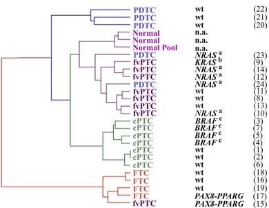
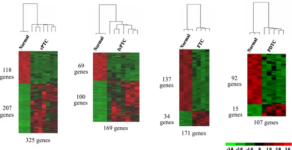
expressão aumentada associados ao ciclo celular, proliferação celular, segregação cromossómica e ao “checkpoint” do fuso mitótico, enquanto que, os genes sub-expressos estavam principalmente relacionados com a adesão celular. Os ATC apresentaram como eventos específicos (ausentes na análise dos PDTC), o aumento de expressão de componentes da via do TGF-β e a sub-expressão de genes associados à função e metabolismo tiroideu, à morfologia epitelial e às junções celulares. A análise não-supervisionada da semelhança génica entre as amostras e a correlação com a pesquisa de alterações do RAS e BRAF, demonstrou que as variantes foliculares de PTC são os precursores mais prováveis dos PDTC com mutação no gene RAS. Por outro lado, a comparação dos genes diferencialmente expressos em cada tipo de tumor em relação ao tecido normal, permitiu avaliar que os ATC partilhavam maior número de genes com os PTC clássicos do que com os restantes tumores, o qual suporta a provável origem dos ATC a partir dos PTC clássicos. A análise dos genes diferencialmente expressos entre cada tipo de tumor e o TN revelou que 307 de 494 (60%) dos genes eram sobre-expressos em PTC, enquanto que 137 de 171 (80%) estavam sub-expressos em FTC, 92 de 107 (86%) estavam sub-sub-expressos em PDTC e 983 de 1333 (74%) estavam com expressão diminuída nos ATC.
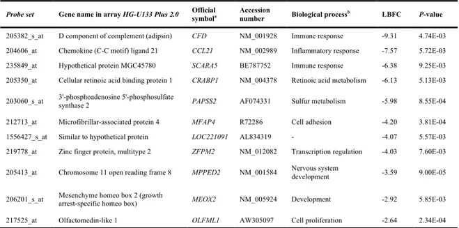
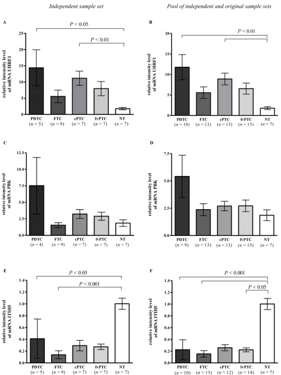
Validaram-se por RT-PCR quantitativo em grupos independentes de tumores, o gene UHRF1, associado à proliferação e identificado como sobre-expressos nos PDTC em relação ao tecido normal e o gene ITIH5, relacionado com a adesão celular e sub-expresso nos PDTC em relação ao tecido normal. Adicionalmente, caracterizou-se o gene CDKN3, o qual codifica um inibidor das cinases dependentes de ciclina (CDK) que foi identificado, na análise de expressão, como o gene mais sobre-expresso nos ATC, tendo-se confirmado a sua expressão aumentada em ATC, bem como em PDTC relativamente ao TN. De acordo com a sua função supressora de tumor, observou-se uma expressão significativa de formas de splicing aberrantes apenas nos ATC. Devido ao papel importante que a activação da via do TGF-β apresenta na promoção da transição epitelial-mesenquimal (EMT) (Huber et al., 2005), validou-se a expressão do gene SNAI2, regulador do EMT, e cuja expressão é induzida por esta via (Xu et al., 2009). Confirmou-se assim, a sobre-expressão específica deste gene em ATC.
Tendo em conta os resultados obtidos nos estudos de expressão génica global dos PDTC e ATC, procedeu-se a uma extensa análise mutacional em genes envolvidos na regulação do ciclo celular como os inibidores de CDK [CDKN1A (p21CIP1), CDKN1B (p27KIP1), CDKN2A
(p14ARF, p16INK4A), CDKN2B (p15INK4B) e CDKN2C (p18INK4C)], genes envolvidos na adesão celular (AXIN1, que codifica um regulador negativo da via WNT) e genes cujo envolvimento
Sumário
xiii
nestes tumores fora previamente descrito (H-, K-, NRAS, BRAF, PIK3CA, TP53 e CTNNB1). A pesquisa de alterações patogénicas em 26 ATC e 22 PDTC revelou que as mutações dos genes TP53 (presentes em 42% dos ATC e 27% dos PDTC) e RAS (presentes em 31% dos ATC e 18% dos PDTC) são as mais frequentes nestes tumores. As mutações nestes genes apresentaram mútua exclusividade (P=0,0354) e a sua presença estava associada a um menor tempo de sobrevida global dos doentes com PDTC e ATC (P=0,0383). No caso dos inibidores de CDK identificou-se, pela primeira vez, que alterações nos genes CDKN2A e CDKN2B podem estar envolvidos em cerca de 25% dos PDTC. Por outro lado, as mutações previamente descritas como frequentes em ATC, tais como BRAF, PIK3CA, AXIN1 ou CTNNB1 (Smallridge et al., 2009), foram identificadas em menos de 8% dos casos.
O método não-disruptivo de marcação de RNA com 4-tiouridina, foi optimizado e utlizado, pela primeira vez, no estudo da estabilidade dos miRNA. A incubação de uma linha celular de tumor desdiferenciado da tiróide (BCPAP), com 200 µM 4-tiouridina durante 24 horas, revelou não ser tóxico para as células, não afectar a expressão dos miRNA e permitiu marcar, purificar e quantificar os miRNA de forma precisa. Recorrendo a “arrays” de expressão de miRNA, obtiveram-se os tempos de semi-vida para 249 miRNA, cujo tempo médio foi de 2,5 dias, variando desde 22 horas (miR-208a e miR-107) até mais de 5,5 dias (miR-1321 e miR-320d). Estes dados revelam que os miRNA apresentam uma maior estabilidade que os RNA mensageiros (Yang et al., 2003; Friedel et al., 2009), sendo comparável à estabilidade proteica (Boisvert et al., 2012). O nosso estudo encontra-se em desenvolvimento mas, pretende-se identificar os factores determinantes para a degradação de miRNA. Em especial, a comparação destes dados com tempos de semi-vida determinados em células normais da tiróide, permitirá identificar miRNA cuja estabilidade se encontra desregulada no processo oncogénico.
Em suma, no presente trabalho identificaram-se várias vias e genes que poderão representar alvos para intervenção terapêutica, com impacto no seguimento clínico e na sobrevivência de doentes com tumores agressivos, como são os PDTC e ATC.
Palavras-‐chave
Carcinoma pouco diferenciado da tiróide; Carcinoma indiferenciado ou anaplásico da tiróide; Desdiferenciação; Expressão génica global; Mutações somáticas; Estabilidade dos microRNA
Abstract
xv
Abstract
Poorly differentiated (PDTC) and anaplastic thyroid carcinomas (ATC) are highly malignant tumours composed by dedifferentiated cells, for which current therapeutic options have been ineffective.
In the present project, the molecular signatures and genetic alterations associated with these tumours were elucidated, by using genome-wide expression analysis as first assessment. The role of the microRNA stability in thyroid tumourigenesis was also evaluated.
The comparison of the expression profiles between PDTC, well-differentiated thyroid carcinomas and normal thyroid tissues, and between ATC and normal thyroid tissues, revealed that PDTC and ATC have common deregulated signatures, indicative of cell adhesion impairment and increased cell cycle, proliferation and chromosomal instability. Additionally, ATC were specifically characterized by loss of epithelial and thyroid-related functions and activation of components from TGF-β pathway. The gene expression data suggested that follicular variants of papillary carcinomas were possible precursors of PDTC, whereas, ATC were more similar to classical papillary carcinomas. Validation by quantitative RT-PCR in independent sample sets, allowed to confirm the significant over-expression of UHRF1 (associated to proliferation) and downregulation of ITIH5 (associated to cell adhesion and invasion) in PDTC relatively to normal tissues. Moreover, CDKN3 gene, which encodes a cyclin-dependent kinase inhibitor, was shown to be significantly upregulated in PDTC and ATC relatively to normal thyroid, and in accordance with its suppressive function, CDKN3 was aberrantly spliced in ATC samples. The over-expression of the epithelial-to-mesenchymal transition factor SNAI2, which is induced by TGF-β, was significant in ATC.
Attending to the deregulated pathways identified, a mutational screening was undertaken in 26 ATC and 22 PDTC, which included the hot-spot regions of RAS, BRAF, TP53, CTNNB1 (β-catenin) and PIK3CA genes, and, for the first time, a comprehensive mutational analysis of cell cycle [CDKN1A (p21CIP1); CDKN1B (p27KIP1); CDKN2A (p14ARF, p16INK4A); CDKN2B (p15INK4B); CDKN2C (p18INK4C)], and cell adhesion regulators (AXIN1). Most mutations were identified in TP53 (42% of ATC; 27% of PDTC) and in RAS (31% of ATC; 18% of PDTC). The alterations in these genes were mutually exclusive (P=0.0354), and were associated with a lower patient survival (P=0.0383). CDKN2A and CDKN2B were found to be mutated, for the first time, in up to 25% of PDTC. Other known recurrent mutations in ATC (in BRAF,
xvi
PIK3CA, CTNNB1 and AXIN1 genes) were rarely detected, undermining their role in ATC development.
The study of the microRNA stability in thyroid tumourigenesis is still ongoing. Turnover rates were determined in a dedifferentiated thyroid carcinoma cell line (BCPAP), under standard conditions. A non-disruptive pulse-labelling method was applied and the decrease in microRNA expression along the time was quantified by two-colour arrays. These results represent the first report for an atlas of human microRNA half-lives. The determined average half-life for 249 microRNA was 2.5 days, a stability time more comparable to proteins than to mRNA. The comparison of these data with decay rates in normal thyroid cells will probably uncover miRNA for which stability is deregulated.
In conclusion, this project presents several genes and molecular events that were found deregulated in PDTC and ATC. More importantly, these represent potential therapeutic targets that can be used for the treatment of these highly aggressive tumours, in the near future.
Keywords
Poorly differentiated thyroid carcinoma; Anaplastic or undifferentiated thyroid carcinoma; Dedifferentiation; Genome-wide expression; Somatic mutations; microRNA stability
Table of contents
xvii
Table of contents
AGRADECIMENTOS V ACKNOWLEDGEMENTS VII SUMÁRIO IX PALAVRAS-‐CHAVE XIII ABSTRACT XV KEYWORDS XVI
TABLE OF CONTENTS XVII
LIST OF FIGURES AND TABLES XXI
CONVENTIONS XXIII
LIST OF ABBREVIATIONS XXIII
1
CHAPTER I
General Introduction
1. MOLECULAR ASPECTS OF THE CARCINOGENIC PROCESS 3
2. THE THYROID GLAND 7
2.1. ORGANOGENESIS AND DIFFERENTIATION 7
2.2. MOLECULAR BASIS OF THYROID MORPHOGENESIS 8
2.3. THYROID HORMONE METABOLISM AND FOLLICULAR CELL REGULATION 8
3. THYROID CANCER 11
3.1. INCIDENCE AND MORTALITY 11
3.2. RISK FACTORS 11
3.3. CHARACTERISATION OF FOLLICULAR CELL-‐DERIVED TUMOURS 12
3.3.1. WELL-‐DIFFERENTIATED THYROID CARCINOMAS 12
3.3.2. POORLY DIFFERENTIATED AND ANAPLASTIC THYROID CARCINOMAS 13
3.4. MULTI-‐STEP MODEL OF THYROID TUMOURS PROGRESSION 14
3.4.1. THE RAS-‐RAF-‐MEK-‐ERK AND PI3K-‐AKT SIGNALLING PATHWAYS 15 3.4.2. MOLECULAR ALTERATIONS INVOLVED IN THYROID CARCINOMA INITIATION 17
3.4.2.a. BRAF mutations 17
3.4.2.b. RET and NTRK1 rearrangements 18
3.4.2.c. N-‐, K-‐ and H-‐RAS mutations 19
3.4.2.d. PAX8-‐PPARG rearrangements 20
3.4.3. MOLECULAR ALTERATIONS INVOLVED IN DEDIFFERENTIATION 20
xviii
3.4.3.b. Loss of thyroid-‐specific functions 22
3.4.3.c. Further alterations in PI3K-‐AKT pathway 22 3.4.3.d. Alterations in cell cycle and in tumour suppressor p53 function 24 3.4.3.e. Deregulated epithelial morphology, E-‐cadherin and β-‐catenin proteins 26
4. REFERENCES 29
55
CHAPTER II
Gene expression profiling associated with the progression to poorly differentiated thyroid carcinomas
1. ABSTRACT AND KEYWORDS 57
2. INTRODUCTION 58
3. MATERIALS AND METHODS 59
4. RESULTS 63 5. DISCUSSION 70 6. ACKNOWLEDGEMENTS 73 7. REFERENCES 74 79 CHAPTER III
Cell cycle deregulation and TP53 and RAS mutations are major events in poorly differentiated and undifferentiated thyroid carcinomas
1. ABSTRACT AND KEYWORDS 81
2. INTRODUCTION 82
3. MATERIALS AND METHODS 83
4. RESULTS 88 5. DISCUSSION 95 6. ACKNOWLEDGEMENTS 100 7. REFERENCES 101 107 CHAPTER IV
Study of microRNA stability in thyroid cancer cells
1. ABSTRACT AND KEYWORDS 109
2. INTRODUCTION 110
3. MATERIALS AND METHODS 114
Table of contents
xix 5. REFERENCES 126 131 CHAPTER V Conclusion 1. FINAL DISCUSSION 133 2. THERAPEUTIC SIGNIFICANCE 138 3. REFERENCES 141 147 CHAPTER VI Annexes
1. LIST OF PRIMERS 148
2. SUPPLEMENTARY INFORMATION OF CHAPTER II 152
3. SUPPLEMENTARY INFORMATION OF CHAPTER III 158
4. SUPPLEMENTARY INFORMATION OF CHAPTER IV 169
List of figures and tables
xxi
List of figures and tables
Figure I.1 The hallmarks and enabling characteristics of cancer. 6
Figure I.2 Main steps in thyroid hormones biosynthesis. 10
Figure I.3 The MAPK-‐ERK and PI3K-‐AKT signalling pathways. 17
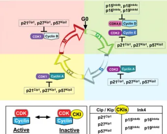
Figure I.4 Cell cycle regulation by cyclins, CDK and CDK inhibitors. 25
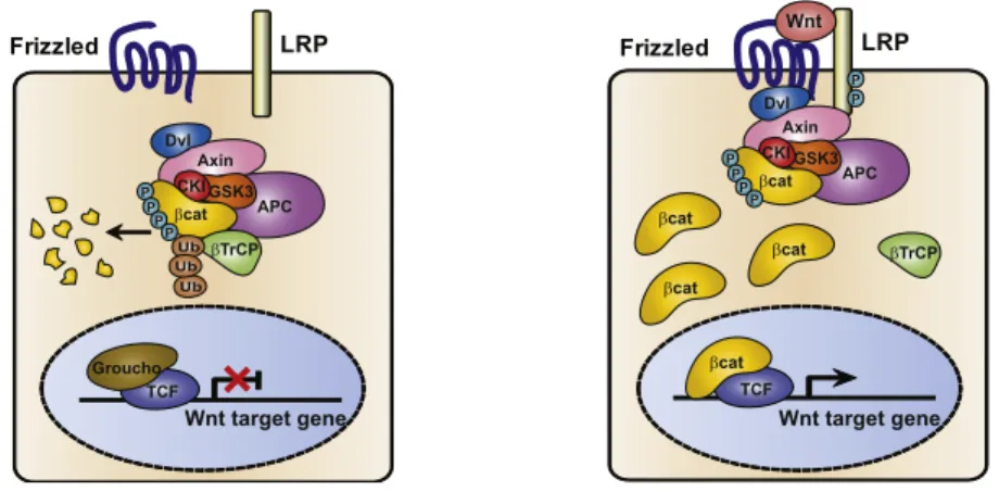
Figure I.5 The canonical WNT/β-‐catenin pathway. 27
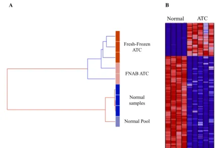
Figure II.1 Global gene expression similarity between samples using the unsupervised
hierarchical clustering method. 64
Figure II.2 Expression profile of the genes differentially expressed between tumours
and normal tissues. 65
Figure II.3 Expression of the UHRF1, PBK and ITIH5 genes in different tumour
histotypes, assessed by quantitative RT-‐PCR. 69
Figure III.1 Analysis of genome-‐wide expression in ATC. 88
Figure III.2 Characterisation of CDKN3 status in thyroid tumours. 91
Figure III.3 SNAI2 expression in different thyroid tumour histotypes and cell lines,
assessed by quantitative RT-‐PCR. 92
Figure III.4 Kaplan-‐Meier estimator for comparison of patients according to the
mutational results. 95
Figure III.5 Molecular alterations involved in the development of PDTC and ATC. 100
Figure IV.1 Schematic overview of the biogenesis and function of human miRNA. 113
Figure IV.2 Cell viability upon treatment with α-‐amanitin. 118
Figure IV.3 Qualitative analysis of primary miRNA expression levels after transcription
shutoff. 119
Figure IV.4 Quantitative RT-‐PCR analysis of 4 mature miRNA following transcription
blockage. 120
Figure IV.5 Quantitative RT-‐PCR analysis of 3 mRNA transcripts following transcription
blockage. 120
Figure IV.6 Cell viability after incubation with 4-‐thiouridine. 121
Figure IV.7 Quantification of 4 miRNA after 4-‐thiouridine-‐specific purification of
labelled RNA. 122
Figure IV.8 Representative scheme of the experimental design for miRNA half-‐lives
determination. 123
Figure IV.9 Graphical comparison of normalisation methods and differential miRNA
expression upon thiouridine labelling. 124
xxii
Figure V.1 Therapeutic approaches available for the identified molecular alterations. 140
Table I.1 Overall frequencies of the main gene alterations reported for PDTC and ATC. 28
Table II.1 Main characteristics of differentially expressed genes in poorly
differentiated tumours. 65
Table II.2 Main characteristics of differentially expressed genes in the four thyroid tumour histotypes versus normal thyroid tissues. 66
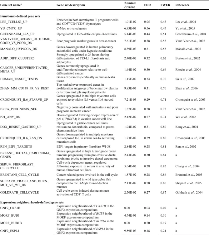
Table II.3 Gene sets enriched in the poorly differentiated versus the
well-‐differentiated groups. 67
Table III.1 Pathway impact analysis of differentially expressed genes between ATC
and normal tissues. 89
Table III.2 Functional profiling of differentially expressed genes between ATC
and normal tissues. 90
Table III.3 Frequency of mutated genes and association with histotype. 93
Table III.4 Comparison of survival distribution by logrank test. 94
Table IV.1 Expressions reported in the literature for miRNA differentially expressed
between ATC and normal thyroid tissues. 111
Table IV.2 Expressions reported in the literature for miRNA differentially expressed
between PDTC and normal thyroid tissues. 112
List of abbreviations
xxiii
Conventions
Human gene names and symbols are in accordance with the Human Genome Organization (HUGO) Gene Nomenclature Committee. Human protein names, shown at the first appearance in the text, are in accordance with the Universal Protein Resource (UniProt) and symbols match the corresponding gene symbol. Exceptions to these conventions are shown at the list of abbreviations. Gene names are presented at the first appearance in the text, inside quotation marks. Human gene symbols are italicised and all letters are in uppercase. Symbols not italicised refer to human proteins. Symbols with the first letter in uppercase and the remaining letters in lowercase indicate mouse/rat genes (in italics) and proteins (not italicised).
List of abbreviations
3’ Downstream
4sU 4-‐thiouridine
5’ Upstream
A Adenosine
ATC Anaplastic (or undifferentiated) thyroid carcinoma
C Cytidine
cAMP 3'-‐5'-‐cyclic adenosine monophosphate
CDK Cyclin-‐dependent kinase
CDKN Cyclin-‐dependent kinase inhibitor
cDNA Complementary DNA
CGH Comparative genomic hybridisation
CIP/KIP Cyclin-‐dependent kinase interacting protein/kinase inhibitory protein
CpG Cytosine and guanine joined by phoshodiester bond
cPTC Classical variant of papillary thyroid carcinoma
del Deletion
DNA Deoxyribonucleic acid
E Glutamic acid or glutamate
E-‐cadherin Cadherin-‐1
EDTA Ethylenediaminetetraacetic acid
xxiv
ERK Extracellular signal-‐regulated kinase
FDR False discovery rate
FFPE Formalin-‐fixed paraffin embedded
FNAB Fine-‐needle aspiration biopsy
fs Frameshift
FTA Follicular thyroid adenoma
FTC Follicular thyroid carcinoma
fvPTC Folicular variant of papillary thyroid carcinoma
FWER Family wise-‐error rate
G Glycine
G Guanosine
G-‐protein Guanine nucleotide-‐binding protein
G1 phase Gap 1 phase of cell cycle
GDP Guanosine 5'-‐diphosphate
Gq Guanine nucleotide-‐binding protein G(q)
GSEA Gene set enrichment analysis
Gsα Guanine nucleotide-‐binding protein G(s) subunit alpha
GTP Guanosine 5'-‐triphosphate
GTPase Guanosine 5'-‐triphosphatase
HEPES 4-‐(2-‐hydroxyethyl)-‐1-‐piperazineethanesulfonic acid
INK4 Inhibitors of CDK4
K Lysine
kDa Kilodalton
Log Logarithm
MAPK Mitogen-‐activated protein kinase
miRISC miRNA-‐induced silencing complex
miRNA MicroRNA
mRNA Messenger RNA
n Number of samples
NADPH Nicotinamide adenine dinucleotide phosphate reduced
p Shorter arm of chromosome
p53 Cellular tumor antigen p53
PBS Phosphate-‐buffered saline
PCA Principal components analysis
List of abbreviations
xxv
PDTC Poorly differentiated thyroid carcinoma
PI3K Phosphatidylinositol 3-‐kinase
PIP3 Phosphatidylinositol-‐3,4,5-‐trisphosphate
Pol RNA polymerase
pre-‐ Precursor
pri-‐ Primary
PTC Papillary thyroid carcinoma
q Longer arm of chromosome
Q Glutamine
R Arginine
RET/PTC Chromosomal rearrangements involving RET gene
RNA Ribonucleic acid
RNAse Ribonuclease
rRNA Ribosomal RNA
RT-‐PCR Reverse-‐transcriptase PCR
S phase Synthesis phase of cell cycle
SEM Standard error of the mean
T Thymidine
T3 Triiodothyronine
T4 Thyroxine
TGF-‐β Transforming growth factor beta
TRH Thyrotropin-‐releasing hormone
Tris Tris(hydroxymethyl)aminomethane
TRK Chromosomal rearrangements involving NTRK1 gene
TSH Thyrotropin or thyroid-‐stimulating hormone
UV Ultraviolet
V Valine
v/v Volume per volume
WDTC Well-‐differentiated thyroid carcinoma
wt Wild-‐type
X Termination codon
CHAPTER I
General Introduction
General Introduction
3
1. Molecular aspects of the carcinogenic process
Described since ancient times, the concept and research of cancer has greatly evolved over the centuries. The oncology field, along with the prolific scientific progression, has drastically changed over the last 200 years. One of the first pioneering steps was Rudolph Virchow’s concept over microscopic examination of tissues that permitted the general acceptance of cells being formed through division of pre-existing cells. Furthermore, Virchow deduced the origin of neoplasia, as growth of new tissue in the course of a pathological process through abnormal division of cells (Virchow, 1859/1863). Later on, the genetic basis of the disease was remarkably noticed by Theodor Boveri in 1914, who proposed that tumours were derived from cells that acquired chromosomal abnormalities (Boveri, 1914/2008). Among the environmental causes for tumour development (hormones, chemical carcinogens, radiation), viral-induced tumours played an extremely important role in cancer research. The first such virus (later on, named Rous sarcoma virus), was discovered in 1911 by Peyton Rous, who reported that cell-free filtrates from avian tumours (containing a “filterable agent”), could transmit the disease to other chickens (Martin, 2004). Research on this avian RNA tumour virus led to the identification of the first retroviral oncogene src (Martin, 1970; Bernstein et al., 1976; Wang et al., 1976), which had a functional cellular precursor (Stehelin et al., 1976; Spector et al., 1978) and protein (Collett et al., 1979; Oppermann et al., 1979) present in several vertebrates, including humans. These meant that normal components of human cells, proto-oncogenes like “v-src sarcoma (Schmidt-Ruppin A-2) viral oncogene homolog (avian)” (SRC), could become mutated and subsequently induce cancer formation/progression. This would set off the search for these cellular genes, culminating in the identification (Der et al., 1982; Parada et al., 1982; Santos et al., 1982) of the first human oncogene, “v-Ha-ras Harvey rat sarcoma viral oncogene homolog” (HRAS), altered by a single point mutation (Reddy et al., 1982; Tabin et al., 1982; Taparowsky et al., 1982). Since then, all three members of RAS family have been found mutated in several types of human cancers and are established as main regulators of the tumorigenesis process (Malumbres and Barbacid, 2003). Oncogenes have been found as excessively or inappropriately activated, promoting ultimately, unrestrained cell proliferation and neoplastic advantage. Oncogene alterations show a dominant trait normally affecting a single allele, and usually involve amplifications, missense mutations and chromosomal rearrangements. Epigenetic events (inheritance of information on gene expression levels by other means than DNA sequence modification) that include DNA methylation, histone modifications,
4
nucleosome remodeling and small non-coding regulatory RNAs, can also be involved in abnormal gene expression. Furthermore, not only mutations at genes controlling these mechanisms can induce epigenetic deregulation, but epigenetic changes of certain genes (like DNA repair genes) can lead to new somatic mutations (You and Jones, 2012).
By 1980s, another class of genes involved in tumorigenesis would be recognised. “retinoblastoma 1” (RB1) and “tumor protein p53” (TP53) were the first two identified tumour suppressor genes, as both alleles were frequently found inactivated in primary tumours (Cavenee et al., 1983; Friend et al., 1986; Fung et al., 1987; Lee et al., 1987; Baker et al., 1989), and wild-type (wt) genes were capable of supressing neoplastic transformation (Huang et al., 1988; Finlay et al., 1989; Baker et al., 1990). Moreover, the findings for RB1 gene confirmed the model previously formulated by Alfred Knudson, based on a two-hit hypothesis (Knudson, 1971), which proposed that retinoblastoma disease was caused by two mutational events. Thus, inactivation of both alleles of a tumour suppressor gene is normally required for tumorigenesis (contrarily to the dominant trait of oncogenes). The Knudson model would explain the mechanism of most tumour suppressor genes, especially in hereditary forms of cancer (Knudson, 2001). The recessive, loss-of-function alterations affecting tumour suppressors genes (responsible for keeping a normal cell proliferation and cell behaviour), includes large chromosomal deletions, missense or nonsense mutations and base insertions and/or deletions. DNA methylation at gene promoters is also recognized as a frequent inactivation mechanism (Jones and Laird, 1999).
Human cancer is believed to derive from a cell that through a multistep acquisition of different alterations is sequentially selected and expanded originating more malignant subclones. This model was initially proposed in 1976 (Nowell, 1976) and later on, was mechanistically applied to colorectal cancer (Fearon and Vogelstein, 1990). The availability of the human genome sequence (Lander et al., 2001; Venter et al., 2001) has allowed the generalization and confirmation of the tumorigenesis model by large-scale sequencing, by DNA copy number analysis and more recently, by next-generation sequencing. Tumours frequently present thousands of mutations and copy number changes (Stephens et al., 2005; Sjoblom et al., 2006; Wood et al., 2007; Parsons et al., 2008; Mardis et al., 2009; Shah et al., 2009; Kan et al., 2010; Lee et al., 2010; Pleasance et al., 2010; Stephens et al., 2012) and show coexistence of diferent cell subclones (Campbell et al., 2008; Anderson et al., 2011; Navin et al., 2011; Notta et al., 2011; Nik-Zainal et al., 2012; Shah et al., 2012), sometimes with convergent phenotypic evolution (Gerlinger et al., 2012), validating cancer as a truly
General Introduction
5
product of Darwinian evolution. The differential selection of each of these clones, as shown to greatly contribute for intra-tumour genetic heterogeneity (Navin et al., 2010; Gerlinger et al., 2012), chemoresistance (Jones et al., 2010; Diaz et al., 2012; Ding et al., 2012; Gerlinger et al., 2012; Misale et al., 2012), relapse (Hunter et al., 2006; Ley et al., 2008; Mullighan et al., 2008; Ding et al., 2012) and metastasis (Shah et al., 2009; Campbell et al., 2010; Ding et al., 2010; Yachida et al., 2010). Among the somatic variants randomly acquired throughout the genome, the majority (“passenger mutations”) do not influence the neoplastic process and thus, are not under selective pressure (Davies et al., 2005; Greenman et al., 2007; Mardis et al., 2009). However, other mutations confer advantage to the neoplastic clone and are termed “driver mutations” (Stratton, 2011). These are responsible for the defects in the regulatory circuits controlling normal cellular properties, providing the means that allow cancer cells to survive, proliferate, and disseminate.
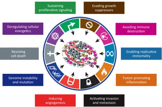
The “hallmarks” of cancer cells necessary for malignancy have been summarized (Figure I.1) (Hanahan and Weinberg, 2000; Hanahan and Weinberg, 2011), although these properties are widely interconnected and not all might be needed for a fully developed tumour. They may be transiently present over time and over different regions of the tumour and not necessary expressed by all neoplastic cells (Floor et al., 2012). For instance, tumour-associated stromal cells are normal cells but can greatly contribute for tumorigenesis. Cancer cells are thus accepted to acquire molecular changes in order to: constantly stimulate proliferation and gain independence from external growth stimuli; override the anti-proliferative cellular mechanisms, which is required to maintain the continuous proliferation; evade apoptosis, which is a major cellular response to neoplastic events (e.g., DNA damage, oncogene signals, insufficient survival factors, hypoxia and loss of adherence); confer limitless replicative potential (immortalization) by upregulation of telomerase; induce and sustain new blood vessels, for oxygen and nutrients supply; invade adjacent tissues, spread and colonize distant sites; reprogramme energy metabolism, mainly to support high glycolytic rates, which allows rapid generation of nucleosides and amino acids; and avoid elimination by immune cells.
Two mechanisms have been proposed as major inducers of the cancer hallmarks: Genomic/chromosomal instability and tumour-promoting inflammation. The former is acquired upon alterations of components involved in DNA damage repair, in genomic integrity surveillance or in chromosomal segregation, and leads to increased mutational rates, which fuels the neoplastic clones evolution to acquire new malignant properties.
6
Inflammation is a recent accepted enhancer of tumorigenesis, given that immune cells are capable of fostering angiogenesis, cell proliferation, and invasiveness, by supplying several survival, proangiogenic and growth factors.
Figure I.1 - The hallmarks and enabling characteristics of cancer (adapted from Hanahan and Weinberg, 2011). Ac#va#ng(invasion( and(metastasis( Inducing( angiogenesis( Resis#ng(( cell(death(
therapies. For example, the deployment of apoptosis-inducing drugs may induce cancer cells to hyperactivate mitogenic signaling, enabling them to compensate for the initial attrition triggered by such treatments. Such considerations suggest that drug development and the design of treatment protocols will benefit from incorporating the concepts of functionally discrete hallmark capabilities and of the multiple biochemical pathways involved in supporting each of them. Thus, in partic-ular, we can envisage that selective cotargeting of multiple core and emerging hallmark capabilities and enabling character-istics (Figure 6) in mechanism-guided combinations will result in more effective and durable therapies for human cancer.
CONCLUSION AND FUTURE VISION
We have sought here to revisit, refine, and extend the concept of cancer hallmarks, which has provided a useful conceptual framework for understanding the complex biology of cancer.
The six acquired capabilities—the hallmarks of cancer—have stood the test of time as being integral components of most forms of cancer. Further refinement of these organizing princi-ples will surely come in the foreseeable future, continuing the remarkable conceptual progress of the last decade.
Looking ahead, we envision significant advances during the coming decade in our understanding of invasion and metastasis. Similarly, the role of aerobic glycolysis in malignant growth will be elucidated, including a resolution of whether this metabolic reprogramming is a discrete capability separable from the core hallmark of chronically sustained proliferation. We remain perplexed as to whether immune surveillance is a barrier that virtually all tumors must circumvent, or only an idiosyncrasy of an especially immunogenic subset of them; this issue too will be resolved in one way or another.
Yet other areas are currently in rapid flux. In recent years, elab-orate molecular mechanisms controlling transcription through chromatin modifications have been uncovered, and there are
Figure 6. Therapeutic Targeting of the Hallmarks of Cancer
Drugs that interfere with each of the acquired capabilities necessary for tumor growth and progression have been developed and are in clinical trials or in some cases approved for clinical use in treating certain forms of human cancer. Additionally, the investigational drugs are being developed to target each of the enabling characteristics and emerging hallmarks depicted inFigure 3, which also hold promise as cancer therapeutics. The drugs listed are but illustrative examples; there is a deep pipeline of candidate drugs with different molecular targets and modes of action in development for most of these hallmarks.
668 Cell 144, March 4, 2011ª2011 Elsevier Inc.
Deregula#ng(cellular( energe#cs( Evading(growth( suppressors( Sustaining( prolifera#ve(signaling( Avoiding(immune( destruc#on( Enabling(replica#ve( immortality( Tumor?promo#ng( inflamma#on( Genome(instability( and(muta#on(
General Introduction
7
2. The thyroid gland
The thyroid gland is the largest endocrine organ in humans. It is located in the front of the trachea, below the cricoid cartilage and is composed of two lobes joined by a thin isthmus (Muro-Cacho and Ku, 2000). The gland is supplied by major arteries (Camacho et al., 2011) and displays a rich lymphatic network, which drains into several lymphatic nodes (Muro-Cacho and Ku, 2000). The main function of this endocrine gland is the production of thyroid hormones that are crucial for development and regulation of the body metabolism and homeostasis.
2.1. Organogenesis and differentiation
The embryonic endoderm is involved in the lining of two structures within the body, the digestive and the respiratory tubes. These share a common chamber in the anterior region of the embryo, the pharynx. Epithelial outpockets from these structure give rise to the thyroid, thymus, and parathyroid glands (Gilbert, 2000).
During human embryonic days 20 to 22, thyroid morphogenesis is initiated by a thickening of the endodermal epithelium in the primitive pharyngeal floor, know as thyroid anlage. The expansion, proliferation and migration of this structure originate the embryonic thyroid in the correct anatomical position and bilobed shape, at embryonic days 45 to 50. The migrated cells give rise to the most numerous cell population in the gland, the thyroid follicular cells. Progenitors from an embryologic structure derived from the neural crest (neuroectodermal origin) that fuses with the primordial thyroid, originate another distinct thyroid cell, the parafollicular or C cells, responsible for calcitonin production (De Felice and Di Lauro, 2004; Fagman and Nilsson, 2010; Braverman and Cooper, 2012).
Thyroid functional differentiation, involves onset of follicle formation, critical structures for storage and regulation of thyroid hormones, around embryonic day 70. For this, thyroid-specific genes (involved in thyrocytes function and thyroid hormone metabolism) are expressed in concerted timing (Szinnai et al., 2007). Iodine uptake and thyroxine hormone (T4) production occur by the tenth to the fourteenth gestational week, during the final stages
8
2.2. Molecular basis of thyroid morphogenesis
Thyroid follicular cell commitment is already hallmarked, at the molecular level, during the formation of the thyroid anlage. Despite being expressed by other embryonic tissues, only thyroid anlage and the derived thyroid primordium express simultaneously 4 transcription factors: homeobox protein NKX-2.1 (NKX2-1), formerly called TTF-1 for thyroid transcription factor-1 (Civitareale et al., 1989; Francis-Lang et al., 1990; Guazzi et al., 1990; Lazzaro et al., 1991); forkhead box protein E1 (FOXE1), formerly known as TTF-2 for thyroid transcription factor-2 (Sinclair et al., 1990; Francis-Lang et al., 1992a; Zannini et al., 1997); paired box protein Pax-8 (PAX8) (Plachov et al., 1990; Poleev et al., 1992); and hematopoietically-expressed homeobox protein HHEX (HHEX) (Bedford et al., 1993; Thomas et al., 1998). The study of knockout mice has demonstrated the crucial role of these factors in the survival and migration of the thyroid cells precursors during embryogenesis (De Felice and Di Lauro, 2004; Fagman et al., 2011). Besides being required during early stages, presence of these factors is a specific and essential feature of adult mature follicular cells, for proper thyroid-specific expressions and functions (Zannini et al., 1992; Damante and Di Lauro, 1994; Pellizzari et al., 2000; De Felice and Di Lauro, 2011).
The hormone released by pituitary, thyrotropin or thyroid-stimulating hormone (TSH), is the main regulator of thyrocytes. However is not involved in early thyroid development and in agreement, TSH is only detectable in the serum approximately at the twelfth gestational week (Thorpe-Beeston et al., 1991). The thyrotropin receptor (TSHR) is a member of G-protein-coupled receptors, with 7 transmembrane domains (Parmentier et al., 1989), whose mice mRNA expression is first detected following migration and before follicles formation. TSH signalling during embryogenesis is only required for proper functional differentiation of cells, since mutant mice and human infants with impaired TSHR exhibit a properly localized gland, but are profoundly hypothyroid (Brown, 2004; De Felice et al., 2004).
2.3. Thyroid hormone metabolism and follicular cell regulation
The functional unit of the thyroid gland is the follicle, formed by a single layer of follicular cells, surrounding a lumen filled with colloid (Figure I.2). This proteinaceous substance contains mainly thyroglobulin (TG), the protein secreted by follicular cells and essential for thyroid hormones formation. The first key steps in synthesis of thyroid hormones involve active transport of iodide across the basolateral membrane into the thyrocyte,