Faculdade de Desporto da Universidade do Porto
Characterization of upper limbs movements of healthy
and poststroke adults
(Caracterização dos movimentos dos membros superiores de adultos saudáveis e pós acidente vascular cerebral)
Thesis submitted in fulfilment of the requirements for the degree of Doctor in Physiotherapy by Faculty of Sport of the University of Porto,
under the terms of Decree-Law nº 74/2006 24th March.
Thesis prepared under the supervision of:
Prof. Doctor Cláudia Isabel Costa da Silva (supervisor)
Department of Physiotherapy, School of Health Sciences, Polytechnic of Porto
Prof. Doctor Ana Rita Vieira Pinheiro (co-supervisor)
Department of Physiotherapy, School of Health Sciences, University of Aveiro
Prof. Doctor Miguel Velhote Correia (co-supervisor)
Department of Electrical and Computer Engineering, Faculty of Engineering, University of Porto
Inês Albuquerque Mesquita
This thesis involved the following entities: Faculty of Sport of the University of Porto (FADEUP); Biomechanics Laboratory of the University of Porto (LABIOMEP); School of Health Sciences, Polytechnic of Porto (ESS - P. Porto); Center for Rehabilitation Research (CIR); Centro Hospitalar Universitário São João, Centros Hospitalares of Porto and Vila Nova de Gaia/Espinho, and Unidade Local de Saúde de Matosinhos.
Mesquita, I. A. (2019). Characterization of upper limbs movements of healthy and poststroke adults. (PhD). Biomechanics Laboratory of the University of Porto (LABIOMEP), School of Health Sciences, Polytechnic of Porto (ESS - P. Porto) and Center for Rehabilitation Research (CIR).
KEYWORDS: STROKE; UPPER EXTREMITIES; MOTOR RECOVERY;
ACADEMIC THESIS
Laboratório de Biomecânica do Porto (LABIOMEP) Faculdade de Desporto
Universidade do Porto
Centro de Investigação em Reabilitação (CIR) Escola Superior de Saúde
Politécnico do Porto
Copyright © 2019 by Mesquita I. A.
All rights reserved. You may copy, distribute, display, and perform the work under the following conditions: Attribution: You must attribute the work in the manner specified by the author or licensor; Non-commercial: You may not use this work for commercial purposes; No Derivative work: You may not alter, transform, or build upon this work.
Notice: For any reuse or distribution, you must make clear to others the license terms of this work. Any of these conditions can be waived if you get permission from the copyright holder.
The web addresses cited in this text were current as of September 2019, unless otherwise noted.
Illustrations: Mesquita I. A. Printer: FADEUP.
Doctoral thesis in Physiotherapy. Porto, 2019
“The important thing is not to stop questioning. Curiosity has its own reason for existing.”
V
ACKNOWLEDGMENTS
I would like to express my gratitude to:My supervisor, Prof. Cláudia Silva, for her continuous support, understanding attitude and encouragement throughout the project, for sharing her expertise in upper limb performance in poststroke subjects and scientific production and for the learning opportunities offered.
My co-supervisor, Prof. Ana Rita Pinheiro, for sharing her expertise in scientific production and neurorehabilitation, and for the meaningful contribution to improve the quality of the scientific articles produced.
My co-supervisor Prof. Miguel Velhote for the receptiveness to interdisciplinary knowledge sharing and contribution to scientific production.
The Laboratory Technician of LABIOMEP, Eng. Pedro Fonseca, for his endless availability for knowledge sharing and support in the recording, processing and analysis of kinematic data, for his critical spirit and significant contribution to scientific production.
Research Assistant of LABIOMEP, Márcio Borgonovo-Santos, for his meticulous critical analysis and expertise on statistics.
The Physiotherapy PhD. students from FADEUP, Fellipe Lima and Edgar Ribeiro that contributed to this project through their support in the data analysis.
Neurology service directors of the hospital centers involved, Dr. Elsa Azevedo, Dr. Joaquim Pinheiro, Dr. Manuel Correia and Dr. Vitor Tedim Cruz, and neurologists, Dr. Ana Campolargo, Dr. Gabriela Lopes, Dr. Miguel Veloso, Dr. Pedro Castro and Dr. Raquel Rocha, for their receptivity and availability to collaborate in the recruitment of poststroke patients and for their contribution to scientific production.
All subjects that participated in the developed studies, without whom these would not have been possible.
VI
The Functional Sciences Department Coordinator, Prof. Pedro Monteiro, and the vice-coordinator, Prof. Cláudia Barrias, from ESS-PP, for their constant support and encouragement.
My colleagues, for their experience, support and encouragement. My patients, for inspiring me every day to do more and better.
My family and friends, for their enthusiastic encouragement and friendship. My parents for their unconditional support and love.
VII
STATEMENT OF ORIGINALITY
I hereby certify that all the work described in this thesis is the original work of the author. Any published (or unpublished) ideas, techniques, or both from the work of others are fully acknowledged by the standard referencing practices.
Inês Albuquerque Mesquita
IX
ETHICAL DISCLAIMER
Ethical approval for the studies mentioned in this thesis has been granted by the Ethics Committee of Faculdade de Desporto da Universidade do Porto, (process: CEFADE 08.2016- Appendix I), by the Hospital Ethics Committee (Appendix I) and by the Comissão Nacional de Proteção de Dados (Appendix I).
All subjects who participated in the studies were free from any physical impairment and signed an informed consent form (Appendix II). All participants were fully informed about the nature and objectives of the studies (Appendix II).
XI
LIST OF PUBLICATIONS
Journals
1- Mesquita, I. A., Pinheiro, A. R. V., Velhote Correia, M. F. P., & Silva, C. I. C. (2019). Methodological considerations for kinematic analysis of upper limbs in healthy and poststroke adults. Part I: A systematic review of sampling and motor tasks. Topics in Stroke Rehabilitation, 26 (2), 142-152. doi:10.1080/10749357.2018.1551953.
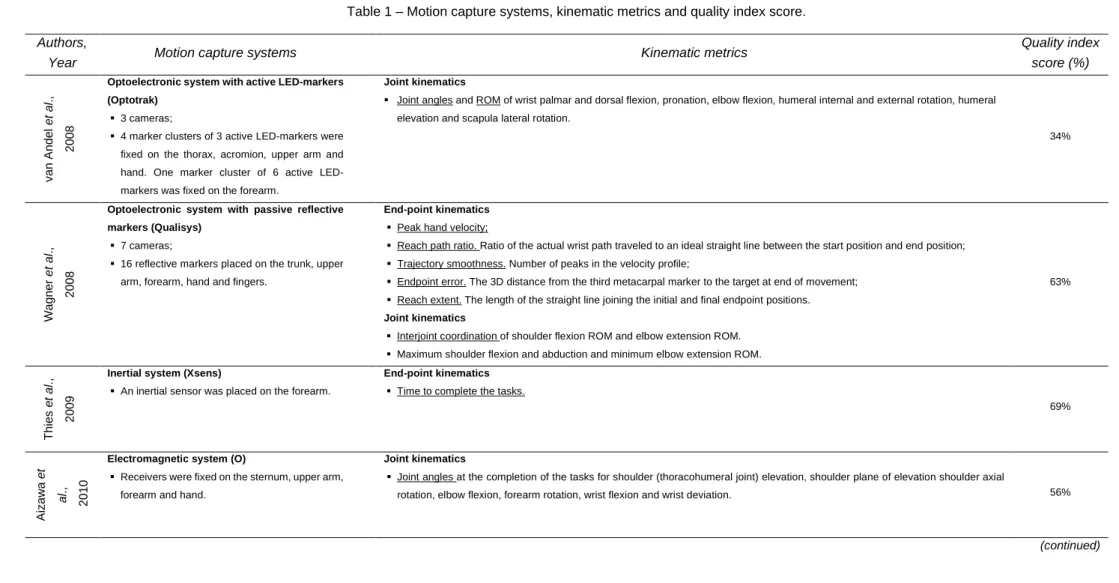
2- Mesquita, I. A., Fonseca, P. F. P., Pinheiro, A. R. V., Velhote Correia, M. F. P., & Silva, C. I. C. (2019). Methodological considerations for kinematic analysis of upper limbs in healthy and poststroke adults Part II: a systematic review of motion capture systems and kinematic metrics. Topics in Stroke
Rehabilitation, 26 (6): 464-472. doi:10.1080/10749357.2019.1611221
3- Mesquita, I. A., Fonseca, P. F. P., Borgonovo-Santos, M., Lima, F. B., Pinheiro, A. R. V., Velhote Correia, M. F. P., & Silva, C. I. C. (2019). A comprehensive kinematic characterization of the drinking task performed by healthy adults. Manuscript Submitted for Publication.
4- Mesquita, I. A., Fonseca, P. F. P., Borgonovo-Santos, M., Ribeiro, E., Pinheiro, A. R. V., Velhote Correia, M. F. P., & Silva, C. I. C. (2019). Comparison of upper limb kinematics in two activities of daily living with different handling requirements. Manuscript Submitted for Publication.
5- Mesquita, I. A.,Fonseca, P. F. P., Borgonovo-Santos, M., Campolargo, A.,
Castro, P., Lopes, G., Rocha, R., Pinheiro, A. R. V., Velhote Correia, M. F. P., & Silva, C. I. C. (2019). Kinematic analysis of both upper limbs after stroke: a case series. Manuscript Submitted for Publication.
XIII
CONTENTS
LIST OF TABLES ... XV LIST OF FIGURES ... XVII LIST OF APPENDICES ... VI ABSTRACT ... VIII RESUMO ... X LIST OF ABREVIATURES ... XII
1. INTRODUCTION ... 1
1.1 The global burden of stroke... 1
1.2 Motor impairment of both upper limbs after stroke ... 2
1.3 Motor recovery after stroke ... 3
1.4 Motor performance quality measurement ... 4
1.4.1 Kinematic analysis ... 5
1.4.2 Importance of kinematic analysis for Physiotherapy in stroke rehabilitation ... 8
1.5 Thesis objectives ... 9
2. THESIS ORGANISATION ... 11
3. DESCRIPTION OF THE WORK DEVELOPED ... 13
4. METHODOLOGICAL CONSIDERATIONS ... 17
4.1. Systematic review ... 17
4.2. Study designs and participants ... 17
4.3. Motion capture system ... 20
4.4. Normalization of the experimental setup to anthropometric characteristics ... 20
4.5. Upper body biomechanical model ... 21
4.6. Kinematic metrics ... 22
4.7 Statistical analysis ... 24
5. ACCEPTED AND SUBMITTED ARTICLES ... 27
Article I - Methodological considerations for kinematic analysis of upper limbs in healthy and poststroke adults. Part I: A systematic review of sampling and motor tasks ... 29
Article II - Methodological considerations for kinematic analysis of upper limbs in healthy and poststroke adults Part II: a systematic review of motion capture systems and kinematic metrics ... 53
Article III - A comprehensive kinematic characterization of the drinking task performed by healthy adults ... 73
XIV
Article IV – Comparison of upper limb kinematics in two activities of daily
living with different handling requirements ... 105
Article V – Kinematic analysis of both upper limbs after stroke: a case series ... 131
5. GENERAL DISCUSSION ... 171
6. CONCLUSIONS AND FUTURE WORK PERSPECTIVES ... 179
7. RELEVANT CONTRIBUTIONS TO OTHER SCIENTIFIC PROJECTS ... 181
8. REFERENCES ... 183 APPENDIX I – Ethical approval ... CXCIII APPENDIX II – Informed consent form ... CC APPENDIX III – Sample characterization questionnaires ... CCVI APPENDIX IV – Fugl-Meyer Assessment for Upper-Extremity ... CCXII APPENDIX V – International Physical Activity Questionnaire... CCXIII
XV
LIST OF TABLES
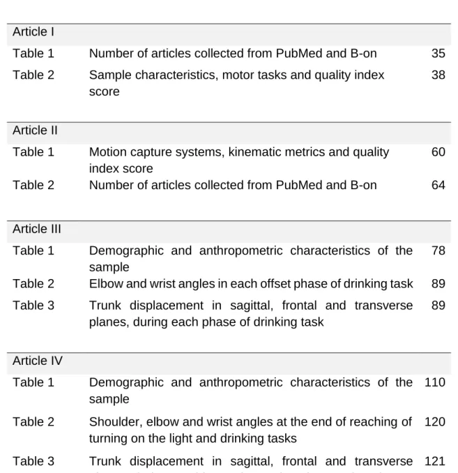
Article ITable 1 Number of articles collected from PubMed and B-on 35
Table 2 Sample characteristics, motor tasks and quality index
score
38
Article II
Table 1 Motion capture systems, kinematic metrics and quality
index score
60
Table 2 Number of articles collected from PubMed and B-on 64
Article III
Table 1 Demographic and anthropometric characteristics of the
sample
78
Table 2 Elbow and wrist angles in each offset phase of drinking task 89
Table 3 Trunk displacement in sagittal, frontal and transverse
planes, during each phase of drinking task
89
Article IV
Table 1 Demographic and anthropometric characteristics of the
sample
110
Table 2 Shoulder, elbow and wrist angles at the end of reaching of
turning on the light and drinking tasks
120
Table 3 Trunk displacement in sagittal, frontal and transverse
planes, during reaching and returning phases of turning on the light and drinking tasks
121
Article V
Table 1 Demographic, anthropometric and stroke characteristics
of the cases
138
Table 2 End-point and joint kinematics of both upper limbs of
studied patients in T1 and T2 and the respective reference 145
XVII
LIST OF FIGURES
Description of the work developedFigure 1 Thesis organization diagram 13
Methodological considerations
Figure 1 Healthy sample diagram 18
Article I
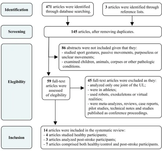
Figure 1 Review selection and exclusion criteria 36
Article II
Figure 1 Review selection and exclusion criteria 65
Article III
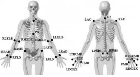
Figure 1 Anatomical marker set used to model the pelvis, trunk
and upper limbs
79
Figure 2 Experimental setup of drinking task 80
Figure 3 An example of phases definition of drinking task 82
Figure 4 Metrics obtained from end-point and joint kinematic
data and the characteristic of movement they represent
83
Figure 5 Normalized time history of hand’s tangential velocity
during drinking task and respective end-point kinematics
86
Figure 6 Normalized time history of shoulder angle during
drinking task and respective joint kinematics
87
Figure 7 Normalized time history of elbow and wrist angles
during drinking task and respective joint kinematics
88
Article IV
Figure 1 Experimental setup of turning on the light and drinking
tasks
XVIII
Figure 2 An example of phases definition of turning on the light
task
113
Figure 3 Metrics obtained from end-point and joint kinematic
data and the characteristic of movement they represent
115
Figure 4 Normalized time history of hand’s tangential velocity
and end-point kinematics
117
Figure 5 Normalized time history of shoulder, elbow and wrist
angles during turning on the light and drinking tasks
119
Article V
Figure 1 Some of the main kinematic alterations of Patient 1 in
T1 and T2
144
Figure 2 Some of the main kinematic alterations of Patient 2 in
T1 and T2
153
Figure 3 Some of the main kinematic alterations of Patient 3 in
T1 and T2
155
Figure 4 Some of the main kinematic alterations of Patient 4 in
T1 and T2
157
Figure 5 Some of the main kinematic alterations of Patient 5 in
T1 and T2
159
Appendix I
Figure 1 Ethical approval of CEFADE CXCIII
Figure 2 Ethical approval of Centro Hospitalar do Porto CXCIV
Figure 3 Ethical approval of U. L. S. - Matosinhos CXCV
Figure 4 Approval of Comissão Nacional de Proteção de
Dados
CXCVI
Figure 5 Ethical approval of Centro Hospitalar de São João CXCVIII
Appendix II
Figure 1 Study information delivered to healthy participants CC
Figure 2 Study information delivered to poststroke participants CCII
XIX
Figure 4 Informed consent delivered to poststroke participants CCV
Appendix III
Figure 1 Healthy sample characterization questionnaire CCVI
Figure 2 Poststroke participants characterization
questionnaire
VI
LIST OF APPENDICES
Appendix IEthical approval CXCIII
Appendix II
Informed consent form CC
Appendix III
Sample characterization questionnaires CCVI
Appendix IV
Fugl-Meyer Assessment for Upper-Extremity CCXII
Appendix V
VIII
ABSTRACT
Introduction: Upper limb (UL) motor impairment affects numerous poststroke
survivors worldwide and its recovery is slow and complex. Evidence of bilateral impairment after stroke is growing, which creates the need to have a healthy reference for the quality of motor performance instead of ipsilesional UL data. Currently, kinematic analysis is considered one of the best ways to improve the understanding about the mechanisms that drive motor recovery, but a set of methodological flaws is hampering this knowledge. Aims: To characterize the ULs movement of healthy and poststroke adults, through kinematic analysis, during the performance of drinking and turning on the light tasks. Methods: 63 healthy adults and 5 poststroke patients were eligible to perform drinking and turning on the light tasks with both ULs. Poststroke patients were assessed in early sub-acute phase and in the beginning of chronic phase. Tasks movements were captured by a 3D motion capture system, end-point and joint kinematics were analysed and comparisons between tasks and healthy and poststroke adults were made. Results: Drinking task has five phases with different motor skills and kinematic strategies that were mainly influenced by age and sex. Turning on the light has a lower handling requirement, when compared to drinking. The different target formats and the different interaction with them seemed to be responsible for differences in kinematic strategies between both tasks performed by healthy adults. Differences were found between the kinematic strategies used by poststroke adults and those of healthy adults. All poststroke patients presented bilateral kinematic alterations in both tasks.
Conclusion: A comprehensive analysis of kinematic strategies of drinking and
turning on the light were made, in order to obtain a reference of the performance of activities of daily living with different handling requirement for poststroke adults. All studied patients showed bilateral kinematic alterations, which supports the implementation of a bilateral assessment and the need to have a healthy reference for the quality of motor performance. Initial severity of stroke and patients’ age appear to have been the most important information to explain the extent of kinematic alterations, but stroke location seemed to have conditioned the specificity of deficits as well as the recovery.
KEY WORDS: STROKE; UPPER EXTREMITIES; MOTOR RECOVERY; MOTOR
X
RESUMO
Introdução: O comprometimento motor do membro superior afeta muitos sobreviventes pós-AVC em todo o mundo e a sua recuperação é lenta e complexa. A evidência de comprometimento bilateral após AVC está a crescer, levando à necessidade de desenvolver uma referência saudável para a qualidade do desempenho motor, em vez dos dados do membro superior ipsilesional. Objetivos: Caracterizar o movimento dos membros superiores de adultos saudáveis e pós-AVC, através da análise cinemática, durante o desempenho das tarefas “beber” e “acender a luz”. Métodos: 63 adultos saudáveis e 5 pacientes pós-AVC foram elegíveis para desempenhar as tarefas “beber” e “acender a luz” com os dois membros superiores. Os pacientes pós-AVC foram avaliados no início da fase sub-aguda e no início da fase crónica. Os movimentos das tarefas foram captados por um sistema de captura de movimento 3D, variáveis cinemáticas da mão e articulares foram analisadas e foram feitas comparações entre tarefas e entre adultos saudáveis e pós-AVC. Resultados: A tarefa beber teve cinco fases com diferentes habilidades motoras e estratégias cinemáticas que foram influenciadas principalmente pela idade e pelo sexo. Acender a luz tem menor exigência manual, quando comparada com o beber. Os formatos diferentes dos alvos e a interação diferente parecem ser responsáveis por diferenças nas estratégias cinemáticas entre as duas tarefas executadas pelos adultos saudáveis. Foram encontradas diferenças entre as estratégias cinemáticas usadas pelos adultos pós-AVC e as usadas pelos adultos saudáveis. Todos os pacientes pós-AVC apresentaram alterações cinemáticas bilaterais em ambas as tarefas. Conclusão: Foi feita uma análise abrangente das estratégias cinemáticas das tarefas beber e acender a luz, de modo a obter uma referência do desempenho de atividades da vida diária com diferentes exigências de manualidade para adultos pós-AVC. Todos os pacientes estudados apresentaram alterações cinemáticas bilaterais, o que suporta a implementação de uma avaliação bilateral e a necessidade de ter uma referência saudável para a qualidade do desempenho motor. A severidade inicial do AVC e a idade dos pacientes parecem ter sido as informações mais importantes para explicar a extensão das alterações cinemáticas, mas a localização do AVC parece ter condicionado a especificidade dos défices, bem como a recuperação. PALAVRAS-CHAVE: ACIDENTE VASCULAR CEREBRAL; MEMBROS SUPERIORES; RECUPERAÇÃO MOTORA; AVALIAÇÃO DA QUALIDADE DA PERFORMANCE MOTORA; ANÁLISE CINEMÁTICA.
XII
LIST OF ABREVIATURES
% Percent < Smaller than > Greater than ° Degrees 3D Three dimensionalADL Activities of daily living
ARAT Action Research Arm Test
BMI Body Mass Index
C7 Processus spinous of the 7th cervical vertebra
cm Centimetres
CNS Central Nervous System
cUL Contralesional Upper Limb
DRINK Drinking
FMA-UE Fugl-Meyer Assessment Scale for Upper Extremity
ICF International Classification of Functioning
IJ Incisura jugularis
iUL Ipsilesional Upper Limb
kg kilogram
LAC Middle part of left acromion
LASIS Left anterior superior iliac spine
LIGHT Turning on the light
LINDEX Distal phalange of left índex
LLELB Lateral epicondyle of left humerus
LLH Lateral side of the head of the second left metacarpal
LMELB Medial epicondyle of left humerus
LMH Medial side of the head of the fifth left metacarpal
LPSIS Left posterior superior iliac spine
LRAD Styloid process of left radius
LTHUMB Distal phalange of left thumb
XIII
m Meters
MANOVA Multifactorial Analysis of Variance
MAS Modified Ashworth Scale
mm Millimeters
NIHSS National Institute of Health Stroke Scale
NMU Number of Movement Units
PRISMA Preferred Reporting Items for Systematic Reviews and
Meta-Analysis PROCESS
PX Processus xiphoideus
RAC Middle part of right acromion
RASIS Right anterior superior iliac spine
RINDEX Distal phalange of right índex
RLELB Lateral epicondyle of right humerus
RLH Lateral side of the head of the second right metacarpal
RMELB Medial epicondyle of right humerus
RMH Medial side of the head of the fifth right metacarpal
ROM Range of Motion
RPSIS Right posterior superior iliac spine
RRAD Styloid process of right radius
RTHUMB Distal phalange of right thumb
RULN Styloid process of right ulna
s Seconds
SD Standard deviation
SRRR Stroke Recovery and Rehabilitation Roundtable
T1 Early sub-acute phase after stroke
T2 Beginning of chronic phase after stroke
UL Upper Limb
WHO World Health Organization
1
1. INTRODUCTION
1.1 The global burden of stroke
Stroke is classically characterized as a neurological deficit attributed to an acute focal injury of the central nervous system (CNS) by a vascular cause, including cerebral infarction, intracerebral hemorrhage, and subarachnoid hemorrhage, and is a major cause of disability and death worldwide (Sacco et al., 2013). It is estimated that stroke affects 17 million people in the world each year (S.A.F.E., 2017), of which 1.1 million occurs in Europe (Bejot, Bailly, Durier, & Giroud,
2016). At the turn of the 21st century, stroke incidence in Portugal was among the
highest in Western European countries: 305/100,000 in rural and 269/100,000 in urban populations (Correia et al., 2004). However, over the last two decades, in Portugal and across Europe, there has been a reduction in the proportion of people having a stroke, due to advances in care quality during acute phase and in primary prevention (Bejot et al., 2016; Correia et al., 2017). Although global stroke incidence is declining, rates observed in young adults are on the rise and because of the ageing population, the absolute number of stroke is expected to dramatically increase in coming years (Bejot et al., 2016).
Most patients with stroke survive the initial illness, and a large proportion of them remain with significant impairments (Peter Langhorne, Bernhardt, & Kwakkel, 2011; Peter Langhorne, Coupar, & Pollock, 2009). The most common and widely recognized impairment caused by stroke is motor impairment, which can be regarded as a loss or limitation of function in muscle control or movement or a limitation in mobility (Peter Langhorne et al., 2009). Motor impairment after stroke typically disturbs the control of movement of the face, upper limb (UL) and leg, and affects about 80% of patients (Peter Langhorne et al., 2009). Consequently, one of the greatest health effects for patients, their families and the economy results from the long-term physical consequences of stroke (Brewer, Horgan, Hickey, & Williams, 2013; Peter Langhorne et al., 2011). Therefore, the recovery and the return to a full life following stroke become the main goals for stroke survivors, caregivers and health professionals (Peter Langhorne et al., 2009; Walker et al., 2017). The focus of stroke rehabilitation,
2
and in particular the work of physiotherapists, is on the recovery of impaired movement and the associated functions (Peter Langhorne et al., 2009).
UL motor recovery seems to be particularly slower and more complex than
that of the lower limb, which could result from thegreater emphasis placed on
retraining gait and mobility in an effort to mobilize the patient as quickly as possible and to minimize costly hospital stays (Duncan et al., 1994). Another plausible explanation is that, unlike lower limbs, ULs perform a wide range of activities of daily living (ADL) involving varied interactions with diverse objects and a complex multi-joint coordination (Levin, Kleim, & Wolf, 2009).
1.2 Motor impairment of both upper limbs after stroke
Motor impairment can be caused by ischaemic or haemorrhagic injury to the motor cortex, premotor cortex, motor tracts, or associated pathways in the cerebrum or cerebellum (Warlow et al., 2008). Motor impairment of contralesional UL (cUL) is particularly worrying as more than 80% of stroke survivors experience acute sensorimotor dysfunction of this limb (Cramer et al., 1997), which becomes chronic for 50% of the patients (Kwakkel, Kollen, van der Grond, & Prevo, 2003). UL dysfunction is responsible for limitation of activities (disability) and reduced participation (handicap) of survivors in everyday life situations (Peter Langhorne et al., 2009), such as feeding, dressing, grooming and handwriting, which affects severely their quality of life (Godwin, Ostwald, Cron, & Wasserman, 2013).
Contralesional impairment increase reliance on the ipsilesional UL (iUL) for function and independence (Wetter, Poole, & Haaland, 2005). However, increasing number of studies (Bustren, Sunnerhagen, & Alt Murphy, 2017; Desrosiers, Bourbonnais, Bravo, Roy, & Guay, 1996; Metrot et al., 2013; Noskin et al., 2008; Nowak et al., 2007; Sunderland, Bowers, Sluman, Wilcock, & Ardron, 1999; Wetter et al., 2005) have reported deficits in iUL after a unilateral stroke.
Several bilateral neural mechanisms may be behind the dysfunction of iUL. A dominant theory suggests that the ipsilesional uncrossed descending corticospinal pathways may play a role in the movement of iUL. In fact, approximately 10% to 15% of the corticospinal pathways from cortex to distal muscles run uncrossed through the spinal cord and therefore can also affect the
3
function of iUL (Shumway-Cook & Woollacott, 2017). Alternatively, a body of evidence supports the importance of interhemispheric transcallosal interactions (Grefkes & Fink, 2011; Shimizu et al., 2002; Ward & Cohen, 2004). In planning and execution of targeted unilateral actions, activity in both hemispheres has been reported (Favre et al., 2014). This suggests that damage in one hemisphere also disturbs the neural processing between the hemispheres. Beyond this, both reticular and vestibular systems innervate body musculature ipsi- and contralaterally (Bassoe Gjelsvik & Syre, 2016). Therefore, a stroke affecting motor pathways on one side of the brain may result in reduced motor control on both sides (Silva et al., 2014).
This is not a new concept in stroke research and rehabilitation, but it continues to be poorly recognized (Kitsos, Hubbard, Kitsos, & Parsons, 2013). Health professionals commonly use iUL (often referred to as “nonaffected”) as a measure of reference and control for recover and research, respectively (Kitsos et al., 2013). However, if iUL is used as reference and control for cUL, cUL impairment may be underestimated (Bustren et al., 2017), and therefore, it is necessary to consider bilateral impairment in UL assessment and to use a healthy reference instead of iUL data.
1.3 Motor recovery after stroke
Recovery after stroke is a heterogeneous and complex process that probably occurs through a combination of spontaneous and learning-dependent processes (Kwakkel, Kollen, & Lindeman, 2004). Spontaneous recovery refers to improvements in recovery of behavior in the absence of a specific, targeted treatment and occurs during a time-sensitive window that begins early after stroke and slowly tapers off (Bernhardt et al., 2017). For UL movement, this window may last from weeks to months after stroke (Nakayama, Jorgensen, Raaschou, & Olsen, 1994). Motor (re)learning would depend on the reacquisition of elemental motor patterns (such as muscle or movement synergies, and learning how to apply them in different combinations to accomplish desired motor tasks) or, in the absence of reacquisition, adaptation of remaining or integration of alternative motor elements (Levin et al., 2009). Currently, it is assumed that, after stroke, changes in motor ability might occur via restitution (which reflects the process
4
toward ‘‘true recovery’’) or compensation (Bernhardt et al., 2017). In accordance with the World Health Organization (WHO) International Classification of Functioning (ICF) framework, Levin et al. (2009), proposed definitions of motor recovery and motor compensation at three different levels: health condition (neuronal), body functions/structure (performance) and activity (functional). In these three areas, motor recovery relates to: restoration of function in neural tissue that was initially lost; restoration of ability to perform movement in the same way as before injury; and successful task completion as typically done by individuals who are not disabled, respectively. Types of motor compensation in these three areas include: the acquisition by neural tissue of a function that it did not have before the injury; performance of a movement in a new way; and successful task completion by use of different techniques, respectively (Levin et al., 2009).
The greater the severity of sensorimotor impairment, the greater the tendency for the development of compensatory movement patterns to improve functional ability (Levin et al., 2009). The use of increased trunk movement to aid in hand positioning/orientation for grasping is an example of adaptive compensatory strategies (Michaelsen, Jacobs, Roby-Brami, & Levin, 2004). However, although compensatory movements may help patients perform tasks in the short term, the presence of compensation may be associated with long-term problems such as reduced range of joint motion and pain (Levin, 1996b). Therefore, the focus of stroke rehabilitation is to improve functional ability by recovery of premorbid movement patterns.
1.4 Motor performance quality measurement
Motor performance quality measures should be selected to distinguish recovery of premorbid movement patterns from alternative movement patterns adopted by or taught to the patient to compensate the loss of these movement patterns (Levin et al., 2009). Many studies use clinical measures to evaluate impairment and functional change after stroke. Impairment scales, such as Fugl-Meyer Assessment Scale (FMA) (Fugl-Meyer, Jääskö, Leyman, Olsson, & Steglind, 1975), measure specific motor aspects that may limit but are not related to task accomplishment (e.g. strength and isolated joint motion), whereas functional
5
scales, such as the Action Research Arm Test (ARAT) (Yozbatiran, Der-Yeghiaian, & Cramer, 2008), measure the level of task success (Levin et al., 2009). Scores on functional scales may improve either when the intervention results in improvements in motor patterns or in increasing compensations and the distinction between them is not clear (Levin et al., 2009). Although impairment scales may offer the clinician an appreciation of specific motor deficits (Levin et al., 2009), they are strongly influenced by the observer’s experience (Patterson, Bishop, McGuirk, Sethi, & Richards, 2011), which makes them subjective. In contrast, the kinematic analysis allows an accurate and objective assessment of the ULs motor functions by providing objective and quantitative parameters (Murphy, Willen, & Sunnerhagen, 2011; Ozturk, Tartar, Huseyinsinoglu, & Ertas, 2016; Patterson et al., 2011; van Dokkum et al., 2014). Therefore, currently, it is considered as one of the best ways for differentiate restitution from compensation and to improve the understanding about the mechanisms that drive motor recovery (Kwakkel et al., 2017).
1.4.1 Kinematic analysis
Kinematic measures of the movement endpoint (hand), whole trajectories and joint angles can be used to address questions about movement quality after stroke (Kwakkel et al., 2017). Based on the theories of UL movement planning, these metrics can be classified into two categories: end-point kinematic metrics and joint kinematic metrics (de los Reyes-Guzmán et al., 2014; Shumway-Cook & Woollacott, 2017). End-point kinematic metrics are widely calculated by three-dimensional (3D) Cartesian coordinates of hand and include several linear metrics, which can characterize for example speed, efficiency, smoothness and control strategy of movement. Joint kinematic metrics include joint range of motion, which can characterize functional range of motion (de los Reyes-Guzmán et al., 2014). Trunk displacement has also been used to quantify compensatory strategies and may also be considered within joint kinematics (Ozturk et al., 2016). Therefore, it is possible to determine whether a given movement is compensatory or becoming more similar to a normal movement (Kwakkel et al., 2017).
6
Kinematic data can be obtained during performance of motor tasks, generally categorized into functional movements (e.g. reaching movements and path drawing) and ADLs (de los Reyes-Guzmán et al., 2014; Ozturk et al., 2016; van Tuijl, Janssen-Potten, & Seelen, 2002). Several authors (de los Reyes-Guzmán et al., 2014; Kim et al., 2014; Murphy et al., 2011) state that the analysis of goal-oriented tasks, such as performing an ADL, increases the validity of studies. It has been reported that movements are smoother, faster, more forceful, and preplanned for the goal-directed tasks in a natural setting than for the tasks in a simulated context (Trombly & Wu, 1999; O. Wu et al., 2015). Therefore, as task constraint and goal affects the movement, if kinematic assessment include purposeful tasks performed within a natural context, it may reflect the specific difficulties of a poststroke adult’s daily life (Shumway-Cook & Woollacott, 2017; C. Wu, Trombly, Lin, & Tickle-Degnen, 1998).
Some authors (Kim et al., 2014; Murphy et al., 2011; Thies et al., 2009) have been selecting the drinking task to kinematically analyse the ULs of poststroke adults. In fact, this seems to be a rich task for kinematic analysis as it includes subtasks such as reaching, grasping, transporting, and manipulating an object, which allows the study of these different motor skills. However, some of these authors (Murphy, Murphy, Persson, Bergstrom, & Sunnerhagen, 2018; Murphy et al., 2011), focused their analysis on the reaching phase without analyzing the pre-shaping that precedes the glass grasping or the other referred motor skills needed in so many other ADLs. On the other hand, the accomplishment of drinking task may become difficult or even impossible in poststroke adults with hand impairment. During pre-shaping for grasping a target, while poststroke adults with mild to moderate impairment may show an extensive opening of the fingers (Nowak et al., 2007), those with more severe impairment usually have problems to open the fingers accurately (Lang et al., 2005), which makes it difficult not only the grasp of the object, but also its release. Consequently, it is necessary to select other less challenging ADLs that these subjects can accomplish. Moreover, considering the great variety of objects with which the human being interacts every day, it is important to know the kinematic strategies used by poststroke adults in other ADLs, to make quality assessment of motor performance more robust and valid (Murphy, 2013).
7
In the last decade, 3D kinematics of ULs of healthy and poststroke adults were studied in order to better understand UL motor complexity, as well as its motor recovery after stroke, respectively (de los Reyes-Guzmán et al., 2014). The knowledge of typical movement (performed by healthy adults) is the foundation for the identification of kinematic alterations presented by poststroke adults. However, in some of these studies, healthy participants were much younger than those belonging to the poststroke groups (Aizawa et al., 2010; Kim et al., 2014); the possible influence of their sex (Aprile et al., 2014; Kim et al., 2014; Murphy, Sunnerhagen, Johnels, & Willen, 2006; Murphy et al., 2011) and handedness (Aprile et al., 2014; Kim et al., 2014; Murphy et al., 2006) were omitted; the anthropometric characteristics were unknown (Maitra & Junkins, 2004; Murphy et al., 2011; Thies et al., 2009) and the experimental setup was not normalized to these characteristics (Aprile et al., 2014; Murphy et al., 2006; Murphy, Willen, & Sunnerhagen, 2013; Murphy, Willén, & Sunnerhagen, 2012; Murphy et al., 2011). In addition to the aforementioned task constraints affecting movement, age, sex, body mass index (BMI), and handedness may also be responsible for variations in kinematic outcomes. Evidence shows that age (Fradet, Lee, & Dounskaia, 2008; Morgan et al., 1994; Pohl, Winstein, & Fisher, 1996; Vrtunski & Patterson, 1985; Welford, 1982; Williams, 1990), sex (Nakatake, Totoribe, Chosa, Yamako, & Miyazaki, 2017), BMI (AlAbdulwahab & Kachanathu, 2016) and handedness (Solodkin, Hlustik, Noll, & Small, 2001) are responsible for differences in postural control, mobility skills and in the functional organization of motor areas. Therefore, the findings of studies conducted so far in healthy adults do not ensure that there is a valid motor performance reference for poststroke adults.
With respect to stroke recovery studies, a lack of a standardized approach has been identified in their methodology, which is hampering the ability to advance understanding of recovery mechanisms, devise better treatments and consolidate knowledge from a body of research using meta-analyses (Bernhardt et al., 2016; Kwakkel et al., 2017; Peter Langhorne et al., 2009). Despite stroke describes a very heterogeneous group of clinical conditions that are unified by a vascular injury, but not by size, location, or impact, stroke research is often designed with a “one size fits all” point of view (Boyd et al., 2017). Furthermore,
8
patient descriptions are not standardized, age groups and gender differences are not often considered, measures are taken at arbitrary time points relative to stroke onset and a core set of kinematic outcomes is not established (Bernhardt et al., 2016). These recognized problems led to the first Stroke Recovery and Rehabilitation Roundtable (SRRR), held in May 2016, with the aim of achieving an agreed approach to the development, conduct and reporting of research (Bernhardt et al., 2016). Consensus was achieved on priority areas, including the standardized measurement of sensorimotor recovery, resulting in the publication of a core set of recommendations for demographic and stroke information collection (Kwakkel et al., 2017). Better knowledge of patients’ profiles not only will help to design better trials in terms of adequate stratification, but also will generate new and better hypotheses about how therapies, including physiotherapy, work and the underlying mechanisms of recovery (Kwakkel et al., 2017).
1.4.2 Importance of kinematic analysis for Physiotherapy in stroke rehabilitation
Stroke rehabilitation involves multidisciplinary teams, in which physiotherapy is a key component (P. Langhorne & Pollock, 2002). As mentioned earlier, the focus of physiotherapy on stroke rehabilitation is the recovery of impaired movement and associated functions. However, the absence of a valid measurement of the quality of motor performance of both ULs is hampering not only the knowledge of typical movement, i.e. which is intended to be recover through physiotherapy, but also knowledge of atypical movement of both ULs caused by stroke. The limitation of this knowledge may hinder the development and validation of effective intervention strategies and procedures in UL motor recovery after stroke. Therefore, kinematic analysis of both ULs of healthy and poststroke adults through an evidence-based approach and respecting the most recent recommendations becomes important to develop up-to-date scientific knowledge regarding typical UL movement patterns and those resulting from a stroke, as well as more effective physiotherapy intervention models. Can physiotherapists play an active role in the development of this knowledge?
9
Physiotherapy is moving beyond the technical application that once dominated the profession (Rutherford & Kozey, 2014). In stroke rehabilitation context, physiotherapists often contribute with in-depth knowledge of movement analysis, which is necessary in order to observe and analyse how poststroke adults use their bodies to reach functional goals in everyday life (Frykberg & Vasa, 2015), as well as to support the clinical reasoning underlying the therapeutic strategies and procedures that are employed in physiotherapy. The need to base the practice of physiotherapy on scientific evidence and make movement analysis more objective, has led to the integration of 3D motion capture systems in research and clinical practice in physiotherapy. Therefore, investigating pathological and non-pathological movement qualities is a recognized role of physiotherapists (Rutherford & Kozey, 2014).
1.5
Thesis objectives
Main objective of this thesis was defined in consideration of: (i) the slow and complex motor recovery of UL affecting numerous poststroke survivors worldwide; ii) the growing but poorly recognized evidence of bilateral impairment after stroke; iii) the need to have a valid healthy reference for the quality of motor performance while performing ADLs; and iv) the requirement to kinematically analyse poststroke adults motor performance according to the most current recommendations.
Thus, the main objective of this thesis was to characterize the ULs movements of healthy and poststroke adults, through kinematic analysis, during the performance of drinking and turning on the light tasks. The specific objectives defined were:
i. To analyse the kinematic strategies used by healthy adults during drinking
task;
ii. To compare the kinematic strategies used by healthy adults in two tasks
with different handling requirement: drinking and turning on the light tasks;
iii. To investigate the kinematic strategies used by poststroke adults during
11
2.
THESIS ORGANISATION
This PhD thesis is organized in six main topics that are structured in sections. Initially, the work developed is described in a topic that translates the rationale between each article (section 3). Subsequently, several methodological considerations that were not detailed in the articles are presented and justified (section 4). The following topic presents five scientific articles already published or submitted to relevant journals in the field, selected for their contribution to the research objectives of this thesis (section 5). This topic is followed by a discussion of the overall research outcomes (section 6). Finally, the main conclusions and future work perspectives are described (section 7), and the relevant contributions to other scientific projects are presented (section 8).
13
3. DESCRIPTION OF THE WORK DEVELOPED
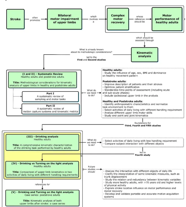
The complex motor recovery of ULs after stroke was the trigger for the elaboration of this thesis. The need to optimize the assessment of motor performance quality after stroke, through kinematic analysis, defined the next steps, namely the elaboration of five scientific articles. Thesis organization is summarized in the figure 1.
14
Our first two studies were two parts of a systematic review of literature,
regardingmethodological considerations for kinematic analysis of upper limbs in
healthy and poststroke adults. The specificities of sampling and motor tasks, as well as the motion capture systems and kinematic metrics used in this specific kinematic analysis were reviewed in the first and second parts, respectively. These two studies were important to define the methodological strategies of the following studies. In our systematic review we analysed articles that studied objectively three-dimension kinematics of ULs of healthy and/or poststroke adults, during the performance of functional movements or ADLs involving the ULs. Usually, healthy participants are young adults and the influence of sex, BMI and dominance on their movement pattern are not analysed; few articles identified anthropometric characteristics and normalized the experimental setup to them; most articles with healthy participants studied simulations of ADL; and most authors analysed joint kinematics or end-point kinematics, mainly related with reaching. From this point on, it became clear the need to: i) study the influence of age, sex, BMI and dominance; ii) identify anthropometric characteristics and normalize the experimental setup to them; iii) select ADLs with greater and lesser difficulty; iv) analyse different UL motor skills; and v) study end-point and joint kinematics. For these reasons, we chose to develop the two following studies.
The third study aimed to describe the kinematic ULs strategies of healthy adults during an ADL, namely drinking task, and to understand if age, sex,
dominance and BMI exerted any effect on the strategies used.We chose drinking
task because it is an ADL that allows the study of various motor skills, such as reaching, concentric and eccentric transporting and hand aperture to grasp and to release. This study was innovative because it included: i) a sample of healthy adults with a wide age range, with both sexes and two categories of BMI (normal and overweight); ii) a comprehensive analysis of drinking task, and its different motor skills, performed by dominant and non-dominant ULs; iii) a wide set of end-point and joint kinematics; iv) the normalization of the base of support and the glass location to the anthropometric characteristics of each participant; and v) the study of the influence of age, sex, BMI and dominance on motor stratetagies.
15
Our fourth study emerged from the need to study other ADLs with less
demanding handling, which poststroke adults with hand impairment could perform. In this study we chose to select the turning on the light ADL, whose interaction with the switch (pressing it) seems to be easier. Although the interaction with the target is clearly different, drinking and turning on the light tasks share two common gestures, which makes them comparable: i) reaching an object and ii) returning to the starting position. Therefore, besides describing the kinematic strategies of ULs to perform turning on the light task, we considered relevant to understand the implications of two different interactions with two different objects on the kinematic variables analysed. Thereby, the main objective of this study was to compare the kinematic strategies used by the ULs of the healthy adults in an ADL with less demanding handling (turning on the light) with those used in a more difficult ADL (drinking). In addition, we studied if turning on the light kinematic strategies were significantly different between dominant and non-dominant UL, as well as between subjects with different age, sex and BMI. To our knowledge, no other study about kinematic analysis of the ULs analysed turning on the light task or an ADL with less demanding handling and compare them with drinking or other ADL with greater difficulty. This and the third study have provided a kinematic reference of the healthy motor performance of two ADLs to poststroke adults (with greater or lesser hand impairment).
In our systematic review we confirmed also that in studies with poststroke adults: i) most of the recommended demographic and stroke information, were not collected; ii) moment of poststroke assessment was chronic phase whose time interval varied greatly; and iii) iUL was not included in the analysis. Therefore, the aim of the fifth study was to analyse the kinematic strategies of both ULs of poststroke adults in the early sub-acute phase and in the beginning of chronic phase, through a case series and with a methodological approach that included: i) the characterization of patients and their stroke, recommended by SRRR (Kwakkel et al., 2017); ii) the selection of drinking and turning on the light tasks; iii) the normalization of tasks environment to the anthropometric characteristics of each patient; and, iv) the bilateral analysis of "end-point kinematics" and "joint kinematics". This approach intended to contribute to: i) the optimization of poststroke patients’ stratification; ii) the inclusion of patients with
16
hand impairment; iii) the improvement of the experimental setup underlying the UL kinematic analysis; and iv) the implementation of bilateral kinematic assessment.
However, as some questions still need to be answered, in future studies, it will be important to replicate our experimental methodologies and analyse the interaction with different objects of daily life; clarify the interpretation of some kinematic measures, such as trunk displacement; study the relation and redundancy between kinematic variables; study more healthy adults, with >70 years old and higher levels of physical activity; explore stroke location influence on motor performance and motor recovery; and develop and validate portable and accurate motion acquisition systems. The guidelines for future studies will be addressed in more detail in section 7 (conclusions and future work perspectives). This section showed the work developed during this PhD project to address the main objectives defined. However, it makes sense to refer some methodological options and discuss their justifications to later describe the articles produced.
17
4. METHODOLOGICAL CONSIDERATIONS
For all research studies, it was necessary to make carefully reflected and evidence-based decisions regarding the methodology performed. Since some of these options were not fully justified in the articles of this thesis, a detailed description of the missing information is presented in the following sub-sections.
4.1. Systematic review
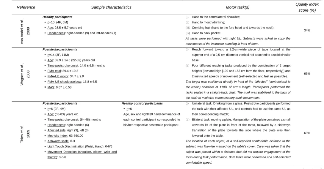
Considering the need to standardize the measurement of poststroke sensorimotor recovery of the ULs based on a valid healthy reference (Kwakkel et al., 2017), and to support our methodological decisions on scientific evidence, we have chosen to conduct a systematic review of methodological considerations for kinematic analysis of ULs in healthy and poststroke adults. Since, according to Bernhardt et al. (2016), insufficient attention has been paid to the recruitment and stratification of poststroke patients, and the influence of factors such as age and sex on healthy movement patterns has been undervalued, we decided that one of the aspects that we would analyse would be the characterization of the healthy and poststroke samples, i.e., the information collected and presented about these participants. For poststroke adults, we analysed whether the information collected was the recommended by SRRR (Kwakkel et al., 2017). Furthermore, as according to Ozturk et al. (2016), there are other major methodological factors upon which the kinematic analysis of the ULs depends, we decided to further analyse these factors: motion capture systems, motor tasks and the kinematic metrics extracted. To make reading easier, we split the review into two parts.
4.2. Study designs and participants
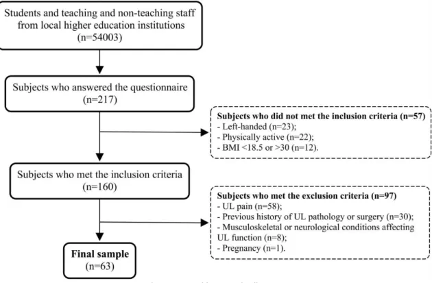
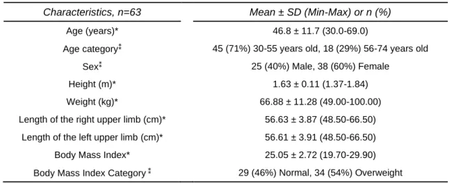
Study design and sample of the third and fourth articles were the same. These studies were cross-sectional observational studies since their goal was to describe kinematic strategies used by healthy adults. Sample was recruited for convenience from a population of students and teaching and non-teaching staff from the Polytechnic of Porto and the University of Porto (n=54003), contacted through e-mail; on this e-mail, people were informed about the study and invited to participate by fulfilling a characterization and inclusion/exclusion criteria
18
selection questionnaire (appendix III). Two hundred and seventeen subjects answered the questionnaire, and sixty-three of whom were recruited since they fit the criteria to participate in this study (a detailed description of this process is displayed in figure 1).
The inclusion and exclusion criteria of healthy adults were defined to meet the objective of studying the influence of age, sex, BMI and dominance factors; to allow that the characteristics of healthy adults matched as much as possible to those of most poststroke adults; but also to ensure the highest quality of movement capture. Thus, as the incidence of stroke in Europe is higher between 40 and 80 years old and the number of young adults having stroke is increasing (Bejot et al., 2016), we considered it appropriate to include healthy adults with ≥30 years old. As most poststroke survivors experience low levels of physical activity (Field, Gebruers, Shanmuga Sundaram, Nicholson, & Mead, 2013; Fini, Holland, Keating, Simek, & Bernhardt, 2017), we chose to select healthy adults with an insufficient physical activity level, according to Sedentary Behaviour Research Network (2017) (Tremblay et al., 2017). We assessed the level of physical activity through the International Physical Activity Questionnaire (IPAQ)
19
(Craig et al., 2003). As there is a higher proportion of right-handed than left-handed subjects worldwide (Llaurens, Raymond, & Faurie, 2009) and handedness may be associated with different patterns of neural activation of motor system (Pool, Rehme, Fink, Eickhoff, & Grefkes, 2014), we chose to include only right-handed adults. To ensure correct identification of anatomical references for placement of movement capture system markers, we excluded individuals with obesity. We excluded adults with current or previous history of UL pathology or surgery, as well as UL pain and pregnancy, since these conditions may affected UL function (Finley, Combs, Carnahan, Peacock, & Van Buskirk, 2012; Kim et al., 2014; Murphy et al., 2011; Patterson et al., 2011).
In the fifth study, we analysed poststroke adults, through a longitudinal observational case series study. We have chosen a case series because it is ideal for studying heterogeneous cases as those of poststroke patients and because it allows better characterization and analysis of their results according to their specific characteristics (Carey & Boden, 2003). Thus, it was possible to include poststroke patients with different demographic characteristics, such as age, and different stroke characteristics, such as initial severity and location, and to raise pertinent questions regarding the possible influence of these various characteristics on UL motor recovery. We chose to analyse motor performance at two key moments of poststroke recovery - the early subacute phase and the beginning of chronic phase - as in the first moment the movement pattern results mainly from the spontaneous recovery process (Carrera & Tononi, 2014; Grefkes & Fink, 2011; Ward & Cohen, 2004), and in the second moment the neural network re-organization processes seems to be already matured (Karnath & Rennig, 2017; Ward & Cohen, 2004). Therefore, the analysis of these two moments may allow observing the evolution of motor performance towards restitution or compensation. We chose to exclude patients with severe UL sensorimotor impairment, as they would have extreme difficulty in performing the proposed motor tasks and this would cause frustration and could negatively affect their recovery.
20
4.3. Motion capture system
Our initial objective was to assess the motor performance of poststroke adults in a hospital or clinical setting. However, when we tested the possibility of using portable motion acquisition systems, we faced difficult problems to solve such as the need to adopt an anatomical position in the calibration process (requirement impossible for poststroke adults), as well as the considerable lack of accuracy in motion detection. For that reason, we decided to use a visual marker-based optoelectronic system (with passive markers), since they are often considered the gold standard in the kinematic analysis because of their high accuracy and reliability (de los Reyes-Guzmán et al., 2014; Ozturk et al., 2016) and they are used as reference for comparisons with other equipments (Vilas-Boas Mdo & Cunha, 2016). However, this decision made it impossible to assess poststroke adults in the clinical setting, due to the large setup volume and consequent difficulty in transportation. Therefore, assessments of both healthy and poststroke adults were performed in the laboratory.
Prior to each recording session the camera system was calibrated with a
measurement volume of approximately 8 m3 and a maximum acceptable error of
0.8 mm.
4.4. Normalization of the experimental setup to anthropometric
characteristics
According to our systematic review (Mesquita, Pinheiro, Velhote Correia, & Silva, 2019) only one of the studies that kinematically analysed the UL between 2007 and 2017 (Kim et al., 2014), adjusted the experimental setup to the anthropometric characteristics of each individual. This adjustment (i.e. normalization) is important to ensure that experimental setup is not the responsible for variability in the kinematic metrics analysed. Both variations in the adopted position and the location of the target can create this variability. There are infinite ways of sitting: straight, diagonally, high or low, far into the seat or toward the edge, etc. Different sitting postures clearly affect motor activity of neck, trunk and scapulae (Caneiro et al., 2010; O’Sullivan et al., 2006; O’Sullivan et al., 2002), which are the biomechanical foundation of UL movement. In this study,
21
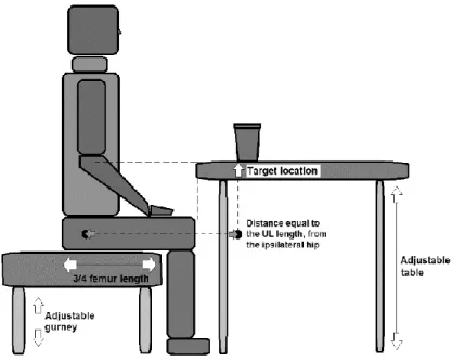
we normalized the height of the hydraulic gurney and the depth of the base of support, considering the length of the leg and femur of each participant, respectively. As for the target location, it is also known that the variation of distance, for example, affects the movement duration (Fitts, 1954) and trunk displacement (Levin, Michaelsen, Cirstea, & Roby-Brami, 2002). Therefore, we normalized the switch and drinking glass position. The lamp and the drinking glass were placed on a table, whose height was adjusted to the olecranon’s height of each patient and at a distance of ipsilateral hip equal to the length between the acromion and the trapezius-metacarpal joint, in the sagittal plane. We chose the hip rather than the acromion as a reference to determine target distance, since hip position in space remains stable throughout the experimental procedures, regardless of the participant's postural control.
4.5. Upper body biomechanical model
To create our upper body biomechanical model comprising both ULs, trunk and pelvis, we used Visual3D v6.01.02 (C-Motion, Inc., Germantown, USA). All modelling was performed according to appropriate C-motion recommendations (C-Motion, 2017). Pelvis was modelled according to the CODA model, with the markers RASIS, LASIS, RPSIS and LPSIS. For the trunk modelling was created a virtual marker (RTA_ORIGIN) whose position was created taking into account the center of mass of the pelvis, with an offset of 100% of the segment length in the posterior position and 5% in the distal (upward, towards the head). This virtual marker was used as the proximal joint center of the trunk, while the right acromial marker (RAC) was used to define the lateral portion of the trunk and the left acromial marker (LAC) the medial portion of the trunk. Markers C7, IJ and PX were used as tracking markers. The right and left shoulder joint center (RT_SHOULDER/LT_SHOULDER, respectively) was approximated as a negative vertical offset of the acromion marker, corresponding to the value of the marker diameter and 17% of the distance between the acromions (RAC and LAC). The humerus was then modelled as having a proximal origin in this virtual marker and a distal limit defined by the lateral and medial markers (RLELB, RMELB, LLELB, LMELB). The elbow joint was defined as the midpoint between the elbow markers (RLELB / RMELB, LLELB / LMELB), which was used to define
22
the proximal point of the radius-ulna complex, and its distal point was laterally defined by the marker of radius (RRAD, LRAD) and medially by the marker of ulna (RULN, LULN). The hand segment was proximally originated from the wrist joint center, defined as the midpoint between the RRAD and RULN markers, and the distal limit was defined laterally by the lateral hand marker (RLH, LLH) and medially by the medial marker (RMH, LMH). In all segments of the ULs, distal anatomical markers were also used as tracking markers as there were no additional markers. In the case of hand, where there were additional markers (RTHUMB and RINDEX), these were not used as tracking markers, since the fingers show movement independent of the hand (defined here as the set of metacarpals).
4.6. Kinematic metrics
To select the set of kinematic metrics, we based on our systematic review and on the literature review of de los Reyes-Guzmán et al. (2014), about quantitative assessment based on kinematic measures of functional impairments during UL movements. In our systematic review (Mesquita, Fonseca, Pinheiro, Velhote Correia, & Silva, 2019) we saw that most authors analysed “end-point kinematics”
or “joint kinematics”, of which “movement time,” “peak velocity,” “number of
movement units (velocity peaks),” “joint angles of shoulder and elbow,” and “trunk displacement” were the most studied. However, we questioned whether their analysis would be sufficient to improve the understanding about the mechanisms driving motor recovery and to differentiate restitution from compensation. Thus, considering that end-point and joint kinematics include a set of metrics that quantify different motion characteristics that may be relevant to this knowledge, we sought to make a selection of metrics that would quantify as many motion characteristics as possible: speed, efficiency, smoothness, control strategy, hand aperture for grasp and release, functional multi-joint angles and compensation. We chose to analyse these metrics in each phase of the tasks, since each phase contains different motor skills.
To quantify speed, we selected absolute and relative durations and mean and peak velocities, according to de los Reyes-Guzmán et al. (2014). Usually, a decrease in absolute and relative durations and an increase in mean and peak
23
velocities are attributed to a better UL function within a given task (de los Reyes-Guzmán et al., 2014).
To quantify movement efficiency, we chose the index of curvature (also known as “hand path ratio”), since, according to Lang et al. (2005) an efficient movement moves directly to the target without extraneous or abnormal trajectories. It is a measure of how directly the hand moves toward the target computed as the ratio between the length of the real subject's hand path and the length of the theoretical or desired trajectory. Although this metric has been frequently used in literature only during reaching movements (de los Reyes-Guzmán et al., 2014), we considered relevant to calculate it also in the other phases.
To quantify smoothness, we selected number of movement units (also known as “number of velocity peaks”) since this metric was applied frequently in poststroke patients (Murphy et al., 2013; Murphy et al., 2012; Murphy et al., 2011; van Dokkum et al., 2014; Wagner, Rhodes, & Patten, 2008). With the presence of movement disorders, the velocity peak number increases resulting in a less smooth movement. If any motor recovery occurs, the velocity profile of the hand movement must present less peaks resulting in a smoother movement (Rohrer et al., 2002).
To quantify control strategy we used time to peak velocity, according to de los Reyes-Guzmán et al. (2014). This measure allows to analyse the duration of the hand's acceleration and deceleration periods toward the target, which are often changed in the presence of movement disorders and are often responsible for the presence of dysmetria (de los Reyes-Guzmán et al., 2014).
To quantify hand aperture for grasp and for release, we select the maximum magnitude of hand aperture and the relative instant of hand aperture, according to Patterson et al. (2011). According to Castiello (2005), the size of pre-shaping of the fingers for grasping an object increases to a maximum and then is reduced to match the size of the object. The moment of maximum hand aperture occurs during the final-slow approach phase (Shumway-Cook & Woollacott, 2017). Although it is recognized that poststroke patients have difficulty in releasing objects (Seo, Rymer, & Kamper, 2009), to our knowledge, no study has kinematically analysed this specific impairment. In order to assess this behavior,
24
we chose to use the same selected kinematic variables for the assessment of the hand aperture to grasp.
To quantify functional multi-joint angles, we chose to analyse joint angles in clinically relevant planes of the main UL joints in each phase transition, to know which joint angles are intended to be achieved in the end of each phase, i.e. in each motor ability. Despite the well-known fact that scapular motion is a vital component of shoulder function, calculation of shoulder joint kinematics using 3D UL motion analysis is usually carried out with the shoulder considered as a virtual thoraco-humeral joint. The main obstacle to performing an individual assessment of scapula-thoracic and gleno-humeral joints is the difficulty in finding a valid and reliable method to record scapular motion, since marker based techniques are subject to inaccuracies relating to the placement of markers or soft tissue artefacts (Lempereur, Brochard, Leboeuf, & Rémy-Néris, 2014). For these reasons, scapula motion was not analysed.
Finally, to quantify compensation, we used trunk displacement since this variable has been widely used for this purpose (Murphy et al., 2013; Murphy et al., 2012; Murphy et al., 2011; Ozturk et al., 2016; Patterson et al., 2011). However, in addition to assessing its displacement in the sagittal plane, we also considered of relevance the study of its displacement in the frontal and transverse planes, considering its three-dimensional movement. Furthermore, we analysed trunk displacement in each phase rather than in the total task, as it is important to understand the behavior of trunk displacement according to the motor skill performed by the UL.
4.7 Statistical analysis
The statistical procedures of this work followed a logical and structured order of importance. Firstly, the descriptive statistics was used to present measures of central tendency and also frequency distribution for probabilities of demographic, anthropometric and kinematic data of healthy and poststroke adults.
Inference tests were used for specific comparisons that could point out possible differences between groups of subjects or factors. In this case, to