309
Radiol Bras. 2012 Nov/Dez;45(6):309–314
Central nervous system malformations and associated defects
diagnosed by obstetric ultrasonography
*
Malformações do sistema nervoso central e malformações associadas diagnosticadas pela ultrassonografia obstétrica
Marcela Leonardo Barros1, Daniel Alvarenga Fernandes2, Enaldo Vieira de Melo3, Roseane Lima
Santos Porto4, Maria Carolina Andrade Maia5, Atilano Salvador Godinho6, Thiago de Oliveira Ferrão7,
Carlos Umberto Pereira8
Objective: To identify and evaluate the prevalence of congenital central nervous system (CNS) malformations and associated defects diagnosed by obstetric ultrasonography. Materials and Methods: Observational, descriptive, cross-sectional study developed in an institution of reference for high-risk pregnancies. Results: Congenital CNS malformations without other associated defects were present in 65.78% of cases, as follows: hydrocephalus (37.5%), myelomeningocele (15%), encephalocele (12.5%), corpus callosum agenesis (12.5%), anencephaly (12.5%), holoprosencephaly (7.5%), Dandy-Walker (7.5%), Arnold-Chiari (5.0%), hydranencephaly (5.0%), meningocele (5.0%), arachnoid cyst (2.5%). Congenital malformations of other systems were associated with such malformations, as follows: craniofacial (73.9%), orthopedic (65.2%), cardiovascular (34.8%), genitourinary (30.4%), gastrointestinal (30.4%), respiratory (8.7%), syndromic (8.7%), ophthalmologic (4.3%). The sonographic sensitivity in the study of CNS malformations was 79.4%. The rate of false-negative results was 20.5%. Oligohydramnios, present in 25% of false-negative studies, stands out among the quantifiable limitations. Conclusion: Obstetric ultrasonography presents good sensitivity in the screening for fetal CNS malformations, specially with the constant improvement and control of specialized methods such as Doppler and volumetric ultrasonography (3D/4D), contributing to consolidate its role as a modality of choice in this routine. Magnetic resonance imaging may play a supplementary role, providing information for an even better perinatal care.
Keywords: Congenital malformations; Central nervous system malformations; Associated malformations; Ultrasonography.
Objetivo: Identificar a prevalência de malformações congênitas do sistema nervoso central (SNC) e malformações asso-ciadas diagnosticadas pela ultrassonografia obstétrica. Materiais e Métodos: Estudo observacional, transversal, des-critivo, em instituição de referência para gestações de alto risco. Resultados: Malformações congênitas do SNC estive-ram presentes sem outras malformações associadas em 65,78%, com a distribuição: hidrocefalia (37,5%), mielomenin-gocele (15%), encefalocele (12,5%), agenesia de corpo caloso (12,5%), anencefalia (12,5%), holoprosencefalia (7,5%), Dandy-Walker (7,5%), Arnold-Chiari (5,0%), hidranencefalia (5,0%), meningocele (5,0%), cisto aracnoideo (2,5%). Mal-formações congênitas de outros sistemas estiveram associadas às do SNC: craniofacial (73,9%), ortopédica (65,2%), cardiovascular (34,8%), geniturinária (30,4%), gastrintestinal (30,4%), respiratória (8,7%), sindrômica (8,7), oftalmoló-gica (4,3%). A sensibilidade ultrassonográfica no estudo de malformações fetais do SNC foi 79,4%. A taxa de falso-ne-gativos foi 20,5%. Dentre as limitações quantificáveis destaca-se o oligodrâmnio, presente em 25% dos falso-nefalso-ne-gativos.
Conclusão: A ultrassonografia obstétrica possui boa sensibilidade no rastreio de malformações fetais do SNC, em espe-cial com o aperfeiçoamento constante e domínio na utilização de métodos espeespe-cializados, como o Doppler e a ultrasso-nografia volumétrica (3D/4D), contribuindo para firmar-se como modalidade de escolha nesta rotina. Complementar ao método, a ressonância magnética pode vir a fornecer subsídios para uma ainda melhor assistência perinatal.
Unitermos: Malformações congênitas; Malformações do sistema nervoso central; Malformações associadas; Ultrasso-nografia.
Abstract
Resumo
* Study developed at Maternidade Nossa Senhora de Lourdes and Hospital Universitário da Universidade Federal de Sergipe (HU/UFS), Aracaju, SE, Brazil.
1. MD, Physician, Department of Medicine, Universidade Fe-deral de Sergipe (UFS), Aracaju, SE, Brazil.
2. MD, Resident in Radiology and Imaging Diagnosis, Hospi-tal Universitário da Universidade Federal de Sergipe (HU/UFS), Aracaju, SE, Brazil.
3. Master, MD, Pediatrician, Assistant Professor, Department of Medicine, Universidade Federal de Sergipe (UFS), Aracaju, SE, Brazil.
Barros ML, Fernandes DA, Melo EV, Porto RLS, Maia MCA, Godinho AS, Ferrão TO, Pereira CU. Central nervous system malformations and associated defects diagnosed by obstetric ultrasonography. Radiol Bras. 2012 Nov/Dez;45(6):309–314.
4. MD, Fellow Master degree, Pediatrician, Hospital Universi-tário da Universidade Federal de Sergipe (HU/UFS), Aracaju, SE, Brazil.
5. Fetal Medicine Specialist, Preceptor of Residency in Radi-ology and Imaging Diagnosis, Hospital Universitário da Universi-dade Federal de Sergipe (HU/UFS), Aracaju, SE, Brazil.
6. MD, Radiologist, Coordinator for Medical Residency in Radi-ology and Imaging Diagnosis, Hospital Universitário da Universi-dade Federal de Sergipe (HU/UFS), Aracaju, SE, Brazil.
7. Fellow PhD degree, Titular Member of Colégio Brasileiro de Radiologia e Diagnóstico por Imagem (CBR), Assistant
Profes-sor, Department of Medicine, Universidade Federal de Sergipe (UFS), Aracaju, SE, Brazil.
8. PhD, Neurosurgeon, Associate Professor, Department of Medicine, Universidade Federal de Sergipe (UFS), Aracaju, SE, Brazil.
Mailing Address: Dr. Daniel Alvarenga Fernandes. Rua Buenos Aires, 726, Ed. América Central, ap. 1404, Jardim das Améri-cas. Cuiabá, MT, Brazil, 78060-634. E-mail: daniel_alvafer@ yahoo.com.br
INTRODUCTION
In some regions of the world, congeni-tal malformations represent the first cause of neonatal deaths(1). Approximately 20%
of gestations with malformed fetuses progress to spontaneous miscarriage, and the remaining 80% will result in stillbirths or live births and, out of the latter, 3% to 5% will result in neonates with congenital anomalies(2,3). In Brazil, such malformations
represent the second cause of infant mor-tality, determining 11.2% of such deaths(4).
Congenital central nervous system (CNS) malformations are highly prevalent, affect-ing 1 to 10:1,000 live newborns(5). Such
statistics may vary seasonally among coun-tries and ethnic groups or among services of prenatal diagnosis and neonatology.
Approximately 21% of congenital mal-formations involve the CNS, constituting one of the most common congenital defects and may occur either isolatedly or in asso-ciation with other malformations of the CNS itself or of other organs or system(6).
Currently, most of congenital anomalies can be diagnosed by means of obstetric ultrasonography and the fetal medicine seeks to establish an intrauterine fetal therapy for some episodes. Considering that an early diagnosis has significant re-percussions on the neonatal prognosis, the present study has proposed to identify the prevalence of CNS malformations and as-sociated malformations diagnosed by ob-stetric ultrasonography.
MATERIALS AND METHODS
Observational, cross-sectional, descrip-tive study developed in a public institution of reference for high-risk gestations. The present article is part of a more comprehen-sive project – Estudo Colaborativo Latino-Americano de Malformações Congênitas
(ECLAMC) (Latin American Collaborative Study of Congenital Malformations) –, a case-control study aimed at investigating epidemiological, clinical and imaging vari-ables of malformed neonates. In Brazil, such network operates in 32 hospitals(7),
and one of them is the institution where the present study was developed. The study project was approved by the Committee for Ethics in Research of the institution,
re-specting all the international principles on research involving humans. The present study was based on data from the ECLAMC. Conflicting interests: none declared.
The present study considered all the records of both stillborns and live new-borns with congenital CNS malformations, either with diagnosis notified on medical records or live birth statements over a six-teen-month period. Neonates born in other hospitals, who for any reason were later assisted in the studied institution were ex-cluded. The cases of congenital CNS mal-formations were identified according to their clinical and imaging presentations, being classified as isolated malformations; syndrome components; or malformations associated with other CNS anomalies or anomalies in other organs and systems.
The postnatal diagnosis was performed by means of physical examination, postna-tal ultrasonography and/or computed to-mography which, in association, were con-sidered as the “gold standard” for a defini-tive diagnosis of congenital malformation, which is defined as an anatomical, physi-cal defect diagnosed at birth and listed on the “Chapter XVII: Congenital malforma-tions, deformations and chromosomal anomalies (Q00–Q99)” of the International Classification of Diseases – 10th revision. Thus, with a definitive diagnosis of malfor-mation documented on medical records, and utilizing the described gold standard, the authors have retrospectively sough to confirm or reject the results of the obstet-ric imaging study, and then verify whether the method could or not detect malforma-tions in neonates, determining the sensitiv-ity and rate of false-negative results of ob-stetric ultrasonography in the study of fe-tal CNS malformations.
The SPSS (Statistical Package for So-cial Sciences) testing version 18.0 was uti-lized for data tabulation and statistical analysis. The categorical variables were described as simple frequencies and per-centages. For the quantitative variables, means and standard deviations were uti-lized. The ultrasonography scans were per-formed by different professionals with more than 5-year experience in the method and with different apparatuses as follows: Voluson 730 Pro 3D/4D, Logic T5 GE and SonoSite MicroMaxx portable
ultrasonog-raphy Machine. Two-dimensional evalua-tion, volumetric evaluation and ultrasound Doppler were utilized.
RESULTS
In the study period, 126 cases of con-genital malformations were evaluated, and the frequency of congenital CNS malfor-mations corresponded to 31.8% (40 cases). The same percentage was found for ortho-pedic malformations, followed by cranio-facial malformations (20%) and other mal-formations (16%).
On average, the birth-weight of the 40 neonates with congenital CNS malforma-tions was 2694.6 ± 872.8 g, ranging be-tween 1000 and 4760 g. The frequency of male newborns was 55.3%, female new-borns, 39.5%, and intersexed newnew-borns, 5.3%. Only one case of stillborn was ob-served among the malformed neonates. The mean age of the puerperas was 27.2 ± 7.7 years, ranging between 13 and 43, and the mean age of the fathers was 31.6 ± 8.7 years, ranging between 18 and 55 years.
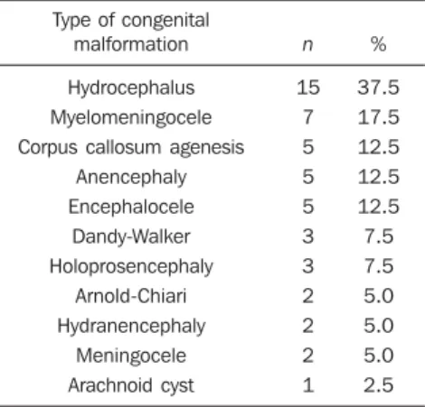
Congenital CNS malformations were isolated in most of cases, while in 37.5% (15 cases) there was association with other congenital CNS malformations. Hydro-cephalus was the most prevalent malforma-tion, followed by myelomeningocele; cor-pus callosum agenesis; anencephaly; and encephalocele. Dandy-Walker syndrome and holoprosencephaly presented a preva-lence of 7.5% each. As regards the other malformations, a lower absolute frequency was observed, as shown on Table 1.
Fig-Table 1 Prevalence of congenital CNS malforma-tions, according to clinical and sonographic diag-nosis.
Type of congenital malformation
Hydrocephalus Myelomeningocele Corpus callosum agenesis
Anencephaly Encephalocele Dandy-Walker Holoprosencephaly Arnold-Chiari Hydranencephaly Meningocele Arachnoid cyst n 15 7 5 5 5 3 3 2 2 2 1 % 37.5 17.5 12.5 12.5 12.5 7.5 7.5 5.0 5.0 5.0 2.5
Figure 2. Proboscis, an anomaly that is also characteristic of trisomy 13, which almost always is associated with holoprosencephaly. Two-dimensional (A) and three-dimensional (B) images.
A B
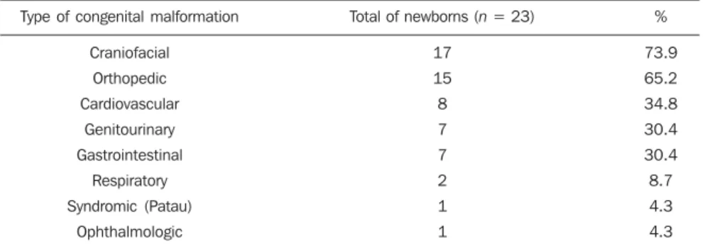
Table 2 Frequency of congenital malformations of other physiological systems in newborns with con-genital CNS malformations.
Type of congenital malformation
Craniofacial
Orthopedic
Cardiovascular
Genitourinary
Gastrointestinal
Respiratory
Syndromic (Patau)
Ophthalmologic
Total of newborns (n = 23)
17
15
8
7
7
2
1
1
%
73.9
65.2
34.8
30.4
30.4
8.7
4.3
4.3
Note: Each newborn has one or more associated malformation.
Figure 1. Cerebellar vermis agenesis with separation of the hemispheres and enlarged cis-terna magna communicating with the fourth ventricle (findings of Dandy-Walker syndrome). Two-dimensional (A) and
three-di-mensional (B) images. A B
ure 1 shows cerebellar vermis agenesis with separation of the hemispheres and en-larged cisterna magna communicating with the fourth ventricle (findings of Dandy-Walker syndrome).
Among the cases of anencephaly, 80% were observed in female newborns. Famil-ial history of malformed newborns was found in 60% of cases of anencephaly and in 23.1% of all the cases with such anteced-ent.
As regards the presence of congenital malformations in other organs or systems, 57.5% of the newborns presented one or more congenital defects in association with CNS malformation. Most prevalent sites of congenital malformations were the follow-ing: craniofacial, followed by orthopedic,
cardiovascular, genitourinary and gas-trointestinal malformations (Table 2). Patau syndrome occurred in one case of holo-prosencephaly. Figure 2 shows proboscis,
lip and palate, single nostril, cyclopia or even microphthalmos.
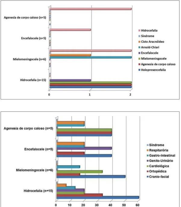
Hydrocephalus was the congenital mal-formation most frequently associated with either myelomeningocele, or corpus callo-sum agenesis or encephalocele (Figure 3). As regards other congenital malforma-tions associated to CNS malformamalforma-tions, craniofacial, followed by orthopedic and cardiovascular malformations were most frequently found (Figure 4).
The sonographic sensitivity in the inves-tigation of fetal CNS malformations was of 79.4%. The rate of false-negative results reached 20.5%. Among quantifiable
limi-tations, oligohydramnios is highlighted, being present in 25% of false-negative sonographic results.
DISCUSSION
The frequency of congenital CNS mal-formations among all the evaluated cases of malformations was 31.8%, a rate simi-lar to the ones reported by some authors(8,9)
and higher than the one found by Noronha et al.(5) and by Pitkin(6), 13% and 21% of
cases, respectively. Differently from the findings reported by Pitkin(6) and by
Vic-tora and Barros(4), congenital CNS
malfor-mations were the most frequent ones, to-gether with orthopedic malformations, fol-lowed by craniofacial malformations. An-other study reports craniofacial and limbs malformations as most frequently found(10).
A case-control study indicates that the etio-pathogenesis of some congenital orthope-dic malformations may involve neurologi-cal factors producing alterations in the spi-nal cord or in nerves(11).
Although some studies report cardio-vascular anomalies as most common mal-formations, marked differences in study populations, as well as in criteria and diag-nostic method utilized may lead to
diagnosis of mild defects. Thus, a higher prevalence of such congenital malforma-tions has been observed as routine echocar-diography is utilized – an unusual practice in most health services(12).
Among congenital CNS malformations, hydrocephalus was the most frequent, fol-lowed by myelomeningocele, as reported by a retrospective study(13). In sequence,
anencephaly together with corpus callosum agenesis and encephalocele were found to be more frequent, in disagreement with the frequencies described by Moore & Per-saud(14). Among neural tube defects,
my-elomeningocele, anencephaly and en-cephalocele were observed in descent or-der. Certain authors report the same ten-dency(9,15).
In cases of anencephaly, female new-borns were affected at a 4:1 ratio that was superior to the ratio observed in some stud-ies(16,17). Three of five cases presented
fa-milial history of malformations, a number that was higher than the one reported by Ramos et al.(18).
Dávila-Gutiérrez reports a relation be-tween hydrocephalus and corpus callosum agenesis(19). On the other hand, other
au-thors report cases of concomitance of hy-drocephalus and myelomeningocele(20,21),
while Levey et al. report coexistence of hydrocephalus and holoprosencephaly(22).
In the present study, such associations were observed with higher frequency.
Congenital craniofacial malformations were more prevalent in cases of hydroceph-alus, as corroborated by Cinalli et al.(23),
and myelomeningocele. Congenital cardio-logic malformations were most frequently present in cases of corpus callosum agen-esis, in agreement with the findings of Mowat et al.(24). On the other hand,
geni-tourinary malformations were most fre-quently found in cases of encephalocele, in agreement with Rittler et al.(25). Patau
syn-drome – a very rare chromosomal abnor-mality, with an incidence of 1/5000 to 1/ 20000 births – occurred in a case of holoprosencephaly. Such association is described in the literature(26).
The sonographic sensitivity in the inves-tigation of fetal CNS malformations was of 79.4%. Randomized studies estimate a sonographic sensitivity of about 80% for detecting CNS malformations in cases of
high-risk gestation(27,28). It is known that
factors such as quality of apparatuses, sound waves interaction with tissues, ex-amination techniques – appropriate adjust-ment of gain waveforms, for example –, dedicated scan time, experience and knowl-edge of the medical sonographer, besides factors such as high maternal body mass índex, fetal statics, advanced gestational age and decreased amniotic fluid index, may change the method sensitivity. Con-stant perfecting and deep knowledge of specialized methods such as Doppler(29,30)
and volumetric (3D/4D) ultrasonography, in association with the increasing techno-logical development with state-of-the-art equipment, have contributed to a good sen-sitivity of the method in the screening for malformations, with prognostic repercus-sions.
The rate of false-negative results was 20.5%. Among the quantifiable limitations, oligohydramnios is highlighted, being present in 25% of false-negative sono-graphic studies. Magnetic resonance imag-ing complementary to ultrasonography, differently from the latter, demonstrates improved diagnostic accuracy with the ges-tational age progress, and is not affected by decreased amniotic fluid levels, maternal obesity or fetal statics (31). Additionally, one
of the main contributions of magnetic reso-nance imaging is to complement the role of ultrasonography in the study of fetal CNS malformations which is complicated at late phases of gestation because of the ad-vanced cranial bones ossification. Even so, it should be stressed that the utilization of magnetic resonance imaging is restricted to complement ultrasonography, considering the limitations of the method such as high cost, fetal motion artifacts, claustrophobia, the recommendation not to perform the method in the first gestational trimester(32),
besides its lesser availability.
Thus, ultrasonography provides early diagnosis with good sensitivity, which in association with the accessibility and avail-ability of the method, has contributed to consolidate its role as the modality of choice in the routine screening for fetal CNS malformations. However, considering the inherent method limitations, continuity of improvements should still be encour-aged in the search for excellence in early
diagnosis, in order to highlight novel tech-nologies to supplement the assessment of the uterine contents. In this context, a wide availability of fetal magnetic resonance imaging for the study population has been sought in order to allow additional diagnos-tic findings, as reported in the literature(32,33).
CONCLUSIONS
Obstetric ultrasonography demonstrates good sensitivity in the screening for fetal CNS malformations, especially with the constant improvements and increased knowledge of specialized methods such as Doppler and volumetric ultrasonography (3D/4D), contributing to consolidate its role as a modality of choice in this routine. As a complementary method, magnetic resonance imaging may be useful, provid-ing information for an even better perina-tal assistance.
REFERENCES
1. Rosano A, Botto LD, Botting B, et al. Infant mortality and congenital anomalies from 1950 to 1994: an international perspective. J Epidemiol Community Health. 2000;54:660–6.
2. Rankin J, Pattenden S, Abramsky L, et al. Preva-lence of congenital anomalies in five British re-gions, 1991-99. Arch Dis Child Fetal Neonatal Ed. 2005;90:F374–9.
3. Nikkilä A, Rydhstroem H, Källén B, et al. Ultra-sound screening for fetal anomalies in southern Sweden: a population-based study. Acta Obstet Gynecol Scand. 2006;85:688–93.
4. Victora CG, Barros FC. Infant mortality due to perinatal causes in Brazil: trends, regional pat-terns and possible interventions. São Paulo Med J. 2001;119:33–42.
5. Noronha L, Medeiros F, Martins VDM, et al. Mal-formações do sistema nervoso central: análise de 157 necrópsias pediátricas. Arq Neuropsiquiatr. 2000;58:890–6.
6. Pitkin RM. Folate and neural tube defects. Am J Clin Nutr. 2007;85:285S–288S.
7. Castilla EE, Orioli IM. ECLAMC: the Latin-American collaborative study of congenital mal-formations. Community Genet. 2004;7:76–94. 8. Moron AF. Diagnóstico pré-natal das
malforma-ções congênitas no contexto do sistema de saúde [tese]. São Paulo: Faculdade de Saúde Pública, Universidade São Paulo; 1995.
9. Costa CMS, Gama SGN, Leal MC. Congenital malformations in Rio de Janeiro, Brazil: preva-lence and associated factors. Cad Saúde Pública. 2006;22:2423–31.
10. Castro MLS, Cunha CJ, Moreira PB, et al. Fre-quency of multiple neonatal malformations in Pelotas, Rio Grande do Sul, Brazil, and associ-ated socio-demographic factors. Cad Saúde Pú-blica. 2006;22:1009–15.
Neurological congenital malformations in a ter-tiary hospital in south Brazil. Arq Neuropsiquiatr. 2009;67:807–11.
12. Goldmuntz E. The epidemiology and genetics of congenital heart disease. Clin Perinatol. 2001;28: 1–10.
13. Pinar H, Tatevosyants N, Singer DB. Central ner-vous system malformations in a perinatal/neona-tal autopsy series. Pediatr Dev Pathol. 1998;1:42– 8.
14. Moore KL, Persaud TVN. Embriologia clínica. Rio de Janeiro, RJ: Guanabara Koogan; 2004. 15. Shurtleff DB, Lemire RJ. Epidemiology, etiologic
factors, and prenatal diagnosis of open spinal dys-raphism. Neurosurg Clin N Am. 1995;6:183–93. 16. Loncarek K, Mustac E, Frkovic A, et al. Preva-lence of anencephaly in the region of Rijeka, Croatia. Eur J Epidemiol. 2001;17:241–4. 17. Verma M, Chhatwal J, Singh D. Congenital
mal-formations – a retrospective study of 10,000 cases. Indian J Pediatr. 1991;58:245–52. 18. Ramos JLA, Laurindo VM, Vaz FAC, et al.
Mal-formações congênitas: estudo prospectivo de dois anos em três maternidades de São Paulo. Pediat (S Paulo). 1981;3:20–8.
19. Dávila-Gutiérrez G. Agenesis and dysgenesis of the corpus callosum. Semin Pediatr Neurol. 2002; 9:292–301.
20. Dicianno BE, Kurowski BG, Yang JM, et al. Re-habilitation and medical management of the adult with spina bifida. Am J Phys Med Rehabil. 2008; 87:1027–50.
21. Shurtleff DB. 44 years’ experience with manage-ment of myelomeningocele: presidential address, Society of Research into Hydrocephalus and Spina Bifida. Eur J Pediatr Surg. 2000;10 Suppl 1:5–8. 22. Levey EB, Stashinko E, Clegg NJ, et al. Manage-ment of children with holoprosencephaly. Am J Med Genet C Semin Med Genet. 2010;154C: 183–90.
23. Cinalli G, Sainte-Rose C, Kollar EM, et al. Hy-drocephalus and craniosynostosis. J Neurosurg. 1998;88:209–14.
24. Mowat DR, Wilson MJ, Goossens M. Mowat-Wilson syndrome. J Med Genet. 2003;40:305– 10.
25. Rittler M, López-Camelo JS, Castilla EE, et al. Preferential associations between oral clefts and other major congenital anomalies. Cleft Palate Craniofac J. 2008;45:525–32.
26. Lehman CD, Nyberg DA, Winter TC 3rd, et al. Trisomy 13 syndrome: prenatal US findings in a review of 33 cases. Radiology. 1995;194:217–22. 27. Crane JP, LeFevre ML, Winborn RC, et al. A ran-domized trial of prenatal ultrasonographic screen-ing: impact on the detection, management, and
outcome of anomalous fetuses. The RADIUS Study Group. Am J Obstet Gynecol. 1994;171: 392–9.
28. Ewigman BG, Crane JP, Frigoletto FD, et al. Ef-fect of prenatal ultrasound screening on perina-tal outcome. RADIUS Study Group. N Engl J Med. 1993;329:821–7.
29. Gabriel ML, Piatto VB, Souza AS. Aplicação clí-nica da ultrassonografia craniana com Doppler em neonatos prematuros de muito baixo peso. Radiol Bras. 2010;43:213–8.
30. Moron AF, Milani HJF, Barreto EQS, et al. Aná-lise da reprodutibilidade do Doppler de ampli-tude tridimensional na avaliação da circulação do cérebro fetal. Radiol Bras. 2010;43:369–74. 31. Prayer D, Brugger PC, Prayer L. Fetal MRI:
tech-niques and protocols. Pediatr Radiol. 2004;34: 685–93.
32. Ximenes RLS, Szejnfeld J, Ximenes ARS, et al. Avaliação crítica dos benefícios e limitações da ressonância magnética como método comple-mentar no diagnóstico das malformações fetais. Radiol Bras. 2008;41:313–8.