Protocol
Bioengineered Temporomandibular Joint Disk Implants: Study
Protocol for a Two-Phase Exploratory Randomized Preclinical
Pilot Trial in 18 Black Merino Sheep (TEMPOJIMS)
David Faustino Ângelo1,2,3, MD; Florencio Gil Monje4, MD, PhD; Raúl González-García4, MD, PhD; Christopher B Little5, MVM, PhD; Lisete Mónico6, PhD; Mário Pinho7, MVM, PhD; Fábio Abade Santos7; Belmira Carrapiço8, MVM; Sandra Cavaco Gonçalves9, MVM; Pedro Morouço2, PhD; Nuno Alves2, MEng, PhD; Carla Moura2, MEng (Biomed Eng), PhD; Yadong Wang10, MEng (Biomed Eng), PhD; Eric Jeffries10, MEng (Biomed Eng), PhD; Jin Gao10, MEng (Biomed Eng), PhD; Rita Sousa3, MD; Lia Lucas Neto3, MD, PhD; Daniel Caldeira3, MD; Francisco Salvado3, MD, PhD
1
Centro Hospitalar de Setúbal, EPE, Stomatology Department, Setúbal, Portugal
2Instituto Politécnico de Leiria, Centre for Rapid and Sustainable Product Development (CDRSP), Leiria, Portugal 3
Faculdade de Medicina, Universidade de Lisboa, Lisboa, Portugal
4University Hospital Infanta Cristina, Badajoz, Spain, Department of Oral and Maxillofacial Surgery, Badajoz, Spain 5
Kolling Institute, Institute of Bone and Joint Research, University of Sydney, Sydney, Australia
6Coimbra University, Coimbra, Portugal 7
Faculdade de Medicina Veterinaria, Interdisciplinary Centre of Research in Animal Health (CIISA), Lisbon University, Lisboa, Portugal
8Faculdade de Medicina Veterinaria, Lisbon University, Lisboa, Portugal 9
Instituto Nacional de Investigação Agrária e Veterinária (INIAV I.P.), Santarém, Portugal
10University of Pittsburgh, Chemical Engineering and Surgery Pittsburgh, University of Pittsburgh, Pittsburgh, PA, United States
Corresponding Author:
David Faustino Ângelo, MD Centro Hospitalar de Setúbal, EPE Stomatology Department
Rua Camilo Castelo Branco Setúbal, 2919-446 Portugal Phone: 351 265 549 00 Fax: 351 265 238 066 Email: [email protected]
Abstract
Background: Preclinical trials are essential to test efficacious options to substitute the temporomandibular joint (TMJ) disk.
The contemporary absence of an ideal treatment for patients with severe TMJ disorders can be related to difficulties concerning the appropriate study design to conduct preclinical trials in the TMJ field. These difficulties can be associated with the use of heterogeneous animal models, the use of the contralateral TMJ as control, the absence of rigorous randomized controlled preclinical trials with blinded outcomes assessors, and difficulties involving multidisciplinary teams.
Objective: This study aims to develop a new, reproducible, and effective study design for preclinical research in the TMJ
domain, obtaining rigorous data related to (1) identify the impact of bilateral discectomy in black Merino sheep, (2) identify the impact of bilateral discopexy in black Merino sheep, and (3) identify the impact of three different bioengineering TMJ discs in black Merino sheep.
Methods: A two-phase exploratory randomized controlled preclinical trial with blinded outcomes is proposed. In the first phase,
nine sheep are randomized into three different surgical bilateral procedures: bilateral discectomy, bilateral discopexy, and sham surgery. In the second phase, nine sheep are randomized to bilaterally test three different TMJ bioengineering disk implants. The primary outcome is the histological gradation of TMJ. Secondary outcomes are imaging changes, absolute masticatory time, ruminant time per cycle, ruminant kinetics, ruminant area, and sheep weight.
Results: Previous preclinical studies in this field have used the contralateral unoperated side as a control, different animal models
ranging from mice to a canine model, with nonrandomized, nonblinded and uncontrolled study designs and limited outcomes measures. The main goal of this exploratory preclinical protocol is to set a new standard for future preclinical trials in oromaxillofacial surgery, particularly in the TMJ field, by proposing a rigorous design in black Merino sheep. The authors also intend to test the feasibility of pilot outcomes. The authors expect to increase the quality of further studies in this field and to progress in future treatment options for patients undergoing surgery for TMJ disk replacement.
Conclusions: The study has commenced, but it is too early to provide results or conclusions.
(JMIR Res Protoc 2017;6(3):e37) doi: 10.2196/resprot.6779 KEYWORDS
temporomandibular joint disorders (TMD); temporomandibular joint bioengineered disk implants; temporomandibular randomized preclinical trial protocol
Introduction
The temporomandibular joint (TMJ) is the most frequently used joint in the human body. The TMJ opens and closes 1500 to 2000 times daily and is essential for everyday functions of the mouth, such as mastication, speech, deglutition, yawning, and snoring, involving special mandatory synergy of both articular sides [1]. The TMJ disk is an essential component in the normal TMJ and has the following functions: (1) it distributes the intra-articular load, (2) it stabilizes the joints during translation, and (3) it decreases the wear of the articular surface [2,3]. The majority of TMJ disorders (TMD) are successfully treated with reversible, conservative, and low-tech treatments such as education and counseling, therapeutic exercises, splint therapy, and pharmacotherapy [4,5].
When the TMJ disk is displaced, malformed, or damaged, it can induce serious internal pathologic processes and/or osteoarthritis [6,7]. Currently, patients suffering from severe TMD have limited validated treatment options. Most surgical approaches, such as TMJ discectomy, do not restore the structural or biological properties of the articulation and disk. This procedure may not be ideal because the TMJ is left without an important functional structure. A variety of interpositional materials have been used to replace the removed disks, including synthetic materials manufactured from silicone, Teflon, polytetrafluoroethylene, and biological interpositional grafts taken from different anatomic sites [8-11]. These interpositional materials do not take in consideration the anatomy and biochemical and biomechanical characteristics of the TMJ native disk [12], and some of them have been associated with serious complications for the patients [8,13,14]. In the late 1980s, Proplast/Teflon TMJ (synthetic interpositional implant) were found to be harmful in many patients. The breakdown of the material, probably caused by TMJ high biomechanical forces, lead to fragmented particles that resulted in an immune foreign body response that caused problems ranging from severe cutaneous inflammatory reaction in the preauricular and cheek areas [15] to severe degenerative joint disease with perforation into the middle cranial fossa [16,17]. The result was a dramatic clinical spectrum of failures for these implants [10]. In December 1991, the US Food and Drug Administration’s Bulletin recommended immediate removal of all previous TMJ Proplast/Teflon implants because of the mechanical failures, many resulting in progressive bone degeneration [18]. In a 1992
workshop, the American Academy of Oral and Maxillofacial Surgery instructed the discontinuation of Proplast/Teflon [18]. The absence of efficacious options to substitute the TMJ disk can be related to difficulties in the translation of animal evidence to the clinical practice in humans. These limitations are likely related to:
1. the use of heterogeneous animal models with conflicting results, possibly due to variable anatomy and intra-articular loading between species [19,20];
2. the use of the contralateral TMJ as control, which may be associated with contralateral overloading [21];
3. the biomaterials used to replace the disk do not account for the morphologic and biomechanical characteristics of the native disk;
4. absence of randomized controlled trials with blinding of outcomes’ assessors; and
5. lack of multidisciplinary teams involved in the project. Preclinical research should promote the effective translation of knowledge into practice. The previously mentioned aspects can limit the effective translation of quality scientific knowledge into clinical practice and these may present potential issues to patients, clinicians, and scientific progress.
The contemporary absence of successful options to substitute the TMJ disk is still a major issue for public health. Little has changed in the past decade regarding study designs for TMJ investigation, and the treatment for patients with severe TMD remains controversial. The main objective of the Temporomandibular Joint Interposal Material Study (TEMPOJIMS) is to develop a new, reproducible, and effective study design for preclinical research in the TMJ field. The second goal is to progress in bioengineering and regenerative medicine evaluating the benefits of a TMJ bioengineering implant to substitute the damaged native TMJ disk. This preclinical exploratory study is divided into two phases. Phase 1 of this study is a blinded randomized preclinical trial, designed to investigate if the TMJ undergoes important injury in bilateral discectomy, bilateral discopexy, and sham surgery. Phase 2 intentions are to evaluate the safety and efficacy of three different TMJ bioengineering implants using the same rigorous method of phase 1.
Methods
Study Design
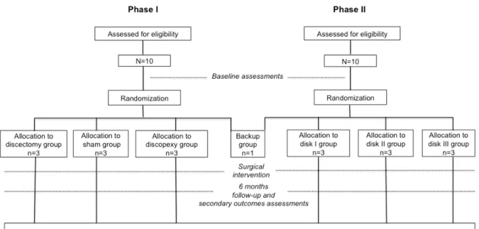
The TEMPOJIMS is a two-phase exploratory randomized controlled preclinical trial planned to gather preliminary information to (1) evaluate a new study design for TMJ investigation; (2) evaluate the black Merino sheep animal model for TMJ investigation; (3) evaluate TMJ behavior under bilateral surgical intervention (discectomy and discopexy) using a histologic primary outcome (microscopic scoring of destructive changes in TMJ using a modified Mankin scoring system [22]), secondary imaging outcome (imaging scoring of TMJ); (4) testing the applicability of pilot secondary outcomes predominantly for ruminant kinetics; and (5) obtain a baseline for interpretation of TMJ disk bioengineering implants results. Phase II is aimed to test safety and efficacy of three different bilateral TMJ bioengineering disk implants (Figure 1). Outcome evaluators and analysts are blinded for surgical assessments.
Major institutions involved in this study are (1) Lisbon Faculty of Medicine for study design, coordination, and statistical analysis; (2) Interdisciplinary Centre of Research in Animal Health in Faculty of Veterinary Medicine for histological preparation and veterinary support of all animals; (3) Centre for Rapid and Sustainable Product Development for bioengineered disk implants (disks I and II); (4) Bioengineering, Surgery, Chemical Engineering, Mechanical Engineering and Materials Science, University of Pittsburgh, for bioengineered disk implants (disk III); (5) Department of Oral and Maxillofacial-Head and Neck Surgery, University Hospital Infanta Cristina, Badajoz, Spain, for surgical support; (6) Institute of Bone and Joint Research-Northern Sydney Local Health District-Sydney Medical School Northern, University of Sydney, Australia, for histological analysis; and (7) Radiology Department of Santa Maria Hospital, Lisbon, Portugal, for imaging analysis.
Figure 1. Study design.
Animal Model
A variety of strains/breeds of sheep have been used in TMJ investigations. To decrease biological variability, the authors recommended black Merino sheep as the animal model to conduct the study [20]. As recommended, the authors proposed to use “sheep skeletally mature” at ≥2 years of age [23]. The inclusion criteria are certified black Merino sheep, adult (age 2-5 years), female, and in good health condition (veterinary check-up is performed on all animals). Regarding the animal ethical considerations, the study design was approved by the Portuguese National Authority for Animal Health registered with number 026618. The study design and organization respect the Animal Research: Reporting of In Vivo Experiments (ARRIVE) guidelines.
Baseline and Follow-Up Evaluation
The baseline and follow-up evaluations are outlined at particular time points (Figure 2). Pilot secondary outcomes and weight are measured at days 11, 10, and 9 before surgery (details on secondary outcomes are reported in outcomes measures). Transportation to surgical facilities is performed 5 days before surgery to avoid animal stress and allow familiarization to the temporary facilities. Head computerized tomography (CT) scan is performed on the day of surgery taking advantage of preanesthesia sedation. Ten days after surgery, animals are transported to TEMPOJIMS main facilities. Days 19, 20, and 21 after surgery, the follow-up secondary outcomes start to be recorded every 30 days for 6 months (Figure 2). At the end, animals are sacrificed and a new CT scan is performed to measure the imaging outcome and to begin the histologic preparation.
Figure 2. Study flowchart.
Randomization, Allocation, and Blinding
The randomization is performed by a statistical group not involved in the outcome assessments, managed by Lisbon Faculty of Medicine. Allocation to each randomized group is performed preoperatively by sealed envelope and separately for phase 1 and phase 2 of the study. The surgical team is not blinded to treatment allocation given the type of intervention; however, surgical team members are not involved in outcome assessments. All outcome evaluators are blinded to intervention. In phase 1, 10 sheep are allocated to the intervention group: sham surgery group (n=3), discectomy group (n=3), discopexy group (n=3), and backup group (n=1). The backup sheep is planned to be used if death occurs due to anesthesia or another complication not related to the surgical intervention. In phase 2, 10 sheep are randomly assigned to disk I group (n=3), disk II group (n=3), disk III group (n=3), and backup group (n=1) (Figure 1).
Intervention Phase
Anesthesia Protocol
Fasting and water restriction are required 24 hours before surgery. Sedation is performed with diazepam (0.5 mg/kg iv), followed by anesthesia induction with ketamine (5 mg/kg iv). Oral intubation is performed and anesthesia is maintained with isoflurane (1.5% to 2%). To assure animal analgesia, meloxicam (0.5 mg/kg iv, bid) is administered on surgery day and during 4 days postoperatively. Antibiotic prophylaxis with amoxicillin and clavulanic acid are used for 5 days.
Surgical Intervention Protocol for Phases 1 and 2
Phase 1
Bilateral discectomy (n=3): under general anesthesia, the surgical team perform a preauricular skin incision and a blunt dissection of the soft tissue covering the joint. The joint area is disclosed and the articular capsule is incised. The disk and its attachments are identified. The medial, anterior, posterior, and lateral disk attachments are detached and discectomy is performed. The wound is closed in layers.
Bilateral discopexy (n=3): under general anesthesia, the surgical team perform a preauricular skin incision and a blunt dissection of the soft tissue covering the joint. The joint area is disclosed and the articular capsule is incised. The disk and its attachments are identified. The lateral and posterior disk attachments are detached and sutured with poly- p-dioxanone (PDS) 3/0. The wound is closed in layers.
Sham surgery (n=3): under general anesthesia, the surgical team will perform a preauricular skin incision and a blunt dissection of the soft tissue covering the joint. The capsule is not incised. The wound is closed in layers.
Phase 2
Disk I (n=3): under general anesthesia, the surgical team perform a preauricular skin incision and a blunt dissection of the soft tissue covering the joint. The joint area is disclosed and the articular capsule is incised. The disk and its attachments are identified. The medial, anterior, posterior, and lateral disk attachments are detached and discectomy is performed. The disk I is introduced into the articular space and sutured in the lateral attachments. The wound is closed in layers. Disk I will be an alternative biomaterial and for intellectual reasons cannot be revealed in this paper.
Disk II (n=3): under general anesthesia, the surgical team perform a preauricular skin incision and a blunt dissection of the soft tissue covering the joint. The joint area is disclosed and the articular capsule is incised. The disk and its attachments are identified. The medial, anterior, posterior, and lateral disk attachments are detached and discectomy is performed. The disk II is introduced into the articular space and sutured in the lateral attachments. The wound is closed in layers. Disk II will be a porous poly(glycerol sebacate) (PGS) scaffold reinforced with polycaprolactone (PCL).
Disk III (n=3): under general anesthesia, the surgical team perform a preauricular skin incision and a blunt dissection of the soft tissue covering the joint. The joint area is disclosed and the articular capsule is incised. The disk and its attachments are identified. The medial, anterior, posterior, and lateral disk attachments are detached and discectomy is performed. The
disk III is introduced into the articular space and sutured in the lateral attachment. The wound is closed in layers. Disk III will be a porous PGS scaffold prepared by a modified salt fusion method. Briefly, ground salt particles (150 mg) with a size range of 25 to 32 µm will be placed into a 3-D printed mold. The mold will be transferred to an incubator at 37°C and 90% relative humidity for 1 hour. The fused templates of salt particles will dry in a vacuum oven at 90°C and 100 millitorr (mTorr) overnight, removing salt cake carefully from the mold before further processing. Fresh-made PGS dissolved in tetrahydrofuran (THF; 20 wt%, 380 µL, salt:PGS=2:1) added to the salt cake, and the THF is allowed to evaporate completely in a fume hood for 30 minutes. The salt cake is transferred to a vacuum oven and cured at 150°C and 100 mTorr for 24 hours. The resultant PGS-impregnated salt templates are soaked in deionized water for 4 hours, and then replaced with water for 4 hours, with water
exchange every 4 hours during the first 12 hours. After the 12-hour water bath, scaffolds are transferred to deionized water for another 24 hours with water exchange every 8 hours. The resultant scaffolds are frozen down at –80°C and then the lyophilization process is applied.
Ten days for recovery is contemplated for wound care and postoperative medication (see Figure 2).
Outcome Measures
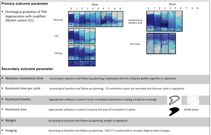
The primary outcome is the microscopic scoring of destructive changes in the TMJ using a modified Mankin scoring system [22]. Secondary outcomes are imaging scoring of TMJ destructive changes, absolute masticatory time, ruminant time per cycle, ruminant kinetics, ruminant area, and sheep body weight. Primary and secondary outcome parameters are outlined in more detail in Figure 3.
Figure 3. Primary and secondary outcome parameters.
Primary Outcome
The goal is to evaluate histologic gradation of TMJ destructive changes. The time point is 6 months following surgical intervention.
Six months after surgery, the TMJ is removed using a necropsy bone oscillatory saw according to the following anatomic references: cranial (cranial aspect of coronoid process in the union region of the zygomatic process), caudal (external to acoustic meatus), dorsal (reference is established to the squamous temporal bone), and ventral (reference is fixed 2 cm below the acoustic meatus in the zone of stylohyoid angle). The joints are fixed in 10% buffered formalin for 24 hours and stored
in 70% ethanol. Decalcification is obtained by immersion in 10% formic acid in 5% formalin for up to 20 days, after which the articulations are cut sagittally through the whole condyle. After decalcifying, TMJ articulations are immersed in three graded methyl salicylate/paraffin mixtures and cut sagittally through the lateral into the central part of the TMJ. Histological sections are sent to Sydney Institute of Bone and Joint Research for histological scoring using a modified Mankin scoring system [22]. This assessment is performed and classified independent by two histologists who will be blinded to intervention. A third histologist will act as arbiter in case of disparity.
Secondary Outcomes
The features evaluated are imaging analysis, absolute masticatory time, ruminant time per cycle, ruminant kinematics, ruminant area, and sheep weight (see Multimedia Appendices
1 and 2). Time point is every month following surgical
intervention for a total of 6 months.
To measure secondary outcomes, a specific cage (see Figure 4) was built with a frontal window and a feeder.
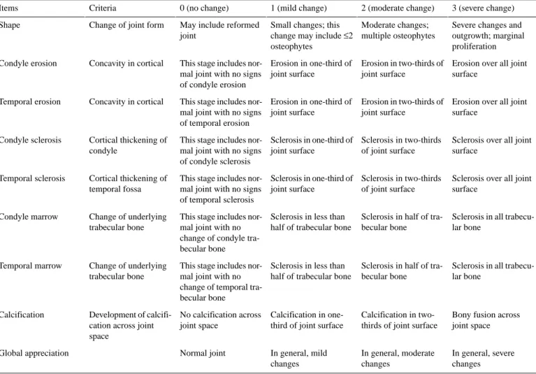
Imaging analysis: preoperative CT is performed on all sheep. After animal sacrifice, TMJ blocks are scanned by CT and imaging evaluation is performed using the criteria and score described in Table 1.
Table 1. TEMPOJIMS imaging evaluation criteria.
3 (severe change) 2 (moderate change) 1 (mild change) 0 (no change) Criteria Items
Severe changes and outgrowth; marginal proliferation Moderate changes;
multiple osteophytes Small changes; this
change may include ≤2 osteophytes
May include reformed joint
Change of joint form Shape
Erosion over all joint surface
Erosion in two-thirds of joint surface
Erosion in one-third of joint surface
This stage includes nor-mal joint with no signs of condyle erosion Concavity in cortical
Condyle erosion
Erosion over all joint surface
Erosion in two-thirds of joint surface
Erosion in one-third of joint surface
This stage includes nor-mal joint with no signs of temporal erosion Concavity in cortical
Temporal erosion
Sclerosis over all joint surface
Sclerosis in two-thirds of joint surface Sclerosis in one-third of
joint surface This stage includes
nor-mal joint with no signs of condyle sclerosis Cortical thickening of
condyle Condyle sclerosis
Sclerosis over all joint surface
Sclerosis in two-thirds of joint surface Sclerosis in one-third of
joint surface This stage includes
nor-mal joint with no signs of temporal sclerosis Cortical thickening of
temporal fossa Temporal sclerosis
Sclerosis in all trabecu-lar bone
Sclerosis in half of tra-becular bone
Sclerosis in less than half of trabecular bone This stage includes
nor-mal joint with no change of condyle tra-becular bone Change of underlying
trabecular bone Condyle marrow
Sclerosis in all trabecu-lar bone
Sclerosis in half of tra-becular bone
Sclerosis in less than half of trabecular bone This stage includes
nor-mal joint with no change of temporal tra-becular bone
Change of underlying trabecular bone Temporal marrow
Bony fusion across joint space Calcification in
two-thirds of joint surface Calcification in
one-third of joint surface No calcification across
joint space Development of
calcifi-cation across joint space Calcification In general, severe changes In general, moderate changes In general, mild changes Normal joint Global appreciation
This assessment is performed and classified independently by two experienced radiologists who will be blinded to intervention. A third radiologist will act as arbiter in case of disparity. Absolute masticatory time: respecting the flowchart (Figure 2), at 9:00 am the animals are placed in individual cages. A dose of 150 grams of dry pellets (Rico Gado A3) are introduced in the feeder and the time until they eat all the pellets is measured with a chronometer (see Multimedia Appendix 1).
Ruminant time per cycle: respecting the timetable (Figure 2), we record 15 ruminatory cycles approximately 4 hours after 150 gram feeding. We use a Canon 7D video camera and images with 25 frames per second. Then, the number of frames per cycle are divided by 25 to obtain time in seconds per cycle (see
Multimedia Appendix 2).
Ruminant kinetics: we use the software Foundry Nuke (2D tracking) to perform the ruminatory tracking and to obtain the ruminatory cycle average. With the software After Effects , we convert the 2-D tracking into a geometric form (see Multimedia Appendix 2).
Ruminant area: we determine the average of 15 cycles and create a geometric form. Using the software Image J , we perform a quantitative measure in pixels of the ruminant area average. Weight: according to the timetable, after eating 150 grams of dry pellets the sheep are weighed (see Multimedia Appendix 1).
All assessments are performed by researchers who are blinded to surgical intervention.
Figure 4. TEMPOJIMS main facilities.
Statistical Analyses
All statistical analyses will be performed using the SPSS version 22 (IBM Corp, Armonk, NY, USA). A cross-sectional analysis will be performed to compare the outcome variables in the three levels of the independent variable before and after the randomized treatment group assignment. In the cross-sectional analyses, one-way analysis of variance (ANOVA) will be performed, after testing all the assumptions. For longitudinal analysis, one-way ANOVA with repeated measures will be performed taking as within-subjects effects observations after surgery (months 1 to 6). Fisher least significant difference will be performed as post hoc tests to check for significant differences for the different treatments.
Reporting of Adverse Events
Adverse events related to the study will be considered, including (1) anesthesia events: idiopathic death, pneumothorax, other complications related to anesthesia; (2) surgical technique: massive bleeding, condylar fracture, other complications related to surgical technique; and (3) postoperative events: TMJ infection, suture dehiscence, decreased appetite, facial paresis, decreased rumination, decreased weight.
Discussion
This study investigates the effects and adverse effects of (1) bilateral discectomy, (2) bilateral discopexy, and (3) bioengineered disk implants. Although this preclinical study
will primarily serve as a pilot study, we expect to gain a better understanding of the morphologic and histologic changes in TMJ and implications in masticatory kinetics.
So far, results on discectomy are conflicting. Previous preclinical studies in this field [24-33] have used the contralateral unoperated side as a control and different animal models ranging from mice to a canine model. Using the contralateral side as a control can be inappropriate considering contralateral overload influence. Theoretically, we expect to reduce this bias using a bilateral approach. Animal variability in the different studies is a warning about the importance of using the same animal model in further studies regarding TMJ implant investigations. Therefore, our group performed a previous study considering black Merino sheep as a promisor animal model for studies regarding TMJ disk implants investigation, TMJ prosthesis, and TMJ osteoarthritis model. To increase the quality of TEMPOJIMS the authors will use a sham surgery control group. We expect to obtain valuable information related to the phase 1 discopexy group regarding if the surgical approach promotes intra-articular damage. This can improve future conclusions about attributing possible damage to the intervention itself instead of the TMJ implant. This question is important considering that a surgical approach to place TMJ implants in phase 2 will be required. Again, using a bilateral intervention could reduce a possible bias.
Most preclinical studies have focused on gross morphological/histological assessments and were not designed
to characterize the fundamental altered joint movement (kinetics) or functional consequences. In this study, we include pilot secondary outcomes to evaluate changes in ruminant kinetics. We expect to correlate the primary with the secondary outcomes to understand if they can be used in future TMJ studies. It may be interesting to understand several items:
1. Are there differences regarding masticatory time in the disk groups versus discectomy and discopexy?
2. Is there a correlation between histologic and imaging and kinetics results?
3. Does the ruminant area and geometry change when performing different interventions?
4. Is there a difference regarding ruminant kinetics in the disk groups versus discectomy and discopexy?
5. Do TMJ implants accelerate osteoarthritis?
Concerning phase 2, the choice of biomaterial is critical. The TMJ implant will be exposed in a mechanical, stressful
environment with a limited blood supply that can limit cell migration and in situ regeneration. Testing three different bioengineering discs in vivo and correlating in vitro with in vivo behavior can seriously improve bioengineering strategies to achieve a safe and efficacious TMJ disk implant for humans. The main strength of this study is the animal model proposed; the conventional and pilot outcomes described; the study design with a randomized, blinded, and placebo control group; and the use of bilateral surgical procedures. Potential limitations of the study include the relatively small sample size. If this study confirms the feasibility of the proposed protocol and initial efficacy of the TMJ disk implants planned, a larger preclinical trial would be warranted to further determine the effectiveness of these discs and promote translation of animal evidence to clinical practice in humans.
Trial Status
At the time of submission, the surgical interventions of phase 1 were ongoing at Faculdade de Medicina Veterinária de Lisboa and TEMPOJIMS facilities in Portugal.
Acknowledgments
This preclinical trial is supported by Faculdade de Medicina Veterinária da Universidade de Lisboa, Instituto Politécnico de Leiria (Centre for Rapid and Sustainable Product Development), Centro Hospitalar de Setúbal, Instituto de Medicina Molecular, Faculdade de Medicina da Universidade de Lisboa. The authors are grateful to Joaquim Ferreira from Lisbon Faculty of Medicine for study design; to Susan Smith from Institute of Bone and Joint Research-Northern Sydney Local Health District-Sydney Medical School Northern, Australia, for histological analysis; to Pedro Nunes from Radiology Department of Centro Hospitalar Lisboa Norte; to Miguel Virgílio for kinematics video recording; and to Joaquim Ângelo and Ermelinda Ângelo for animal logistics control. This study was granted by Portuguese Grunenthal Foundation and by Secção Regional Oeste da Ordem dos Médicos. This publication was supported by the Portuguese Foundation for Science and Technology (FCT) through the following projects: UID/Multi/04044/2013 and PTDC/EMS-SIS/7032/2014.
Authors' Contributions
The contributors, with input from the other investigators, conceived this study protocol. JF, RF, NG, AT, NG, and DA developed the protocol and study materials with input from all investigators. NG, AT, and DA participated in the randomization process. LM will conduct the statistical analyses. FM, RG, and SF will participate in the surgical interventions. CB and SC are the coordinators of the veterinary staff and responsible for the animal anesthesia and animal welfare. DC participated in organization support and was study advisor. PM, NA, and MC are dedicated to disk implants 1 and 2. WY, JE, and GJ are dedicated to disk implant 3. SR will coordinate the imaging evaluation. MP and FB are responsible for processing the histologic samples and preparing sections. LC group will coordinate histologic scoring system. All authors read and approved the final manuscript.
Conflicts of Interest
None declared.
Multimedia Appendix 1
Outcomes assessments in TEMPOJIMS main facilities, absolute masticatory time and weight. [MP4 File (MP4 Video), 230MB-Multimedia Appendix 1]
Multimedia Appendix 2
Outcomes assessments in TEMPOJIMS main facilities. After recording 15 ruminant cycles with a Canon 7D Video Camera we used the software Foundry Nuke (2D tracking) to make the ruminant tracking to obtain the ruminant cycle average in each time period.
[MP4 File (MP4 Video), 4MB-Multimedia Appendix 2]
1. Bae Y, Park Y. The effect of relaxation exercises for the masticator muscles on temporomandibular joint dysfunction (TMD). J Phys Ther Sci 2013;25(5):583-586. [doi: 10.1589/jpts.25.583]
2. Allen K, Athanasiou K. Tissue engineering of the TMJ disc: a review. Tissue Eng 2006;12(5):1183-1196. [doi:
10.1089/ten.2006.12.1183]
3. Tanaka E, Sasaki A, Tahmina K, Yamaguchi K, Mori Y, Tanne K. Mechanical properties of human articular disk and its influence on TMJ loading studied with the finite element method. J Oral Rehabil 2001 Mar;28(3):273-279. [Medline:
11394374]
4. Martins-Júnior RL, Palma Antônio JC, Marquardt EJ, Gondin TM, Kerber FD. Temporomandibular disorders: a report of 124 patients. J Contemp Dent Pract 2010 Oct 14;11(5):71-78. [Medline: 20978727]
5. Navrátil L, Navratil V, Hajkova S, Hlinakova P, Dostalova T, Vranová J. Comprehensive treatment of temporomandibular joint disorders. Cranio 2014 Jan;32(1):24-30. [doi: 10.1179/0886963413Z.0000000002] [Medline: 24660643]
6. Mehra P, Wolford LM. The Mitek mini anchor for TMJ disc repositioning: surgical technique and results. Int J Oral Maxillofac Surg 2001 Dec;30(6):497-503. [doi: 10.1054/ijom.2001.0163] [Medline: 11829231]
7. Al-Baghdadi M, Durham J, Araujo-Soares V, Robalino S, Errington L, Steele J. TMJ disc displacement without reduction management: a systematic review. J Dent Res 2014 Jul;93(7 Suppl):37S-51S [FREE Full text] [doi:
10.1177/0022034514528333] [Medline: 24659775]
8. Estabrooks L, Fairbanks C, Collett R, Miller L. A retrospective evaluation of 301 TMJ Proplast-Teflon implants. Oral Surg Oral Med Oral Pathol 1990 Sep;70(3):381-386. [Medline: 2216373]
9. Henry CH, Wolford LM. Treatment outcomes for temporomandibular joint reconstruction after Proplast-Teflon implant failure. J Oral Maxillofac Surg 1993 Apr;51(4):352-358; discussion 359. [Medline: 8450350]
10. Mercuri LG, Giobbie-Hurder A. Long-term outcomes after total alloplastic temporomandibular joint reconstruction following exposure to failed materials. J Oral Maxillofac Surg 2004 Sep;62(9):1088-1096. [Medline: 15346359]
11. Morouço P, Ângelo D, Francisco L, Moura C, Alves N. Tissue engineering for temporomandibular joint disc repair and regeneration: a methodological perspective. Adv Cell Mol Otolaryngol 2017 Jan 17;4(1):33709. [doi: 10.3402/acmo.v4.33709] 12. Lypka M, Yamashita DR. Exuberant foreign body giant cell reaction to a teflon/proplast temporomandibular joint implant: report of a case. J Oral Maxillofac Surg 2007 Sep;65(9):1680-1684. [doi: 10.1016/j.joms.2006.09.030] [Medline: 17719383] 13. Spagnoli D, Kent JN. Multicenter evaluation of temporomandibular joint Proplast-Teflon disk implant. Oral Surg Oral Med
Oral Pathol 1992 Oct;74(4):411-421. [Medline: 1408011]
14. Fricton JR, Look JO, Schiffman E, Swift J. Long-term study of temporomandibular joint surgery with alloplastic implants compared with nonimplant surgery and nonsurgical rehabilitation for painful temporomandibular joint disc displacement. J Oral Maxillofac Surg 2002 Dec;60(12):1400-1411; discussion 1411. [doi: 10.1053/joms.2002.36091] [Medline: 12465000] 15. Kulber DA, Davos I, Aronowitz JA. Severe cutaneous foreign body giant cell reaction after temporomandibular joint
reconstruction with Proplast-Teflon. J Oral Maxillofac Surg 1995 Jun;53(6):719-722; discussion 722. [Medline: 7776062] 16. Chuong R, Piper MA. Cerebrospinal fluid leak associated with proplast implant removal from the temporomandibular joint.
Oral Surg Oral Med Oral Pathol 1992 Oct;74(4):422-425. [Medline: 1408012]
17. Berarducci JP, Thompson DA, Scheffer RB. Perforation into middle cranial fossa as a sequel to use of a Proplast-Teflon implant for temporomandibular joint reconstruction. J Oral Maxillofac Surg 1990 May;48(5):496-498. [Medline: 2329399] 18. [No authors listed]. Recommendations for management of patients with temporomandibular joint implants.
Temporomandibular Joint Implant Surgery Workshop. J Oral Maxillofac Surg 1993 Oct;51(10):1164-1172. [Medline:
8410459]
19. Herring S. TMJ anatomy and animal models. J Musculoskelet Neuronal Interact 2003 Dec;3(4):391-394; discussion 406 [FREE Full text] [Medline: 15758330]
20. Angelo D, Morouço P, Alves N, Viana T, Santos F, González R. Choosing sheep (Ovis aries) as animal model for temporomandibular joint research: Morphological, histological and biomechanical characterization of the joint disc. Morphologie 2016 Jul:223-233 [FREE Full text] [doi: 10.1016/j.morpho.2016.06.002] [Medline: 27450042]
21. Leiggener CS, Erni S, Gallo LM. Novel approach to the study of jaw kinematics in an alloplastic TMJ reconstruction. Int J Oral Maxillofac Surg 2012 Sep;41(9):1041-1045. [doi: 10.1016/j.ijom.2012.06.014] [Medline: 22819692]
22. Little CB, Smith MM, Cake MA, Read RA, Murphy MJ, Barry FP. The OARSI histopathology initiative - recommendations for histological assessments of osteoarthritis in sheep and goats. Osteoarthritis Cartilage 2010 Oct;18 Suppl 3:S80-S92 [FREE Full text] [doi: 10.1016/j.joca.2010.04.016] [Medline: 20864026]
23. Cake MA, Appleyard RC, Read RA, Smith MM, Murrell GA, Ghosh P. Ovariectomy alters the structural and biomechanical properties of ovine femoro-tibial articular cartilage and increases cartilage iNOS. Osteoarthritis Cartilage 2005
Dec;13(12):1066-1075 [FREE Full text] [doi: 10.1016/j.joca.2005.07.001] [Medline: 16154775]
24. Cohen W, Servais J, Polur I, Li Y, Xu L. Articular cartilage degeneration in the contralateral non-surgical temporomandibular joint in mice with a unilateral partial discectomy. J Oral Pathol Med 2014 Feb;43(2):162-165. [doi: 10.1111/jop.12113] [Medline: 24044578]
25. Ahtiainen K, Mauno J, Ellä V, Hagström J, Lindqvist C, Miettinen S, et al. Autologous adipose stem cells and polylactide discs in the replacement of the rabbit temporomandibular joint disc. J R Soc Interface 2013 Aug 06;10(85):20130287 [FREE Full text] [doi: 10.1098/rsif.2013.0287] [Medline: 23720535]
26. Brown B, Chung W, Almarza A, Pavlick M, Reppas S, Ochs M. Inductive, scaffold-based, regenerative medicine approach to reconstruction of the temporomandibular joint disk. J Oral Maxillofac Surg 2012 Nov;70(11):2656-2668. [doi:
10.1016/j.joms.2011.12.030]
27. Xu L, Polur I, Lim C, Servais J, Dobeck J, Li Y, et al. Early-onset osteoarthritis of mouse temporomandibular joint induced by partial discectomy. Osteoarthritis Cartilage 2009 Jul;17(7):917-922. [doi: 10.1016/j.joca.2009.01.002] [Medline:
19230720]
28. Lai WT, Tsai Y, Su S, Su C, Stockstill JW, Burch JG. Histological analysis of regeneration of temporomandibular joint discs in rabbits by using a reconstituted collagen template. Int J Oral Maxillofac Surg 2005 May;34(3):311-320. [doi:
10.1016/j.ijom.2004.05.003] [Medline: 15741041]
29. Sato S, Goto S, Koeda S, Motegi K. Changes of the elastic fibre network of the rabbit temporomandibular joint following discectomy. J Oral Rehabil 2002 Sep;29(9):847-852. [Medline: 12366539]
30. Sato S, Goto S, Motegi K. Changes of the collagen fibre arrangement of the rabbit temporomandibular joint following discectomy. J Craniomaxillofac Surg 2002 Jun;30(3):178-183. [Medline: 12220997]
31. Bjørnland T, Haanaes HR. Discectomy of the temporomandibular joint: an experimental study in monkeys. J Craniomaxillofac Surg 1999 Apr;27(2):113-116. [Medline: 10342149]
32. Ogi N, Kurita K, Ishimaru JI, Goss AN. Short-term effect of the use of a frozen-stored disc allograft for repair of the osteoarthritic sheep temporomandibular joint: a preliminary report. J Oral Maxillofac Surg 1999 Feb;57(2):139-144; discussion 144. [Medline: 9973121]
33. Hinton R, Stinson J. Effect of postoperative diet on condylar cartilage response to discectomy. J Oral Maxillofac Surg 1997 Nov;55(11):1259-1264. [Medline: 9371117] Abbreviations CT: computerized tomography PCL: polycaprolactone PDS: poly-p-dioxanone PGS: poly(glycerol sebacate)
TEMPOJIMS: Temporomandibular Joint Interposal Material Study THF: tetrahydrofuran
TMD: temporomandibular joint disorders TMJ: temporomandibular joint
Edited by G Eysenbach; submitted 10.10.16; peer-reviewed by W Cohen, A Almarza, P Boffano; comments to author 08.12.16; accepted 16.12.16; published 02.03.17
Please cite as:
Ângelo DF, Monje FG, González-García R, Little CB, Mónico L, Pinho M, Santos FA, Carrapiço B, Gonçalves SC, Morouço P, Alves N, Moura C, Wang Y, Jeffries E, Gao J, Sousa R, Neto LL, Caldeira D, Salvado F
Bioengineered Temporomandibular Joint Disk Implants: Study Protocol for a Two-Phase Exploratory Randomized Preclinical Pilot Trial in 18 Black Merino Sheep (TEMPOJIMS)
JMIR Res Protoc 2017;6(3):e37
URL: http://www.researchprotocols.org/2017/3/e37/
doi: 10.2196/resprot.6779
PMID: 28254733
©David Faustino Ângelo, Florencio Gil Monje, Raúl González-García, Christopher B Little, Lisete Mónico, Mário Pinho, Fábio Abade Santos, Belmira Carrapiço, Sandra Cavaco Gonçalves, Pedro Morouço, Nuno Alves, Carla Moura, Yadong Wang, Eric Jeffries, Jin Gao, Rita Sousa, Lia Lucas Neto, Daniel Caldeira, Francisco Salvado. Originally published in JMIR Research Protocols (http://www.researchprotocols.org), 02.03.2017. This is an open-access article distributed under the terms of the Creative Commons Attribution License (http://creativecommons.org/licenses/by/2.0/), which permits unrestricted use, distribution, and reproduction in any medium, provided the original work, first published in JMIR Research Protocols, is properly cited. The complete bibliographic information, a link to the original publication on http://www.researchprotocols.org, as well as this copyright and license information must be included.