Bone Physiology as Inspiration for Tissue Regenerative Therapies
Diana Lopes
1,a, Cláudia Martins-Cruz
1,a, Mariana B. Oliveira
1,*João F. Mano
1,*1
Department of Chemistry, CICECO – Aveiro Institute of Materials, University of Aveiro,
3810 193 Aveiro, Portugal.
a
Authors contributed equally to this work.
*
Corresponding author. E-mails: M.B. Oliveira:
[email protected]
; J.F. Mano:
[email protected]
Abstract
The development, maintenance of healthy bone and regeneration of injured tissue in the human body comprise a set of intricate and finely coordinated processes. However, an analysis of current bone regeneration strategies shows that only a small fraction of well-reported bone biology aspects has been used as inspiration and transposed into the development of therapeutic products. Specific topics that include inter-scale bone structural organization, developmental aspects of bone morphogenesis, bone repair mechanisms, role of specific cells and heterotypic cell contact in the bone niche (including vascularization networks and immune system cells), cell-cell direct and soluble-mediated contact, extracellular matrix composition (with particular focus on the non-soluble fraction of proteins), as well as mechanical aspects of native bone will be the main reviewed topics. In this Review we suggest a systematic parallelization of (i) fundamental well-established biology of bone, (ii) updated and recent advances on the understanding of biological phenomena occurring in native and injured tissue, and (iii) critical discussion of how those individual aspects have been translated into tissue regeneration strategies using biomaterials and other tissue engineering approaches. We aim at presenting a perspective on unexplored aspects of bone physiology and how they could be translated into innovative regeneration-driven concepts.
1. Introduction
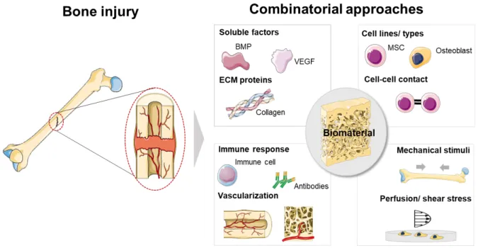
Bone physiology involves the coordinated regulation of a myriad of biological processes that lead tissue development, homeostasis and repair upon trauma [1]. Bone regenerative processes can thus be highly complicated to emulate, partially due to the numerous and multifactorial cues contributing for the regulation of its niche. Those include the tissue’s complex composition and intricate associated pathways involving aspects like bone’s soluble microenvironment, extracellular matrix (ECM) insoluble proteins and glycoprotein composition and renewal, cell-cell and cell-ECM interactions mechanical stimulation, or the role of microRNAs. The understanding of the individual and coordinated role of each intervenient on bone tissue physiology, as well as their interconnected actions, has inspired the design of a plethora of biomimetic and/or bioinspired bone regeneration approaches. In fact, there is an urgent need for the development of new, effective and compatible treatments for bone-related injuries. Age-related fractures are expected to increase in the United States from 2.1 million in 2005 to over 3 million fractures in 2025, solely considering the elderly/aging population at risk [2]. In Europe, the annual number of fractures is estimated to rise 28% from 2010 to 2025, with an absolute increase from 3.5 million to 4.5 million injuries, respectively [3]. The application of concepts learnt from nature for the emulation of the structure and physiology of healthy bone, however, requires insightful and careful approaches. Although mimicking bone’s structure and function based on its physiology is an attractive idea, the implementation of effective reproducible therapies is most often dependent on achieving a balance between sufficient complexity to warrant function, along with ease/speed of manufacturing and regulatory compliance.
The study of individual factors in biologically non-representative environments in most available literature concerning bone regeneration may be hindering the disclosure of unprecedented valuable results. The effects of (bio)chemical (e.g. presence of soluble cytokines, as bone morphogenic proteins – BMPs), structural and chemical properties (e.g. different biomaterials chemistries and architectures), or externally applied mechanical stimuli (e.g. compressive stress or flow perfusion) on bone regeneration strategies have been explored in a unifactorial fashion, focusing on single stimuli for the design of biomaterials and other regenerative approaches.
Interestingly, though, a growing body of knowledge comprising both fundamental and applied studies focusing on bone’s response to engineered cues have undoubtedly proven that bone is a complex and dynamic system, in which different biological processes and structural characteristics play complementary roles towards the successful regeneration and maintenance of bone’s healthy behavior [1]. The advent of stem cells biology, as well as the progressive know-how on the structural, biophysical and biochemical role of the ECM components and the scrutiny of immune cells crosstalk supported recent advances in the design of biomaterials targeting bone regeneration [4]. The knowledge of such complexity may be an effective manner to pave the way for the design of multifactorial strategies targeting bone regeneration and disease treatment [5]. Some high-throughput studies have approached multifactorial aspects of bone regeneration as crosstalk between different cell types (including mineral-forming cells, reabsorption cells, immune cells and vascular cells) [6, 7], the combinatorial role of ECM proteins [8, 9], the effect of physical factors as biomaterials bioactivity, stiffness and viscoelasticity [10, 11], as well as the role of extrinsic mechanical forces actuating in the native bone (e.g., compression and shear stress) [12]. Although some key aspects of bone physiology are well studied and have led the development of therapies, several aspects still remain poorly understood and explored as potential added-value assets to enhance regeneration. Some examples include the crosstalk of bone resident cells with immune cells, the role of hematopoietic stem cells (HSC) on regenerative phenomena, or the tight control of time- and space-coordinated biochemical and biophysical signaling [13].
This Review sets the objective of establishing an unprecedented correlation between (i) well-known and recently reported bone physiology phenomena and (ii) the use of this know-how for the development of well-established or proof-of-concept bone-healing therapeutic approaches. A thorough analysis is performed to identify aspects of bone biology lacking exploitation for regenerative therapies, which may represent a source of innovative ideas for novel and impactful future developments on the field.
1.1. The need for regenerative medicine therapies – analysis of critical aspects to achieve bone regeneration
While bone tissue trauma normally heals by itself, the so-called “critical” defects - with average diameters of 2 cm or higher in humans - do not show this spontaneous ability [14]. Such defects with poor healing ability often derive from tumor ablation, serious injury and orthopedic diseases [15]. Specifically considering physically-caused injuries, including a range of fractures from standard injury to open tibial defects, bone healing repair failure percentage can go from 10 to 50% [16]. Failure on bone healing will ultimately culminate in the suppression of blood supply to the tissue, which will result in the non-union of the bone due to ischemia, osteonecrosis and bone loss [17].
Efforts to repair bone defects excluding the ones that specifically target bone regeneration can be divided in two main segments: (i) implantation of bone grafts (of autologous or allogenic origin) or (ii) development of synthetic permanent bone substitution grafts [18]. Limitations have been reported for both therapeutic approaches. Although commonly applied in clinics and known to foster bone repair, autologous bone grafts, inflict morbidity of the donor’s extraction site [18, 19]. Allogeneic bone grafts may potentially be rejected due to host-to-graft immune response. Moreover, the implantation of allografts requires a complex implantation technique that involves the achievement of constant vascular supply to the site, as well as a maintenance of an adequate mechanical environment to promote vessels formation [20]. Permanent substitution grafts, manufactured from non-degradable materials, have been associated with unwanted side effects including bone resorption, poor osseointegration and triggering of adverse (e.g. allergenic) reactions on the host [18]. Current strategies based on synthetic grafts for bone healing are out of the scope of this Review. Thorough reviews of this important topic related to bone repair therapies can be found in References [21, 22].
Well-reported problems associated with the current bone repair approaches show that there is a dire need for the development and marketplace implementation of new and more efficient strategies. In opposition to previously mentioned techniques focused on tissue repair, tissue engineering seeks the complete regeneration of damaged tissues through the in vitro and/or in
vivo-synthesis of novel biological matrices with equivalent properties to those of the original healthy
tissue. Four main pillars may be used separately or in combined design strategies to promote bone regeneration: (i) biomaterials, (ii) biomolecules, (iii) cells and (iv) externally-applied stimuli. The following sections of this Review will exploit bioinspired rationales behind the use of different tissue engineering players through a parallelization strategy with native bone’s physiological phenomena.
2. An Inter-Scale View of Bone Structure
Bone is the anatomic structure responsible for the movement, protection, maintenance of mineral homeostasis and structural support of the human body. A fully-grown adult’s skeleton is composed of 206 individual bones. Human bones are divided in five major categories, which include long (aimed at supporting body weight; e.g. clavicles, raddi, metacarpals, tibiae, phalanges, femurs, humeri, metatarsals, fibulae and ulnae), short (providing movement and stability; e.g. tarsal and carpal), flat (targeted at internal organs protection; e.g. skull, sternum, mandible, ribs and scapulae), irregular (e.g. vertebrae, coccyx, sacrum and hyoid) and sesamoid bones (embedded at tendons; e.g. patella) [23].
During tissue formation, two bone types can be identified: (i) the woven/primary bone, which appears during the embryonic development and fracture repair, and consists of osteoid (unmineralized ECM) with collagen fibers that show little or no determined orientation in the three-dimensional (3D) space, along with a random distribution of cells [24, 25]. This is a transient structure, which is later replaced by mature lamellar bone; and (ii) the lamellar/secondary bone, which constitutes the adult skeleton and consists of highly organized sheets of mineralized osteon [26]. This structure is stronger and more rigid, and less elastic than the woven bone [24].
Lamellar bone is constituted by both cortical/compact and trabecular/cancellous bone. The first one is dense, solid and located in the most outer part of the tissue; on its turn, cancellous bone contains a sponge-like structure with interconnecting cavities and is located at the internal section of bone. Both cortical and cancellous bones are composed of osteons. The ratio of both bone types varies according to the anatomical site (e.g. femoral head with a 50:50 cortical to trabecular ratio; vertebra with 25:75; radial diaphysis with 95:5; and overall, the human skeleton with a 80:20
ratio) [1]. On a structural perspective, in cortical bone the lamellae with about 3 µm thickness, is organized into concentric circles, surrounding a vertical Haversian canal containing blood vessels and nerves. This entire structure is designated the osteon or Haversian system, and is the functional unit of bone. The system is formed when osteoclasts remodel existing bone, leaving cylindrical cavities that are subsequently filled with osteoblasts that produce lamellae around the Haversian canal [24, 27]. Osteocytes are located between lamellae, within small cavities known as lacuna, interconnected by a series of tunnels called canaliculi [1, 24]. The trabecular bone is composed of large spaces, with a honeycomb-like network of trabecular plates. The matrix consists of a 3D network of fine columns that crosslink to form the trabeculae [1, 24]. This results in a light and porous bone that is strong against multidirectional forces, and crucial to enable body movement. The spaces between trabeculae are filled with bone marrow. A fibrous connective tissue layer, called periosteum, surrounds the external surface of cortical bone, while in the inner section, a membranous structure - the endosteum - covers the surface of cortical and trabecular bone. The latter is also in contact with the bone marrow space, blood vessels canals and is composed by blood vessels, osteoblasts and osteoclasts [1]. Both periosteum and endosteum have inspired the development of two-dimensional (2D) membranes to mimic these anatomical structures and assist tissue regeneration processes [28].
On a nanoscale perspective, bone is constituted by a plethora of structural proteins and polysaccharides, which main composition is collagen fibrils with diameters between 35 and 60 nm, and up to 1 μm length, organized with a 67 nm periodicity and 40 nm gaps [29-31]. These fibrils are mineralized by the anisotropic and extremely stiff inorganic component - hydroxyapatite crystals [32] - that lay in the collagen gaps [31]. Interestingly, despite the variations on bone structural shapes depending on bone types and throughout different species, the mineralized collagen fibrils observed in humans are highly conserved across species and bone types [33]. Bone organic and inorganic phases are thought to have an interplay that allows load transfer. Gupta et al. [34] showed that both components deform elastically during initial loading, although to different degrees. This specific deformation pattern is hypothesized to culminate in fibril-matrix decoupling targeting the protection of the brittle mineral phase, improving the effective
redistribution of strain energy in the tissue. Nanostructured scaffolds based on nanomaterials capable of closely resembling the native ECM structure have been designed. According with some authors, such constructs demonstrate a beneficial effect concerning the formation of functional tissue due to an improvement of the interactions at the cellular and protein level [29, 30]. Although the macro- and microstructure of bone closely replicated using porous scaffolds, the emulation of the osteon organization is dependent on a nanometric control of materials distribution. In fact, a precise combination of micro- and nanometric aspects of the bone structure is fundamental for the development of truly mimetic structured biomaterials. Nonetheless, the effective need for highly organized osteon/adult bone interscale mimetic biomaterials is not unanimous. Effective biomaterials capable of triggering osteogenic differentiation and bone repair have been often based on biophysical and biochemical cues inspired on bone developmental processes, instead of adult bone structural features. These strategies are composed of much simpler units than the adult completely formed bone itself. The role of nanostructured biomaterials as cue providers targeting bone regeneration must not be overlooked. Platforms exhibiting, for example, topographic, pro-differentiation and pro-mineralization cues through techniques as nanopatterning, electrospinning, and development of nanocomposites [30] has led to some of the most interesting outputs in the field. Complete reviews correlating (i) nanomaterials and their similarity with the native bone niche and (ii) their bone regeneration outputs can be found in References [30, 35-38].
3. Bone Development Mechanisms
During mammals’ fetal development and natural bone repair upon injury, bone formation is achieved through two processes: intramembranous and endochondral ossification [1]. The primary structure for these two mechanisms is the woven (or immature hollow) bone, that is readily replaced by the lamellar/secondary bone (parallel fibrils deposited in opposite directions) [25]. The formation of lamellar bone occurs at a much slower rate than that of woven bone [39]. This structure does not appear only in the fetal life, but every time a bone suffers a non-critical injury [25].
3.1. Intramembranous bone formation
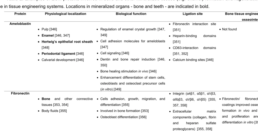
Intramembranous ossification is the most primitive form of ossification, with the first proof of its existence dating to 500 million years BP, in opposition to endochondral ossification, with first reported case with 100 million years BP [40]. In intramembranous ossification, mesenchymal stem cells (MSCs) present in mesenchyme or in the medullary cavity caused by a bone injury -differentiate into osteoblasts. In fetal development, this process is mainly responsible for the formation of the flat skull bones and some parts of the clavicles [25, 41]. Unlike in the endochondral ossification process, in intramembranous ossification the bone is formed without a cartilaginous intermediate. The formation of a nidus – a cluster of undifferentiated MSCs – is the starting point for the intramembranous ossification process. Cells in these clusters stop their proliferation and develop into the osteoprogenitor phenotype, and then eventually differentiate into osteoblasts, through an intermediate pre-osteoblast lineage [41, 42]. Runx2 is one of the most important early transcription factors responsible for osteoblastic differentiation [43, 44]. The expression of Runx2 is dependent on the Wnt signaling, which leads to high levels of β-catenin in MSCs. In turn, Runx2 induces the later expression of the transcription factor gene Osterix, also involved in the differentiation of MSCs to osteoblasts [45]. After full differentiation, osteoblasts produce a non-mineralized type I collagen-rich fibrillar ECM: the osteoid. While entrapped in this matrix, osteoblasts differentiate into mature osteocytes, and the matrix is further mineralized. The described mechanism is also considered by most authors as the backbone for the formation of subperiosteal bone and, thus, the process behind the woven and lamellar bone type formation in this region [46]. However, this concept has been challenged, and the mechanism behind periosteum surface ossification was suggested as developmentally distinct [45]. A schematic representation of the intramembranous ossification process can be found in Figure 1a.
3.2. Endochondral ossification
During endochondral ossification chondrocytes from surrounding cartilage tissues initially form a matrix template, the growth plate, and then differentiate into bone structures [43]. This ossification
process drives the embryonic formation of long bones. When chondrocytes’ morphology is round, these cells synthesize type II collagen [47] and subsequently form a columnar layer, becoming pre-hypertrophic. They eventually differentiate into post-mitotic hypertrophic cells, which release type X collagen, mineralizing the surrounding matrix, leading the formation of the bone structure [47]. During bone formation various cycles of death of hypertrophic chondrocytes occur, which is accompanied by the invasion of blood vessels, leading to the replacement of the initially collagenous matrix by trabecular bone, also known as primary spongiosa [48]. As the process continues, trabecular bone is resorbed, and its center is split into different plates. While chondrocytes are present in the plates the previous process continues [48]. The adequate differentiation of chondrocytes into the hypertrophic phenotype is of extreme importance for the genesis and proliferation of bone tissue [48]. A schematic representation of the endochondral ossification process can be found in Figure 1b.
For endochondral ossification-driven bone formation, some biochemical factors must be present in specific moments of chondrocyte-to-osteoblast differentiation. Those include:
The Sox Trio: Sox9/5/6. These molecules are responsible for the differentiation of MSCs into the chondrogenic phenotype, as well as for the regulation of the expression of critical genes for the formation of cartilaginous matrix [48-50].
Expression of the fibroblast growth factor (FGF) receptor 3 and a membrane-spanning tyrosine kinase receptor by chondrocytes. These cells contain a domain that binds to extracellular ligands, including FGFs, initiating the receptor’s autophosphorylation, as well as the stimulation of the tyrosine kinase activity, leading to the inhibition of proliferation and growth of chondrocytes [51, 52];
Presence of BMPs, which are responsible for the formation of mesenchymal condensations and of joints in initial stages of endochondral ossification. After condensation, when long bones are already formed, BMP-2, -3, -4, -5 and -7 are released in the perichondrium. BMP-2 and -6 are produced by hypertrophic chondrocytes, and BMP-7 by proliferative chondrocytes. BMPs positively regulate chondrocyte proliferation and negatively modulate chondrocyte terminal differentiation [51, 52];
High level expression of parathyroid hormone-related peptide (PTHrP) by hypertrophic chondrocytes. This peptide binds and activates the receptor parathyroid hormone (PTH)/PTHrP, also activated by PTH (main regulator of calcium/phosphate metabolism and remodulation of the bone). In fact, the PTH/PTHrP complex is the main regulator of bone development and mineral ion homeostasis. The PTH peptide acts by maturing the immature chondrocytes to a hypertrophic phenotype. When the chondrocytes express PTHrP or an activated form of the receptor, a decrease on the cartilage maturation and increase in bone formation is observed [53, 54]. For a successful bone formation, hypertrophic chondrocytes must express high levels of alkaline phosphatase (ALP), osteonectin, osteopontin, bone sialoprotein and osteocalcin [55-60].
Indian hedgehog homolog (Ihh). This protein, present in the embryogenic patterning, controls the endochondral bone formation by inhibiting the differentiation of hypertrophic chondrocytes, therefore delaying the mineralization of the matrix. The control of growth plate elongation is not a chondrocyte property, but a property of the growth plate module arising from the interaction with chondrocytes involved in the negative feedback loop of Ihh/PTHrP. Ihh also acts as a chondrocyte proliferation stimulator, through a PTHrP-independent pathway [52, 61].
Runx2, Runx3 and core-binding factor beta subunit (CBFβ). These three transcription factors have been described in the literature as promoters of chondrocytic hypertrophy, complementing each other in the process [62, 63];
The proteins hypoxia-inducible factor-1 (HIF-1α) and vascular endothelial growth factor (VEGF). These two factors are essential for bone vascularization, in which HIF-1α acts by mediating hypoxic responses, allowing the survival of chondrocytes, and targets VEGF. VEGF is responsible for the stimulation of angiogenesis and vasculogenesis, as well as restoration of the oxygen supply in hypoxic conditions. It is hypothesized that these two proteins act together in a pathway that regulates chondrocytes survival [64, 65].
3.3. Development-mimetic strategies to engineer bone tissue
A close look at the embryonic development pathways of bone has served as inspiration for the design of finely tuned regenerative approaches, in strategies addressed as “developmental
engineering” [66]. Although endochondral ossification is the pathway that gives rise to most of human bones [24], approaches to differentiate stem cells into functional bone cells (namely, osteoblasts) are mostly based on external stimuli provided to undifferentiated cells that include mineralized/mineralizable platforms, which resembles the intramembranous ossification process [67]. This is a much simpler process when compared to endochondral ossification, thus easier to be carried out in vitro and easier to trace overtime; however, it often results in poor vascularization and limited-area bone regeneration. Consequently, endochondral ossification has been hypothesized as advantageous over intramembranous process for tissue engineering due to its inherent ability to form vascularized bone due to the release of VEGF and MMPs by hypertrophic chondrocytes, which allow overcoming associated hypoxia in the tissue [68]. Despite the successful generation of bone tissue reported for endochondral ossification-mimetic strategies, the implantation of tailored mineralized biomaterial matrices has also enabled high quality bone regeneration, in which the final tissue recapitulates key characteristics of the native precursor, including vascular networks. Examples of tissue engineering strategies focused on both intramembranous and endochondral developmental pathways will be reviewed in the following Sections 3.3.1 and 3.3.2.
3.3.1. Regenerative strategies based on intramembranous ossification: the role of mineralized biomaterial matrices
Mineralized biomaterials have been reported as effective promoters of intramembranous ossification-analogous pathways [69-71]. Although in initial approaches their utility was mostly reported exclusively for the treatment of small scale injuries due to their inability to autonomously induce MSCs differentiation, seminal work by Yuan et al.,in 2010, introduced physicochemical and structural tailoring of calcium phosphate-based ceramics as a way to achieve osteoinductive biomaterials capable of promoting the full regeneration of large-scale defects in sheep and dog ectopic and orthotopic models [72]. Calcium phosphates with different chemical compositions – hydroxyapatite, tricalcium phosphate (TCP), and mixtures of both (biphasic calcium phosphates -BCP) were exposed to different post-synthesis sintering temperatures, so different microstructural
features could be obtained (smaller grains for 1150ºC, and larger ones for 1300ºC). Those materials were tested for their ability to induce in vitro MSCs osteogenic differentiation, as well as
in vivo bone formation. TCP showed the highest osteoinductive effect on in vitro cultured MSCs
and the strongest ability to induce bone formation, with outputs similar to the implantation of autografts or treatment with recombinant human BMP-2. Moreover, the implantation of TCP avoided the formation of fibrous tissue when compared to the autograft strategy, and promoted a more defect-localized bone formation when compared to BMP-2 administration. An overall analysis of the study by Yuan et al. suggested that elevated specific area achieved through a reduction on grain size accompanied by resorbability features may be the key to process efficient bioceramics targeting bone regeneration [72].
Although bioceramics were proven to promote the regeneration of mineralized tissue on bone defects, the analysis of the de novo formed tissue is often restricted to bone-specific genes and proteins. However, the formation of a vascular network in bone is of utmost importance to achieve highly functional regenerated tissues. Recently, Díaz et al. (2018) evaluated a series of mineralized biomaterials and their ability to induce bone healing in a major cranial defect in the complete absence of growth factors and endogenous cells [73]. Moreover, the invasion of the implanted biomaterials by endothelial cells and the formation of blood vessels was also assessed. Macroporous poly(ethylene glycol)‐diacrylate‐co‐N‐acryloyl 6‐aminocaproic acid (PEGDA‐co‐ A6ACA), poly(ethylene glycol)‐diacrylate (PEGDA) and acryloyl 6 aminocaproic acid (A6ACA) hydrogels were mineralized in vitro through immersion in a Ca2+/PO
43- solution and in simulated
body fluid (m-SBF). The in vivo performance of the hydrogels was tested before and after the mineralization step. Although endogenous cell proliferation and infiltration and blood vessels formation could be observed in both mineralized and non-mineralized porous biomaterials, the presence of bone forming cells, osteoclast precursors and hard tissue formation was only observed in mineralized biomaterials, suggesting the indispensable role of mineral environments for the promotion of osteogenic differentiation using cell-free and growth factor-free biomaterials [73]. Despite the significant advances concerning the application of calcium phosphates as osteoinducers, their interaction with stem cells and the bone defect moiety is still not completely
unravelled [69]. The hypothesis that microarchitectural features act as key drivers for osteogenesis led by calcium phosphates gained momentum during the last decade [74, 75]. Moreover, free ions – specifically calcium - possibly released from these materials to the surrounding environment also showed the ability to induce osteogenesis on MSCs through the stimulation of BMP-2 expression [76]. The full elucidation of the pathways driving bone cells invasion of synthetic mineralized biomaterials, mechanisms leading MSCs osteogenic differentiation and the stimulation of neoangiogenesis in bone defects treated with these materials is in great need to promote the design of rationally tailored mineralized/mineralizable bone regenerative matrices.
3.3.2. Regenerative strategies based on endochondral ossification
In 1998, Bianco et al. [77] discussed bone formation through the endochondral ossification pathway, by modulating the terminal differentiation of what the authors called the “borderline chondrocyte”. The authors asked whether there was a future for hypertrophic chondrocytes as primary modules for bone regeneration, as these cells induce the differentiation of neighbor MSCs
in vivo [77]. It has been later hypothesized that the regeneration of bones natively formed by
endochondral ossification would benefit from undergoing the same pathway for their regeneration. With the rise of stem cells as important players on regenerative medicine strategies, the discussion about the selection of the most beneficial way to differentiate cells into functional osteoblasts, and even to fully functional tissues, has gained momentum. Ten years after Bianco and co-workers inquired about the pertinence of using hypertrophic prone-to-mineralization chondrocytes as precursors for bone formation, Jukes et al. [78] introduced the endochondral ossification into the stem cells world for bone tissue engineering by inducing the formation of ectopic bone on animal models using murine embryonic stem cells (ESCs) previously stimulated through the endochondral pathway in vitro. Despite this breakthrough, the validation of the endochondral route as an effective way to promote bone formation was still restricted to ESCs, which are prone to ethical concerns and considered of poor clinical relevance for that reason [78-80]. Only in 2010, Scotti et al. [81] reported the use of clinically relevant and widely available bone marrow-derived MSCs (BMMSCs) to undergo osteogenic differentiation through the remodeling of MSC-originated hypertrophic
cartilage templates and generate ectopic bone tissue when implanted in nude mice (Figure 1c). Hypertrophic cartilage in more advanced maturation stages accelerated bone formation, although the implantation of precursor hypertrophic tissues in all stages of maturation rendered bone formation in vivo. Interestingly, gene, protein and structural analysis of the developed tissues showed that morphogenesis occurred with a high level of parallelism with the well-known developmental processes observed for endochondral bone formation in embryos, which included the early activation of Ihh signaling and the in vivo subsequent development of a bony collar, its vascularization and osteoclastic remodeling of cartilaginous precursors.
The main advantage associated with the recapitulation of endochondral ossification is the possibility of engineering a fully functional bone organ, containing a mature vascularized mineralized matrix, as well as a hematopoietic bone marrow component. Indeed, the implantation of different progenitor and stem cell types has led the in vivo recreation of functional hematopoietic niches. Ectopically implanted CD146+ human skeletal progenitor cells were able to induce the formation of a hematopoietic compartment in mice [75], and the formation of a mature HSC niche after embryonic MSCs implantation was reported to be dependent on the endochondral ossification process [83]. The suppression of directly involved factors on the endochondral ossification process, including VEGF and Osterix, inhibited the generation of such hematopoietic niche [83]. The application of tissue engineered constructs as templates for endochondral ossification capable of promoting not only mineralized tissue formation, but also the development of bone-like ossicles featuring vascularization and functional HSCs niche compartments was hypothesized by Scotti et
al. [84] in 2013. Human BMMSCs seeded onto type I collagen porous scaffolds, cultured in vitro for
3 weeks in serum-free chondrogenic medium, and for additional 2 weeks in pro-hypertrophic medium, which contained IL-β1 aimed at the acceleration of cartilage mass remodeling [85]. Indeed, pre-treatment with this cytokine results in higher accumulation of matrix metalloproteinase 13 (MMP-13) and DIPEN (an aggrecan epitope exposed after its degradation) after 5 weeks of implantation. After pre-conditioning, the constructs were implanted into nude mice and, extensive remodeling was indeed observed after 12 weeks. At that stage, the formed tissues were similar to native bone’s structure, with an outer layer resembling cortical bone and inner parts with
cancellous bone features. Regions identified as hypertrophic cartilage in the first weeks of culture developed into bone marrow and densely mineralized bone tissue. Impressively, mouse sequential bleeding after 1, 2 and 3.5 months after transplantation confirmed the functionality of the ossicle-derived HSCs, capable of multilineage reconstitution.
The achievement of ossicle structures using other sources of adult stem cells, including human adipose-derived stem cells (ASCs), in endochondral ossification-mimetic strategies has been challenging. However, the application of stem cells from abundant and easily retrievable sources is potentially highly valuable. This hypothesis is reinforced by the failure of primary chondrocyte lineages - including fully mature nasal chondrocytes induced in vitro for an hyperthrophic phenotype – on being capable of leading in vivo endochondral ossification; in opposition, implanted tissues prepared from hyperthrophic nasal chondrocytes reverted their phenotype into a hyaline status [86]. In 2016, ASCs assembled as 3D cellular micrometric pellets or adhered onto collagen scaffolds were cultured in chondrogenic cell culture media supplemented with early and, optionally, with late hyperthropic supplements administered on later times of in vitro culture [87]. Those constructs were implanted in female nude mice, and both early and late endochondral ossification templates underwent cartilaginous remodeling and developed functional bone marrow-specific features incorporated in the newly formed ossicles. Reprogrammed cells may also represent a breakthrough in the future obtaining of scalable cell sources capable of undergoing endochondral ossification. Specifically, dermal fibroblasts directly reprogrammed through into the chondrogenic lineage doxycycline-inducible human Sox9 were capable of promoting endochondral ossification in
vivo [88, 89].
Despite the clear promise represented by stem or precursor cells as in vitro-modulated templates for in vivo bone formation mostly in immunologically suppressed animal models, the achievement of effective protocols to directly drive endochondral differentiation in immunocompetent models, avoiding the existing in vitro long-term pre-conditioning protocols, would benefit the translational steps needed for the implementation of these techniques into widely available regenerative therapies. Co-culture strategies targeting the understanding of different cell types on hypertrophic cartilage formation and endochondral ossification processes have been a recent matter of interest.
Todorov et al. [90] addressed the effect of the presence of monocytes committed to osteoclastogenesis as possible enhancers of tissues’ remodeling through chemotaxis of skeletal and vascular cells. However, the presence of such monocytes did not lead to any improvement of cellular chemotaxis in vivo. Future studies may elucidate the role of different cells types on the successful induction of endochondral ossification as a bone regeneration targeting system.
4. Adult bone physiology 4.1. General aspects
Bone undergoes longitudinal and radial growth, modeling and remodeling during the whole life of adult (i.e. non-embryonic) individuals [1]. Longitudinal and radial growth occur during childhood and adolescence period. On its turn, bone modeling, an anabolic process involving new bone deposition in response to physiological or mechanical factors is less frequent in adults than remodeling. Contrarily to bone remodeling, where osteoclasts and osteoblasts work sequentially in the same bone remodeling unit, in bone modeling bones are shaped or reshaped by the independent action of osteoblasts and osteoclasts, i. e. the activity of both cells may not be coupled anatomically or temporally [91]. This is achieved by the action of bone osteoprogenitor-derived cells: osteoblasts and osteoclasts [92]. Bone morphogenesis regulated by exposure to mechanical challenges is reviewed in more detail in Section 6. Also, the role of different bone resident or migrating cell types, as well as their crosstalk during bone healthy state maintenance and during injury/repair processes are reviewed in Section 5 and further ahead on this section. Remodeling occurs continuously in human adults to form and maintain the complex and functional skeleton structure [1]. This process helps counter the effects of increasing bone fragility throughout life, allowing for the maintenance of the structural stability of the human body [23, 93]. Bone remodeling is increased during adults’ middle age and happens in four stages: activation (activation and recruitment of osteoprogenitor cells), resorption (resorption of the osteoprogenitor cells by osteoclasts), reversal (transitional phase from bone resorption to bone formation) and formation (matrix synthesis by osteoblasts) [1, 94]. A detailed explanation of the previously
mentioned phases is provided by Clarke [1]. In brief, during bone resorption osteoclasts act by removing the “old bone” packets; afterwards, new synthesized matrix is created, along with mineralized tissue. Bone formation and degradation are tightly kept in equilibrium throughout humans’ life by the bone homeostasis and remodeling [1]. Osteoclasts (the promotors of bone resorption) and osteoblasts (involved in bone formation) are the main mediators of this process [1]. The bone unit responsible to maintain this equilibrium is the basic multicellular unit (BMU), composed by osteoclasts, osteoblasts, connective tissue, nerves and blood vessels [6].
4.2. Bone Healing: tissue response upon injury
The maintenance of a fully functional bone system is indispensable for body structural maintenance and organ protection. For that, it is important to have a system that guaranties its integrity [23] through the activation of mechanisms of healing upon fracture avoiding the formation of scar tissues [93]. Fractures are the most common large-organ traumatic injuries in humans. As discussed previously, the repair of bone fractures is a postnatal regenerative process that recapitulates many of the ontological events of embryonic skeletal development [95]. Although fracture repair processes usually restore the damaged skeletal organ to its pre-injury cellular composition, structure and biomechanical function, critical fractures will not heal normally [95]. A complete schematic representation of the process occurring after trauma – bone healing – can be found in Reference [95]. This phenomenon endows several stadia that occur in a sequential manner: (i) inflammation, (ii) soft and hard callus formation and (iii) bone remodeling, which are reviewed in the following topics:
The (i) inflammatory phase, which is characterized by the proliferation and migration of mesenchymal progenitor and blood cells to the healing fracture site [96]. Blood cells that reside in the defect area form a hematoma [97]. Several pro-inflammatory cytokines and growth factors (including tumor necrosis factor-alpha (TNF-α), interleukin-1 (IL-1), IL-6, IL-11 and IL-18) are targeted to the defect site in a temporally and spatially controlled manner [95, 98]. These signals recruit inflammatory cells and promote angiogenesis. At this stage, platelets are recruited and activated in the defect site and produce transforming growth factor-β1 (TGF-β1) and platelet
derived growth factor (PDGF) [98]. Simultaneously, recruited osteoprogenitor cells produce BMPs which, in coordination with other factors, promote the local recruitment and osteogenic differentiation of MSCs [96, 98].
The (ii) soft callus formation. After the formation of the blood hematoma, blood cells, fibroblasts and immune cells are recruited to the injury site, forming the granulation tissue [99]. Bone is formed in the peripheric regions of the fractures sites via intramembranous ossification after 7 to 10 days after injury, generating the periosteum [96, 99] The inner parts of the fracture (mechanically less stable [100] and contain the granulation tissue) are subsequently replaced by fibrous tissue (mainly composed of fibroblast cells), fibrocartilage and later cartilage of the soft callus. This structure provides a cartilaginous scaffold within the bone fracture site, that acts as both a fixation and stabilization structure and a template for subsequent mineralization. Within soft callus construct, the differentiation of mesenchymal cells into chondrocytes takes place [99]. These cells then proliferate until complete differentiation into a mature hypertrophic phenotype [99]. At this stage, TGF-β2 and -β3, as well as BMPs, mediate cell differentiation and proliferation at the injury site [99-101]. By the process of endochondral ossification, the soft callus is transformed in the hard callus, with a mineralized matrix produced by osteoblasts. At this stage, the formation of the woven bone is initiated [99, 100].
The formed primary bone is gradually replaced by secondary (lamellar) bone, in the (iii) bone remodeling process and osteocytes undergo apoptosis in a reestablishment of the normal bone physiology [99, 100], reaching a physiological status indistinguishable from the pre-fractured condition.
Understanding the fundamental components that make up the ECM and cell components of native bone tissue is vital for the creation of engineered regenerative strategies able to recapitulate the stages of intramembranous and endochondral ossification. Also, unraveling regulatory factors that drive soft callus formation, a key intermediate stage in endochondral ossification, is important when considering strategies to mimic its ECM or in priming its progenitor cells.
A plethora of immune system cells take part of the bone healing process, and their role has been thoroughly reviewed [102, 103]. Macrophages are highly influential of this process, and several
studies point to their presence in the healing cascade [98, 99, 104-106]. Their absence in the healing place have been associated to a complete depletion on the regeneration injured tissues [107, 108]. In bone healing, an optimal balance between macrophages with proinflammatory phenotypes - usually addressed as M1 - and pro-healing phenotypes - usually addressed as M2 – is required for an adequate regeneration process. M1 macrophages initiate the inflammatory response [108] and secrete pro-inflammatory cytokines [105], and M2 macrophages are responsible for tissue remodeling, with a phenotype induced by IL-4 and -13, and secreting IL-10 [109]. These two macrophage types work together to start and finish the immune response in an interlocked chain of events. M1 macrophages, not only initiate the inflammatory response but also secrete factors that stimulate the beginning of the angiogenic process [109, 110]. These are gradually substituted by pro-healing M2 macrophages, which promote ECM synthesis, cell proliferation and vessel maturation on the healing site [109, 110]. An unbalance corresponding to a long M1 macrophages permanence at the defect site may lead to an excessive inflammation, that may compromise fracture healing [101]. Spiller and coworkers developed a bone regeneration system based on the controlled release of interferon-gamma (IFN-γ) and IL-4 through a decellularized bone scaffold to reproduce the in vivo transition of M1 to M2 macrophages that ultimately could improve the vascularization of the construct [112]. The rapid release of IFN-γ caused early M1 polarization of macrophages, while the sustained release of IL-4 caused M2 polarization, in vitro [112]. This temporal modulation of macrophage phenotype could be advantageous to improve the vascularization of the scaffolds in vivo. In fact, 3D printed silicate-β-tricalcium phosphate scaffolds loaded with IFN-γ were able to drive the sequential activation of M1 and M2 macrophage polarization states in a temporally-controlled manner [113]. Through the combined action of released silicate and IFN-γ, timely induction of M1 phenotypes in early time points (one day after implantation) and pro-healing polarization (seven days after implantation) triggered enhanced vascularization of the implanted scaffolds [113].
Other cell types are also present in the fracture location, which include monocytes, neutrophils and natural killer (NK) cells [114]. These cells produce cytokines that are responsible for the recruitment and activation of other cells with differentiation and proliferation potential to regenerate
the tissue (e.g. osteoprogenitor MSCs) [114]. When osteoprogenitor cells are recruited to the fracture place, their osteogenic differentiation is partially induced by immune cells present at the injury site [111]. T-lymphocytes are also part of the regenerative process: they act by inhibiting the healing process through the action of cytokines IFN-γ and TNF-α [115-118]. Conversely, MSCs have been reported to affect the immune response in a plethora of ways, through suppression or inhibition mechanisms. This response is coordinated by the cellular microenvironment and the MSCs-to-T-lymphocytes ratio, with a high ratio inhibiting the immune response, and a low ratio inducing it [114, 119, 120]. The full elucidation of MSCs/T-cells crosstalk is still dependent on further studies. Interactions between immune cells and bone cells are reviewed in Section 5.3.5, and their applications in biomaterial-based tissue regeneration strategies is summarized in Section 5.4.1 and in Table 1.
5. The adult bone niche
5.1. Bone primary stem cell niche
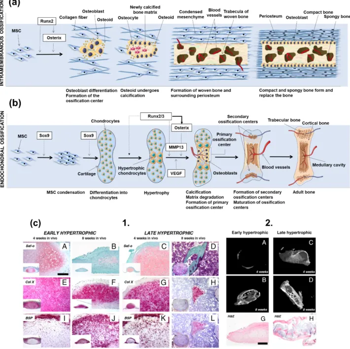
The bone tissue comprises two primary niches: the osteoblastic and the vascular niche [13]. Two stem cells types - HSCs and MSCs – reside in the bone cavity, which is filled with bone marrow and blood vessels [13]. HSCs, which are surrounded by stromal cells in the bone marrow, are responsible for the formation of the immune and blood system, as well as osteoclasts [13, 121]. MSCs also reside in the bone marrow and intervene in the formation of the mesenchymal lineage cells, which include osteoblasts, adipocytes, chondrocytes, fibroblast and other stromal cells. Together, both stem cell types maintain the normal bone homeostasis and cellular generation [13]. Unlike what was thought until recent years, HSCs are not located on the inner surface of the bone. Instead, HSCs were recently described to be on the perivascular niche where they are regulated by growth factors, chemokines and cytokines (e.g.: stem cell factor, chemokine stromal cell-derived factor 1 (CXCL-12) and angiopoientin-I), secreted through CXCL-12-abundant reticular cells, endothelial cells and MSCs [122].
MSCs, characterized by the expression of PDGFRα, CD51, nestin, CD139, interferon-induced GTP-binding protein MX1 (Mx1), leptin receptor (Lepr) and periaxin (Prx), give rise to osteoprogenitor
cells that form the osteoblastic niche. Thereafter, the factors referred previously are released to promote a correct HSC maintenance (reviewed by Yin and Li [13]). The maintenance of a functional microenvironment in the bone niche is dependent on the precise level of the hierarchical lineages of the HSCs and MSCs, so osteoblastogenesis and hematopoiesis can maintain a correct balance of osteoblast and osteoclast production. Importantly, N-cadherin positive osteoblasts interact with HSCs and help the anchoring of these cells to the osteoblastic niche [123].
Cell signaling inside the bone niche, for instance between osteoblasts and B lymphocyte precursors, is well known to determine features of the immune system [114]. Immunogenesis will not be discussed in this Review, and bone niche interactions will be described in the scope of the correct function of bone tissue itself. Nonetheless, the importance of a proper function of bone niches for successful bone development must be emphasized due to its crucial contribution for the continuous exportation of immune cells and tissue progenitor cells to the peripheral immune system, thus sustaining tissue repair and regeneration (Figure 2) [124].
5.2. Bone resident cells 5.2.1. Osteoblasts
The main function of osteoblasts is to synthetize new bone matrix [1]. Different sub-populations of osteoblasts have shown to respond differently to several signals (mechanical, hormonal and from cytokines) [1]. As discussed on Section 3.1, under physiological conditions, MSCs undergo the Wnt/β-catenin pathway to differentiate into the osteoblastic phenotype. Osteoblasts have a cuboidal morphology while proliferating on the bone matrix surfaces, unlike their precursors cells (preosteoblasts), which show a spindle shape [1]. Mature osteoblasts secrete bone ECM proteins, such as type I collagen. Typical gene indicators of osteoblast differentiation are Runx2, distal-less homeobox 5 (Dlx5), Osterix, alias core-binding factor alpha1 (Cbfa1), osteoblast specific factor 2 (Osf2) and ColIA1 [125, 126].
Osteoblasts residing in bone tissue can be divided in two types: mesenchymal (MOBL) or surface osteoblasts (SOBL) [25]. In the bone matrix, the undifferentiated MSCs start to differentiate into MOBL, which secrete collagen throughout the matrix, forming a woven structure. After the creation
of sufficient woven bone to form a platform-like structure, SOBL secrete collagen fibrils in a parallel way onto the previously made bone structure, creating the highly oriented lamellar bone. Once this process finishes, osteoblasts are matured into osteocytes surrounded by collagen matrix [25] (Figure 2a).
5.2.1.1. Stem cells differentiation into osteoblasts: exploiting physiological sources
Stem cells osteogenic differentiation, which is commonly targeted in tissue engineering strategies, is divided in three main stages: (i) peak in cell number; (ii) cellular differentiation, initiated with the expression and transcription of ALP; (iii) and a terminal step: production of osteocalcin and osteopontin [92]. In the human body, BMMSCs reside in a specific niche composed of a large variety of support cells that include hematopoietic progenitors; osteoclasts, immune cells and blood cells. The osteogenic differentiation of MSCs is known to be influenced by factors secreted by osteoblasts and osteocytes [6]. This phenomenon occurs through a communication network amongst osteoblasts and osteocytes that enhance a response in the MSCs, when these two bone cell types are in contact [6]. In vitro, the co-culture of MSCs with osteocytes showed greater osteogenic differentiation than the ones with osteoblasts, indicating that osteocytes induce MSCs’ osteogenesis more effectively than osteoblasts. On the other hand, osteoblasts helped the proliferation of the MSCs [6]. The differentiation of MSCs recruited to injured sites is not solely driven by contact with residing cells. Aspects of the fracture environment known to regulate MSCs fate include the control over the mineralized environment, respective release of ionic cues and the fine temporal variation of mechanical properties of the generated ECM during regeneration. Examples of tissue engineered approaches based on the use of in vivo-like mineralized synthetic biomaterials are described in Section 3.3.1. Other aspects such as control over biophysical cues generated throughout the bone repair process, also mimicked through the application of biomaterials with adequate and dynamic mechanical properties, are reviewed in Section 6.2.
Induced osteogenesis of MSCs may provide an important tool for the development of tissue engineering strategies focusing the treatment of large bone defects, which is currently challenging. Stem cells can derive from adult or embryonic sources, or from reprogramed adult cells (human
induced pluripotent stem cells; hiPSCs) [127, 128]. Adult human sources of MSCs proven to enable osteogenic differentiation include bone marrow and adipose tissue [129, 130]. Stem cells from postnatal sources can be obtained from placenta, umbilical cord, cord blood and amnion [127, 128]. BMMSCs have been the most commonly used cell type for bone engineering. However, due to the complex and invasive isolation process, the limited cell number and the reduction of differentiation potential with donors age, researchers have been trying to surpass these disadvantages by using other cell sources [131]. Nonetheless, efforts to promote their efficient expansion using non-conventional 3D microcarriers under bioreactor configurations must be addressed. The in vitro expansion of MSCs and their use in combined strategies using microcarriers as cell growth supports and implantable scaffolding materials was recently reviewed in detail by Bunpetch et al. [132]. Human umbilical cord and adipose tissue are routinely discarded as clinical waste and, in the case of adipose tissue, may be used as noncontroversial MSCs sources [133].
hiPSCs represent a promising tool for bone regeneration [134]. These pluripotent cells closely resemble human embryonic stem cells; however, they are obtained through the reprograming of human somatic cells [135]. Using hiPSCs instead of stem cells derived from other sources, can be advantageous since they can be obtained directly from the patient (patient-specific) and overcome any ethical and immunological issues [134]. Their ability to differentiate into the three germ layers enable them to be reprogrammed into different bone cells, namely osteoblasts and osteoclasts, which highlights their potential to be used in cell-based therapies of bone defects and injuries [136, 137]. Methods commonly employed to differentiate ESCs into the osteogenic lineage have been adapted for iPSCs differentiation [138]. Wu et al. recently provided an updated review on the methods used for iPSCs differentiation targeting bone repair [139], which are classified according to (i) their dependence on the production of intermediate embryonic body structures, (ii) the direct generation of iPSCs-derived mesenchymal precursors, and (iii) the direct differentiation of iPSCs into osteoblasts without intermediate steps. The classical protocol for the osteogenic differentiation of iPSCs, based on a protocol established for embryonic stem cells [140], involves the initial formation of embryonic bodies followed by the harvest of iPSC-derived MSCs present in those structures and, finally, their differentiation into osteoblasts using osteogenic media. This protocol,
however, was proven as low-yield, and approaches based on the direct seeding of dissociated embryonic bodies onto osteoinductive biomaterials have risen as effective ways of achieving regenerative systems for bone defects using iPSCs [136, 137, 141-143]. Alternatively to the formation of embryonic bodies, the adaptation of a protocol for embryonic cells differentiation [144] rendered iPSCs differentiation into MSC-like cells (often named iPSC-MSCs), which may be potentially differentiated into any mesenchymal lineage – including the osteogenic one. iPSC-MSCs were proven as valuable for the tissue regeneration field [145-147]. It is known that iPSC-MSCs are less responsive to the traditional chemically induced osteogenic differentiation protocol applied to MSCs (comprising ascorbic acid, β-glycerophosphate and dexamethasone), which has led researchers to find alternative differentiation methods [148]. In 2013, de Peppo et al. targeted the use of iPSC-MSCs as components of a tissue engineered system comprising decellularized bone as 3D scaffolds and a perfusion bioreactor. Perfusion conditions led to increasing expression of osteogenic markers, which were kept stable after the subcutaneous implantation of the iPSC in nude mice for 12 weeks [136]. Most of the studies reporting biomaterial-driven osteogenic differentiation of iPSC-MSCs rely on the use of mineralized matrices, either using decellularized bone or synthetic calcium phosphates. In 2018, a strategy to promote localized iPSC-MSCs in situ differentiation through localized BMP-2 recruiting was suggested by the co-injection of cells and a BMP-2 antibody in alginate beads [149]. This strategy avoided the formation of ectopic bone, commonly reported for BMP-2 releasing systems, and has the potential to surpass drawbacks associated to growth factors that include short half-life
The use of iPSCs has not been restricted to their differentiation into osteoblasts. A recent study using iPSC-MSCs also brought to light the potential of iPSCs to be differentiated into cell types other than osteoblasts with extreme relevance for the formation of functional bone, such as osteoclasts. Jeon et al. [7] used iPSC-MSCs differentiated into osteoblasts and osteoclasts differentiated from iPSCs-derived macrophages. iPSC-MSCs and iPSCs-derived macrophages were co-cultured and differentiated in PLGA/PLLA/hydroxyapatite porous scaffolds, rendering de novo-formed bone tissue upon implantation in nude mice.
Although iPSCs osteogenic differentiation relying on mesenchymal-like precursors has received a great deal of attention, the direct differentiation of iPSCs into osteoblasts has also been reported. In a precursor study, Levi et al. used biomaterials with the ability to concomitantly release osteogenic cytokines – BMP-2 - and provide biomineralization cues as boosting agents for in vivo bone formation through iPSCs differentiation [150]. Subsequent studied used methods based on the use of small molecules [151,152], osteogenic scaffolds [153-154], and gene modification to promote the fast, safe and high-yield differentiation of iPSCs and their application in bone regeneration strategies.
In general, protocols targeting the direct differentiation of iPSCs into osteoblasts depend on the use of cytokines, multi-step approaches using different supplements overtime, or in situ cell differentiation seeded onto specific biomaterials. In 2016, Kang et al. reported a breakthrough one-step protocol based on the use of adenosine, a natural occurring nucleoside, as a cell culture medium supplement to directly converse hiPSCs into functional osteoblasts [152]. Adenosine-treated cells seeded onto 3D microporous matrices rendered the successful repair of a critical bone defect, which included the formation of vascularized neobone capable of undergoing resorption. Despite the clear promise represented by iPSCs as easily obtained cells for tissue regeneration, the field is still not free of challenges that include the delayed or low osteogenic differentiation of some cells that are exposed to differentiation protocols developed so far [148, 155], which may culminate in the long-term formation of dangerous teratoma tissues. Comparison between differentiation protocols, namely a robust parallelization between MSC precursor-based methods and direct differentiation protocols, are still in need.
5.2.2. Osteocytes
Osteocytes are fully matured and differentiated osteoblasts which descend from the mesenchymal lineage (Figure 2a). They comprise 90% to 95% of the whole bone cells in adult bone and may live up to decades in their mineralized environment exhibiting a dendritic configuration. Their function is to support the skeleton and bone metabolism. As osteoblasts differentiate into osteocytes, ALP production decreases, while osteocalcin raises [1] (Figure 2a). Other expressed markers, including
phosphate-regulating protein with homologies to endopeptidases expressed by genes of the X chromosome (PHEX), matrix extracellular phosphoglycoprotein (MEPE), dentin matrix protein 1 (DMP-1), FGF-23, sclerostin, and oxygen regulated protein (ORP143), are thought to protect osteocytes against hypoxia [156]. These cells were considered as “passive placeholders in bone” in the past. However, they were proven to have numerous functions including bone remodeling -through the activation of both osteoclasts and osteoblasts -, as well as in endocrine cell functioning [156]. The interactions between osteocytes and osteoblasts/osteoclasts are reviewed in Section 5.4. Osteocytes are also responsible for the excretion of proteins such as CD44, galectin 3 and osteocalcin. These proteins have the function to promote cell adhesion and the regulation of mineral exchange in the bone. Osteocytes also produce Runx2 and Osterix, which are required for osteoblast differentiation, and are followed by ALP and collagen, necessary for the formation of the osteoid. Several soluble molecules produced by osteocytes positively interfere with biomineralization (e.g. PHEX, MEPE and DMP-1) [156]. The communication between osteocytes, which occurs mainly by gaps composed of connexin 43 [157, 158], are required for their survival, maturation and correct activity. The diverse roles of osteocytes also encompass their phagocytic activity, during osteolysis, since they have lysosomes in their constitution. One of the most remarkable functions of osteocytes is mechanosensing by translating stress factors into biologic signals [156]. Vazquez et al. [159] developed a 3D in vitro co-culture systems to assess the effect of mechanical loading on the interaction between osteocytes and osteoblasts. They demonstrated that osteocytes subjected to mechanical appropriate mechanical cues act upon osteoblasts towards bone formation [159].
5.2.3. Osteoclasts
Osteoclasts, generated in the bone marrow from mononuclear monocyte-macrophage precursors derived from the hematopoietic lineage [160], are the only cells capable of resorbing bone and, consequently, play an essential role in bone remodeling. The receptor activator of nuclear factor kappa-β ligand (RANKL) and macrophage colony stimulating factor (M-CSF) are reported to drive osteoclasts’ proliferation, differentiation and survival [160, 161]. Bone resorption occurs in the presence of factors secreted by osteoclasts: hydrogen ions that lead the acidification of the
resorption compartment dissolving the mineral bone matrix, and cathepsin K, an enzyme responsible for the digestion of the insoluble fraction of the matrix (mainly type I collagen). These cells are bound by the bone matrix through integrins (β1 for collagen, laminin and fibronectin, and αvβ3 for osteopontin and bone sialoprotein) [162]. Such binding polarizes osteoclasts, creating an actin ring that seals the periphery of the ligation of the osteoclasts to the matrix, and a ruffled border in the resorbing surface, which leads to the secretion of H+ ions, followed by the exocytosis of
enzymes from the acidified vesicles [162].
5.3. Heterotypic cell-cell interactions in bone
It is well established that bone cells interact in adult bone to regulate homeostasis process, supporting the balance between bone resorption and formation that allows the maintenance of the tissue’s integrity [162]. We review the interactions between the main reported cells that constitute healthy bone - osteoblasts, osteoclasts and osteocytes -, and their crosstalk with the vascular system that irrigates and also constitutes functional bone. Due to their relevance in bone healing, interactions between immune cells and bone resident cells are also reviewed.
5.3.1. Osteocytes-osteoblasts
Bone formation is regulated by several signaling mechanisms, with particular importance for the Wnt/β-catenin pathway [1]. The activation of canonical Wnt signaling in early osteoblasts promotes osteoblast differentiation and bone formation [163], with opposing effects observed when Wnt signaling is disrupted [164]. Osteocytes secrete Wnt antagonists, which include sclerostin and the LRP5/6 inhibitor Dickkopf-related protein 1 (DKK1). Both molecules inhibit osteoblast differentiation and bone formation [165, 166]. Moreover, the in vivo loss of secreted frizzled-related protein 1 (SFRP1), which is a competitive antagonist of Wnt ligand, resulted in increased bone mass and mineral density, as well as in in vitro enhancement of osteoblast proliferation and differentiation into osteocytes [167]. With their capacity to interfere in canonical Wnt signaling, therefore affecting osteoblasts differentiation, osteocytes play a regulatory role in bone formation.
5.3.2. Osteoblasts-osteoclasts
Signaling between osteoblasts and osteoclasts is crucial for osteoclast maturation [1]. It is known that osteoblasts and stromal cells produce RANKL, M-CSF, and osteoprotegerin (OPG), while early osteoclast precursors produce c-Fms (M-CSF receptor) and receptor activator of nuclear factor κ B (RANK) - a receptor for RANKL. RANKL and M-CSF stimulate osteoclast differentiation, while OPG is an inhibitor of RANKL, and competes with RANKL for RANK [1]. Low levels of OPG lead to accelerated osteoclast development, which culminates in osteoporosis: a disease characterized by the disruption of bone resorption/formation balance, and in which bone resorption exceeds bone growth [160]. Although osteoblasts were thought to be the main providers of RANKL to osteoclasts, osteocytes have proven to have an extremely relevant role in this mediation [168].
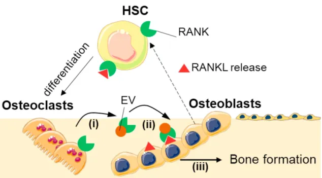
Besides the role of osteoblasts and osteocytes on osteoclastogenesis, the reverse role of osteocytes on osteoblasts’ function has also been a matter of interest. In 2011, osteoclasts were reported as being uncapable of stimulating RANKL reverse signalling in osteoblasts through direct cell interactions [169]. However, in 2018, Ikebuchi et al. presented a breakthrough mechanism driven by extracellular vesicles, in which osteoclasts modulate osteoblasts function and, therefore, bone formation [170]. In the suggested setting, RANKL receptors in osteoblasts are responsible for the reverse RANK-RANKL modulation. Osteocytes release extracellular vesicles containing RANK on their surface, which bind to RANKL on the surface of osteoblasts (and hypothetically osteocytes), triggering intracellular signalling. The authors proved that mTOR pathway was activated, triggering the production of Runx2, and leading to bone formation.
5.3.3. Osteocytes-osteoclasts
Osteocytes – both healthy and apoptotic at microdamage sites – have been reported to recruit osteoclasts to the bone remodeling sites and were shown to send bone resorption cues to those cells [171]. The expression of the RANKL during the dendritic process associated with osteocytes maturation was associated with the osteocyte-led bone resorption [171]. Upon injury, right after damage, pro-apoptotic molecules are released by osteocytes; contrarily, anti-apoptotic molecules are produced at 1-2 mm distance from the cracks [171]. The promotion of a defective performance