Review Article
Biology of Bone Tissue: Structure, Function,
and Factors That Influence Bone Cells
Rinaldo Florencio-Silva,
1Gisela Rodrigues da Silva Sasso,
1Estela Sasso-Cerri,
2Manuel Jesus Simões,
1and Paulo Sérgio Cerri
21Department of Morphology and Genetics, Laboratory of Histology and Structural Biology, Federal University of S˜ao Paulo,
04023-900 S˜ao Paulo, SP, Brazil
2Department of Morphology, Laboratory of Histology and Embryology, Dental School, Universidade Estadual Paulista (UNESP),
14801-903 Araraquara, SP, Brazil
Correspondence should be addressed to Paulo S´ergio Cerri; [email protected] Received 3 December 2014; Revised 30 April 2015; Accepted 4 May 2015
Academic Editor: Wanda Lattanzi
Copyright © 2015 Rinaldo Florencio-Silva et al. his is an open access article distributed under the Creative Commons Attribution License, which permits unrestricted use, distribution, and reproduction in any medium, provided the original work is properly cited.
Bone tissue is continuously remodeled through the concerted actions of bone cells, which include bone resorption by osteoclasts and bone formation by osteoblasts, whereas osteocytes act as mechanosensors and orchestrators of the bone remodeling process. his process is under the control of local (e.g., growth factors and cytokines) and systemic (e.g., calcitonin and estrogens) factors that all together contribute for bone homeostasis. An imbalance between bone resorption and formation can result in bone diseases including osteoporosis. Recently, it has been recognized that, during bone remodeling, there are an intricate communication among bone cells. For instance, the coupling from bone resorption to bone formation is achieved by interaction between osteoclasts and osteoblasts. Moreover, osteocytes produce factors that inluence osteoblast and osteoclast activities, whereas osteocyte apoptosis is followed by osteoclastic bone resorption. he increasing knowledge about the structure and functions of bone cells contributed to a better understanding of bone biology. It has been suggested that there is a complex communication between bone cells and other organs, indicating the dynamic nature of bone tissue. In this review, we discuss the current data about the structure and functions of bone cells and the factors that inluence bone remodeling.
1. Introduction
Bone is a mineralized connective tissue that exhibits four types of cells: osteoblasts, bone lining cells, osteocytes, and osteoclasts [1, 2]. Bone exerts important functions in the body, such as locomotion, support and protection of sot tissues, calcium and phosphate storage, and harboring of bone marrow [3,4]. Despite its inert appearance, bone is a highly dynamic organ that is continuously resorbed by osteo-clasts and neoformed by osteoblasts. here is evidence that osteocytes act as mechanosensors and orchestrators of this bone remodeling process [5–8]. he function of bone lining cells is not well clear, but these cells seem to play an important role in coupling bone resorption to bone formation [9].
Bone remodeling is a highly complex process by which old bone is replaced by new bone, in a cycle comprised of
three phases: (1) initiation of bone resorption by osteoclasts, (2) the transition (or reversal period) from resorption to new bone formation, and (3) the bone formation by osteoblasts [10, 11]. his process occurs due to coordinated actions of osteoclasts, osteoblasts, osteocytes, and bone lining cells which together form the temporary anatomical structure called basic multicellular unit (BMU) [12–14].
Normal bone remodeling is necessary for fracture healing and skeleton adaptation to mechanical use, as well as for calcium homeostasis [15]. On the other hand, an imbalance of bone resorption and formation results in several bone diseases. For example, excessive resorption by osteoclasts without the corresponding amount of nerformed bone by osteoblasts contributes to bone loss and osteoporosis [16], whereas the contrary may result in osteopetrosis [17]. hus, the equilibrium between bone formation and resorption is
Oc
Ot
Ob Ob
B
B
(a)
BV
BV
B B
(b)
B
B
B
Ob Ob
(c)
B B
B
(d)
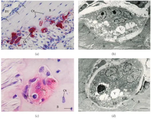
Figure 1: (a)–(d) Light micrographs of portions of alveolar bone of rats. (a) HE-stained section showing a portion of a bony trabecula (B). Polarized osteoblasts (Ob) and giant multinucleated osteoclasts (Oc) are observed in the bone surface; osteocyte (Ot) surrounding bone matrix is also observed. (b) Section subjected to immunohistochemistry for osteocalcin detection and counterstained with hematoxylin. Note osteocalcin-positive osteoblasts (arrows) on the surface of a bony trabecula (B). BV: blood vessel. (c) Undecalciied section subjected to the Gomori method for the detection of alkaline phosphatase, evidencing a portion of bone matrix (B) positive to the alkaline phosphatase (in brown/black). Ob: osteoblasts. (d) Undecalciied section subjected to the von Kossa method for calcium detection (brown/dark color). von Kossa-positive bone matrix (B) is observed; some positive granules (arrow) can also be observed on the surface of the bone trabeculae. Scale bar: 15�m.
necessary and depends on the action of several local and systemic factors including hormones, cytokines, chemokines, and biomechanical stimulation [18–20].
Recent studies have shown that bone inluences the activity of other organs and the bone is also inluenced by other organs and systems of the body [21], providing new insights and evidencing the complexity and dynamic nature of bone tissue.
In this review we will address the current data about bone cells biology, bone matrix, and the factors that inluence the bone remodeling process. Moreover, we will briely discuss the role of estrogen on bone tissue under physiological and pathological conditions.
2. Bone Cells
2.1. Osteoblasts. Osteoblasts are cuboidal cells that are located
along the bone surface comprising 4–6% of the total resident bone cells and are largely known for their bone forming function [22]. hese cells show morphological characteristics of protein synthesizing cells, including abundant rough endo-plasmic reticulum and prominent Golgi apparatus, as well
as various secretory vesicles [22,23]. As polarized cells, the osteoblasts secrete the osteoid toward the bone matrix [24] (Figures1(a),1(b), and2(a)).
Osteoblasts are derived from mesenchymal stem cells (MSC). he commitment of MSC towards the osteopro-genitor lineage requires the expression of speciic genes, following timely programmed steps, including the synthesis of bone morphogenetic proteins (BMPs) and members of the Wingless (Wnt) pathways [25]. he expressions of Runt-related transcription factors 2, Distal-less homeobox 5 (Dlx5), and osterix (Osx) are crucial for osteoblast diferentiation [22,26]. Additionally,Runx2is a master gene of osteoblast diferentiation, as demonstrated by the fact that Runx2-null mice are devoid of osteoblasts [26, 27].Runx2has demon-strated to upregulate osteoblast-related genes such asColIA1,
ALP,BSP,BGLAP, andOCN[28].
Once a pool of osteoblast progenitors expressingRunx2
andColIA1has been established during osteoblast
B Ob Otd Otd
B
Ob Ob
B
(a)
B N N
BLC
Otd
Otd
(b)
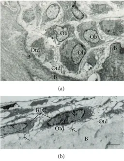
Figure 2: Electron micrographs of portions of alveolar bone of rats. (a) Oteoblasts exhibiting abundant rough endoplasmic reticulum are observed adjacent to the bone (B) surface. A layer of bundles of collagen ibrils situated between osteoblasts (Ob) and calciied bone surface (B) constitutes the osteoid (Otd). Scale bar: 2.7�m. (b) Bone lining cells (BLC) exhibiting scarce cytoplasm are situated on the osteoid surface (Otd). Bone lining cells (BLC) extend some thin cytoplasmic projections (arrows) towards the osteoid (Otd). Scale bar: 2�m. N: nucleus.
increase in the expression of Osx and in the secretion of bone matrix proteins such as osteocalcin (OCN), bone sialopro-tein (BSP) I/II, and collagen type I. Moreover, the osteoblasts undergo morphological changes, becoming large and cu-boidal cells [26,29–31].
here is evidence that other factors such as ibroblast growth factor (FGF), microRNAs, and connexin 43 play important roles in the osteoblast diferentiation [32–35]. FGF-2 knockout mice showed a decreased bone mass coupled to increase of adipocytes in the bone marrow, indicating the participation of FGFs in the osteoblast diferentiation [34]. It has also been demonstrated that FGF-18 upregulates osteoblast diferentiation in an autocrine mechanism [36]. MicroRNAs are involved in the regulation of gene expression in many cell types, including osteoblasts, in which some mic-roRNAs stimulate and others inhibit osteoblast diferentia-tion [37, 38]. Connexin 43 is known to be the main con-nexin in bone [35]. he mutation in the gene encoding con-nexin 43 impairs osteoblast diferentiation and causes skeletal malformation in mouse [39].
he synthesis of bone matrix by osteoblasts occurs in two main steps: deposition of organic matrix and its subse-quent mineralization (Figures1(b)–1(d)). In the irst step, the osteoblasts secrete collagen proteins, mainly type I collagen, noncollagen proteins (OCN, osteonectin, BSP II, and osteo-pontin), and proteoglycan including decorin and biglycan, which form the organic matrix. hereater, mineralization of bone matrix takes place into two phases: the vesicular
and the ibrillar phases [40,41]. he vesicular phase occurs when portions with a variable diameter ranging from 30 to 200 nm, called matrix vesicles, are released from the apical membrane domain of the osteoblasts into the newly formed bone matrix in which they bind to proteoglycans and other organic components. Because of its negative charge, the sulphated proteoglycans immobilize calcium ions that are stored within the matrix vesicles [41,42]. When osteoblasts secrete enzymes that degrade the proteoglycans, the calcium ions are released from the proteoglycans and cross the calcium channels presented in the matrix vesicles membrane. hese channels are formed by proteins called annexins [40].
On the other hand, phosphate-containing compounds are degraded by the ALP secreted by osteoblasts, releasing phos-phate ions inside the matrix vesicles. hen, the phosphos-phate and calcium ions inside the vesicles nucleate, forming the hydroxyapatite crystals [43]. he ibrillar phase occurs when the supersaturation of calcium and phosphate ions inside the matrix vesicles leads to the rupture of these structures and the hydroxyapatite crystals spread to the surrounding matrix [44,45].
Mature osteoblasts appear as a single layer of cuboidal cells containing abundant rough endoplasmic reticulum and large Golgi complex (Figures2(a)and3(a)). Some of these osteoblasts show cytoplasmic processes towards the bone matrix and reach the osteocyte processes [46]. At this stage, the mature osteoblasts can undergo apoptosis or become osteocytes or bone lining cells [47,48]. Interestingly, round/ ovoid structures containing dense bodies and TUNEL-pos-itive structures have been observed inside osteoblast vac-uoles. hese indings suggest that besides professional phago-cytes, osteoblasts are also able to engulf and degrade apoptotic bodies during alveolar bone formation [49].
2.2. Bone Lining Cells. Bone lining cells are quiescent
lat-shaped osteoblasts that cover the bone surfaces, where neither bone resorption nor bone formation occurs [50]. hese cells exhibit a thin and lat nuclear proile; its cytoplasm extends along the bone surface and displays few cytoplasmic organelles such as proiles of rough endoplasmic reticulum and Golgi apparatus [50] (Figure2(b)). Some of these cells show processes extending into canaliculi, and gap junctions are also observed between adjacent bone lining cells and between these cells and osteocytes [50,51].
Ot Ot
BV
BLC Ob
BLC
B BV Ob
B
(a)
* *
Ot Ot
(b)
Ot
Ot B
(c)
Ot N
La
Ca B
(d)
Figure 3: Light (a and b) and electron micrographs of portions of alveolar bone rats. (a) a semithin section stained with toluidine blue showing a portion of a bony trabecula (B). Osteoblasts (Ob) and bone lining cells (BLC) are present on bone surface while osteocytes (Ot) are observed entrapped in the bone matrix. BV: blood vessels. Scale bar: 15�m. (b) Section subjected to the silver impregnation method. Note the cytoplasmic processes (arrows) of the osteocytes (Ot) connecting them with each other. Scale bar: 15�m. (c) Scanning electron micrograph showing two osteocytes (Ot) surrounded by bone matrix (B). Note that the cytoplasmic processes (arrows) are observed between the osteocytes (Ot) forming an interconnected network. Scale bar: 2�m. (d) Transmission electron micrograph showing a typical osteocyte (Ot) inside a lacuna (La) in the bone matrix (B), with its cytoplasmic processes (arrows) inside the canaliculi (Ca). Scale bar: 2�m. N: nucleus.
2.3. Osteocytes. Osteocytes, which comprise 90–95% of the
total bone cells, are the most abundant and long-lived cells, with a lifespan of up to 25 years [54]. Diferent from oste-oblasts and osteoclasts, which have been deined by their respective functions during bone formation and bone resorp-tion, osteocytes were earlier deined by their morphology and location. For decades, due to diiculties in isolating osteocytes from bone matrix led to the erroneous notion that these cells would be passive cells, and their functions were misinterpreted [55]. he development of new technologies such as the identiication of osteocyte-speciic markers, new animal models, development of techniques for bone cell isolation and culture, and the establishment of phenotypically stable cell lines led to the improvement of the understanding of osteocyte biology. In fact, it has been recognized that these cells play numerous important functions in bone [8].
he osteocytes are located within lacunae surrounded by mineralized bone matrix, wherein they show a dendritic morphology [15,55,56] (Figures3(a)–3(d)). he morphology of embedded osteocytes difers depending on the bone type. For instance, osteocytes from trabecular bone are more
rounded than osteocytes from cortical bone, which display an elongated morphology [57].
Osteocytes are derived from MSCs lineage through oste-oblast diferentiation. In this process, four recognizable stages have been proposed: osteoid-osteocyte, preosteocyte, young osteocyte, and mature osteocyte [54]. At the end of a bone formation cycle, a subpopulation of osteoblasts becomes osteocytes incorporated into the bone matrix. his process is accompanied by conspicuous morphological and ultrastruc-tural changes, including the reduction of the round osteoblast size. he number of organelles such as rough endoplasmic reticulum and Golgi apparatus decreases, and the nucleus-to-cytoplasm ratio increases, which correspond to a decrease in the protein synthesis and secretion [58].
similarly to other cell types with dendritic morphology such as podocytes, type II lung alveolar cells, and cells of the choroid plexus [59]. It has been suggested that E11/gp38 uses energy from GTPase activity to interact with cytoskeletal components and molecules involved in cell motility, whereby regulate actin cytoskeleton dynamics [60, 61]. Accordingly, inhibition of E11/gp38 expression in osteocyte-like MLO-Y4 cells has been shown to block dendrite elongation, suggesting that E11/gp38 is implicated in dendrite formation in osteo-cytes [59].
Once the stage of mature osteocyte totally entrapped within mineralized bone matrix is accomplished, several of the previously expressed osteoblast markers such as OCN, BSPII, collagen type I, and ALP are downregulated. On the other hand, osteocyte markers including dentine matrix pro-tein 1 (DMP1) and sclerostin are highly expressed [8,62–64]. Whereas the osteocyte cell body is located inside the lacuna, its cytoplasmic processes (up to 50 per each cell) cross tiny tunnels that originate from the lacuna space called canaliculi, forming the osteocyte lacunocanalicular system [65] (Figures 3(b)–3(d)). hese cytoplasmic processes are connected to other neighboring osteocytes processes by gap junctions, as well as to cytoplasmic processes of osteoblasts and bone lining cells on the bone surface, facilitating the intercellular transport of small signaling molecules such as prostaglandins and nitric oxide among these cells [66]. In addition, the osteocyte lacunocanalicular system is in close proximity to the vascular supply, whereby oxygen and nutrients achieve osteocytes [15].
It has been estimated that osteocyte surface is 400-fold larger than that of the all Haversian and Volkmann systems and more than 100-fold larger than the trabecular bone surface [67,68]. he cell-cell communication is also achieved by interstitial luid that lows between the osteocytes pro-cesses and canaliculi [68]. By the lacunocanalicular system (Figure3(b)), the osteocytes act as mechanosensors as their interconnected network has the capacity to detect mechanical pressures and loads, thereby helping the adaptation of bone to daily mechanical forces [55]. By this way, the osteocytes seem to act as orchestrators of bone remodeling, through regulation of osteoblast and osteoclast activities [15, 69]. Moreover, osteocyte apoptosis has been recognized as a chemotactic signal to osteoclastic bone resorption [70–73]. In agreement, it has been shown that during bone resorption, apoptotic osteocytes are engulfed by osteoclasts [74–76].
he mechanosensitive function of osteocytes is accom-plished due to the strategic location of these cells within bone matrix. hus, the shape and spatial arrangement of the osteocytes are in agreement with their sensing and signal transport functions, promoting the translation of mechanical stimuli into biochemical signals, a phenomenon that is called piezoelectric efect [77]. he mechanisms and components by which osteocytes convert mechanical stimuli to biochemical signals are not well known. However, two mechanisms have been proposed. One of them is that there is a protein complex formed by a cilium and its associated proteins PolyCystins 1 and 2, which has been suggested to be crucial for osteocyte mechanosensing and for osteoblast/osteocyte-mediated bone formation [78]. he second mechanism involves osteocyte
cytoskeleton components, including focal adhesion protein complex and its multiple actin-associated proteins such as paxillin, vinculin, talin, and zyxin [79]. Upon mechanical stimulation, osteocytes produce several secondary messen-gers, for example, ATP, nitric oxide (NO), Ca2+, and pros-taglandins (PGE2and PGI2,) which inluence bone physiol-ogy [8,80]. Independently of the mechanism involved, it is important to mention that the mechanosensitive function of osteocytes is possible due to the intricate canalicular network, which allows the communication among bone cells.
2.4. Osteoclasts. Osteoclasts are terminally diferentiated
multinucleated cells (Figures 4(a)–4(d)), which originate from mononuclear cells of the hematopoietic stem cell lineage, under the inluence of several factors. Among these factors the macrophage colony-stimulating factor (M-CSF), secreted by osteoprogenitor mesenchymal cells and oste-oblasts [81], and RANK ligand, secreted by osteoblasts, osteocytes, and stromal cells, are included [20]. Together, these factors promote the activation of transcription factors [81,82] and gene expression in osteoclasts [83,84].
M-CSF binds to its receptor (cFMS) present in osteo-clast precursors, which stimulates their proliferation and inhibits their apoptosis [82, 85]. RANKL is a crucial fac-tor for osteoclastogenesis and is expressed by osteoblasts, osteocytes, and stromal cells. When it binds to its recep-tor RANK in osteoclast precursors, osteoclast formation is induced [86]. On the other hand, another factor called oste-oprotegerin (OPG), which is produced by a wide range of cells including osteoblasts, stromal cells, and gingival and periodontal ibroblasts [87–89], binds to RANKL, pre-venting the RANK/RANKL interaction and, consequently, inhibiting the osteoclastogenesis [87] (Figure 5). hus, the RANKL/RANK/OPG system is a key mediator of osteoclas-togenesis [19,86,89].
he RANKL/RANK interaction also promotes the expression of other osteoclastogenic factors such as NFATc1 and DC-STAMP. By interacting with the transcription factors PU.1, cFos, and MITF, NFATc1 regulates osteoclast-speciic genes includingTRAP and cathepsin K, which are crucial for osteoclast activity [90]. Under the inluence of the RANKL/RANK interaction, NFATc1 also induces the expression of DC-STAMP, which is crucial for the fusion of osteoclast precursors [91,92].
Despite these osteoclastogenic factors having been well deined, it has recently been demonstrated that the osteo-clastogenic potential may difer depending on the bone site considered. It has been reported that osteoclasts from long bone marrow are formed faster than in the jaw. his diferent dynamic of osteoclastogenesis possibly could be, due to the cellular composition of the bone-site speciic marrow [93].
Oc Ot
BV B
Oc
(a)
B N
N Oc
V V RB
RB V
(b)
B Ot Oc
Ap
Oc1
(c)
Oc N N
B V
Ap
CZ CZ
RB
(d)
Figure 4: Light (a and c) and electron micrographs (b and d) of portions of alveolar bone of rats. In (a) tartrate-resistant acid phosphatase (TRAP) activity (in red color) is observed in the cytoplasm of osteoclasts (OC) adjacent to the alveolar bone (B) surface. Note that in the opposite side of the bony trabecula B is covered by large and polarized osteoblasts (Ob). Ot, osteocytes (Ot); BV: blood vessel. Bar: 40�m. (b) Multinucleated osteoclast (OC) shows evident ruled border (RB) adjacent to the excavated bone surface (arrows). Several vacuoles (V) are observed in the cytoplasm adjacent to ruled border (RB). N: nucleus. Bar: 4�m. (c) Portions of TRAP-positive osteoclasts (Oc and Oc1) are observed in a resorbing bone lacuna. A round cell (Ap) with condensed irregular blocks of chromatin, typical apoptotic cell, is observed inside a large vacuole of the Oc1. B: bone matrix; Ot: osteocyte. Bar: 15�m. (d) An osteoclast (Oc) showing ruled border (RB) and clear zone (CZ) is in close juxtaposition to the excavation of the bone surface (arrows), that is, Howship lacuna. Vacuoles (V) with varied size are present next to the ruled border (RB); one of them contains a round cell with masses of condensed chromatin (Ap), typical of cell undergoing apoptosis. B: bone matrix; N: nucleus. Bar: 3�m.
cytoskeleton, in which an F-actin ring that comprises a dense continuous zone of highly dynamic podosome is formed and consequently an area of membrane that develop into the ruled border is isolated. It is important to mention that these domains are only formed when osteoclasts are in contact with extracellular mineralized matrix, in a process which
�v�3-integrin, as well as the CD44, mediates the attachment
of the osteoclast podosomes to the bone surface [96–99]. Ultrastructurally, the ruled border is a membrane domain formed by microvilli, which is isolated from the surrounded tissue by the clear zone, also known as sealing zone. he clear zone is an area devoid of organelles located in the periphery of the osteoclast adjacent to the bone matrix [98]. his sealing zone is formed by an actin ring and several other proteins, including actin, talin, vinculin, paxillin, tensin, and actin-associated proteins such as �-actinin, imbrin, gelsolin, and dynamin [95]. he�v�3-integrin binds to
non-collagenous bone matrix containing-RGD sequence such as bone sialoprotein, osteopontin, and vitronectin, establishing a peripheric sealing that delimits the central region, where the ruled border is located [98] (Figures4(b)–4(d)).
he maintenance of the ruled border is also essen-tial for osteoclast activity; this structure is formed due to intense traicking of lysosomal and endosomal components. In the ruled border, there is a vacuolar-type H+-ATPase (V-ATPase), which helps to acidify the resorption lacuna and hence to enable dissolution of hydroxyapatite crystals [20, 100, 101]. In this region, protons and enzymes, such as tartrate-resistant acid phosphatase (TRAP), cathepsin K, and matrix metalloproteinase-9 (MMP-9) are transported into a compartment called Howship lacuna leading to bone degradation [94, 101–104] (Figure5). he products of this degradation are then endocytosed across the ruled border and transcytosed to the functional secretory domain at the plasma membrane [7,95].
CZ
CZ RB
Cp TRAP
CAII RANKL RANK
OPG
Sclerostin, DKK-1 PGE2, NO, IGF-1
RANK RANKL OPG
Sema4D
Sema3A Eph2 Eph4
Cx3
Oc
N N
Ob
Ot
Col1 OCN OSN OSP BSP BMP
BLC
Cx3
HL
Cx3
Ot
B B
5
HCO3−
Cl−
Cl−
Cl−
CO2+H2O
H+H+
H+
MMP-9
HCO3+H
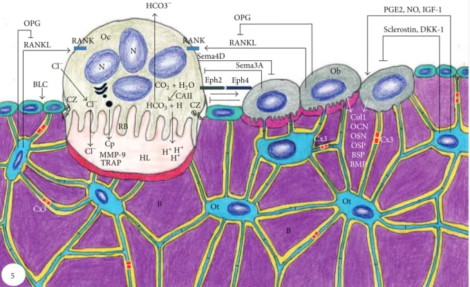
Figure 5: Schematic summary of bone tissue showing bone cells and the relationships among them and with bone matrix (B). Osteoclast (Oc) activation occurs ater binding of RANKL to its receptor RANK, present in the membrane of osteoclast precursors. hen, osteoclast becomes polarized through its cytoskeleton reorganization; the ruled border (RB) and clear zone (CZ) are membrane specializations observed in the portion of the osteoclast juxtaposed to the bone resorption surface, Howship lacuna (HL). Dissolution of hydroxyapatite occurs in the bone surface adjacent to the ruled border (RF) upon its acidiication due to pumping of hydrogen ions (H+) to the HL. H+ and ions bicarbonate (HCO3−) originate from the cleavage of carbonic acid (H2CO3) under the action of carbonic anhydrase II (CAII).
Ater dissolution of mineral phase, osteoclast (Oc) releases cathepsin (Cp), matrix metalloproteinase-9 (MMP-9), and tartrate-resistant acid phosphatase (TRAP) that degrade the organic matrix. EphrinB2 (Eph2) present in osteoclast membrane binds to ephrinB4 (Eph4) in osteoblast (Ob) membrane, promoting its diferentiation, whereas the reverse signaling (ephrinB4/ephrinB2) inhibits osteoclastogenesis. Sema4D produced by osteoclasts inhibits osteoblasts, while Sema3A secreted by osteoblasts inhibits osteoclasts. Osteoblasts (Ob) also produce receptor activator of nuclear factor KB (RANKL) and osteoprotegerin (OPG), which increase and decrease osteoclastogenesis, respectively. Osteoblasts (Ob) secrete collagenous (Col1) and noncollagenous proteins such as osteocalcin (OCN), osteopontin (OSP), osteonectin (OSN), bone sialoprotein (BSP), and bone morphogenetic proteins (BMP). Osteocytes (Ot) are located within lacunae surrounded by mineralized bone matrix (B). Its cytoplasmic processes cross canaliculi to make connection with other neighboring osteocytes processes by gap junctions, mainly composed by connexin 43 (Cx3), as well as to cytoplasmic processes of osteoblasts (Ob) and bone lining cells (BLC) on bone surface. RANKL secreted by osteocytes stimulates osteoclastogenesis, while prostaglandin E2(PGE2), nitric oxide (NO), and insulin-like growth factor (IGF) stimulate osteoblast activity. Conversely, osteocytes produce OPG that inhibits osteoclastogenesis; moreover, osteocytes produce sclerostin and dickkopf WNT signaling pathway inhibitor (DKK-1) that decrease osteoblast activity.
In periodontitis, a disease of the periodontium caused by bac-terial proliferation [107,108] induces the migration of inlam-matory cells. hese cells produce chemical mediators such as IL-6 and RANKL that stimulate the migration of osteo-clasts [89,109,110]. As a result, an abnormal increased bone resorption occurs in the alveolar bone, contributing to the loss of the insertions of the teeth and to the progression of periodontitis [89,111].
On the other hand, in osteopetrosis, which is a rare bone disease, genetic mutations that afect formation and tion functions in osteoclasts lead to decreased bone resorp-tion, resulting in a disproportionate accumulation of bone
mass [17]. hese diseases demonstrate the importance of the normal bone remodeling process for the maintenance of bone homeostasis.
2.5. Extracellular Bone Matrix. Bone is composed by inor-ganic salts and orinor-ganic matrix [113]. he organic matrix con-tains collagenous proteins (90%), predominantly type I colla-gen, and noncollagenous proteins including osteocalcin, oste-onectin, osteopontin, ibronectin and bone sialoprotein II, bone morphogenetic proteins (BMPs), and growth factors [114]. here are also small leucine-rich proteoglycans includ-ing decorin, biglycan, lumican, osteoaderin, and seric pro-teins [114–116].
he inorganic material of bone consists predominantly of phosphate and calcium ions; however, signiicant amounts of bicarbonate, sodium, potassium, citrate, magnesium, car-bonate, luorite, zinc, barium, and strontium are also present [1, 2]. Calcium and phosphate ions nucleate to form the hydroxyapatite crystals, which are represented by the chem-ical formula Ca10(PO4)6(OH)2. Together with collagen, the noncollagenous matrix proteins form a scafold for hydrox-yapatite deposition and such association is responsible for the typical stifness and resistance of bone tissue [4].
Bone matrix constitutes a complex and organized frame-work that provides mechanical support and exerts essential role in the bone homeostasis. he bone matrix can release several molecules that interfere in the bone cells activity and, consequently, has a participation in the bone remod-eling [117]. Once loss of bone mass alone is insuicient to cause bone fractures [118], it is suggested that other factors, including changes in the bone matrix proteins and their modiications, are of crucial importance to the understanding and prediction of bone fractures [119]. In fact, it is known that collagen plays a critical role in the structure and function of bone tissue [120].
Accordingly, it has been demonstrated that there is a variation in the concentration of bone matrix proteins with age, nutrition, disease, and antiosteoporotic treatments [119,
121,122] which may contribute to postyield deformation and fracture of bone [119]. For instance, in vivo and in vitro
studies have reported that the increase in hyaluronic acid synthesis ater parathyroid hormone (PTH) treatment was related to a subsequent bone resorption [123–127] suggesting a possible relationship between hyaluronic acid synthesis and the increase in osteoclast activity.
2.6. Interactions between Bone Cells and Bone Matrix. As
previously discussed, bone matrix does not only provides support for bone cells, but also has a key role in regu-lating the activity of bone cells through several adhesion molecules [117,128]. Integrins are the most common adhesion molecules involved in the interaction between bone cells and bone matrix [129]. Osteoblasts make interactions with bone matrix by integrins, which recognize and bind to RGD and other sequences present in bone matrix proteins including osteopontin, ibronectin, collagen, osteopontin, and bone sialoprotein [130,131]. he most common integrins present in osteoblasts are�1�1,�2�1, and�5�1[132]. hese proteins
also play an important role in osteoblast organization on the bone surface during osteoid synthesis [129].
On the other hand, the interaction between osteoclasts and bone matrix is essential for osteoclast function, since as previously mentioned, bone resorption occurs only when
osteoclasts bind to mineralized bone surface [97]. hus, during bone resorption osteoclasts express�v�3 and �2�1
integrins to interact with the extracellular matrix, in which the former bind to bone-enriched RGD-containing proteins, such as bone sialoprotein and osteopontin, whereas�1
inte-grins bind to collagen ibrils [133,134]. Despite these bindings, osteoclasts are highly motile even active resorption and, as migrating cells, osteoclasts do not express cadherins. How-ever, it has been demonstrated that cadherins provide inti-mate contact between osteoclast precursors and stromal cells, which express crucial growth factors for osteoclast diferenti-ation [135].
Integrins play a mediating role in osteocyte-bone matrix interactions. hese interactions are essential for the mechan-osensitive function of these cells, whereby signals induced by tissue deformation are generated and ampliied [136]. It is still not clear which integrins are involved, but it has been suggested that�3and�1integrins are involved in
osteocyte-bone matrix interaction [137,138]. hese interactions occur between osteocyte body and the bone matrix of the lacuna wall as well as between canalicular wall with the osteocyte processes [137].
Only a narrow pericellular space illed by a luid separates the osteocyte cell body and processes from a mineralized bone matrix [58]. he space between osteocyte cell body and the lacunar wall is approximately 0.5–1.0�m wide, whereas the distance between the membranes of osteocyte processes and the canalicular wall varies from 50 to 100 nm [139]. he chemical composition of the pericellular luid has not been precisely deined. However, a diverse array of macro-molecules produced by osteocytes such as osteopontin, osteo-calcin, dentin matrix protein, proteoglycans, and hyaluronic acid is present [136,140,141].
he osteocyte and their processes are surrounded by a nonorganized pericellular matrix; delicate ibrous connec-tions were observed within the canalicular network, termed “tethers” [139]. It has been suggested that perlecan is a possible compound of these tethers [141]. Osteocyte proc-esses can also attach directly by the “hillocks,” which are protruding structures originating from the canalicular walls. hese structures form close contacts, possibly by means of�3-integrins, with the membrane of osteocyte processes [137, 142]. hus, these structures seem to play a key role in the mechanosensitive function of osteocytes, by sensing the luid lux movements along with the pericellular space, provoked by mechanical load forces [143]. In addition, the luid lux movement is also essential for the bidirectional solute transport in the pericellular space, which inluences osteocyte signaling pathways and communication among bone cells [144,145].
2.7. Local and Systemic Factor hat Regulate Bone
Home-ostasis. Bone remodeling is a highly complex cycle that is
the bone cells besides factors of the bone matrix that are released during bone resorption [46, 146]. he systemic factors which are important to the maintenance of bone homeostasis include parathyroid hormone (PTH), calci-tonin, 1,25-dihydroxyvitamin D3(calcitriol), glucocorticoids, androgens, and estrogens [16,147–150]. Similar to PTH, PTH related protein (PTHrP), which also binds to PTH receptor, has also been reported to inluence bone remodeling [147].
Estrogen plays crucial roles for bone tissue homeostasis; the decrease in estrogen level at menopause is the main cause of bone loss and osteoporosis [16]. he mechanisms by which estrogen act on bone tissue are not completely understood. Nevertheless, several studies have shown that estrogen maintains bone homeostasis by inhibiting osteoblast and osteocyte apoptosis [151–153] and preventing excessive bone resorption. he estrogen suppresses the osteoclast formation and activity as well as induces osteoclast apop-tosis [16, 76,104, 154]. It has been suggested that estrogen decreases osteoclast formation by inhibiting the synthesis of the osteoclastogenic cytokine RANKL by osteoblasts and osteocytes. Moreover, estrogen stimulates these bone cells to produce osteoprotegerin (OPG), a decoy receptor of RANK in osteoclast, thus inhibiting osteoclastogenesis [19, 155–
159]. In addition, estrogen inhibits osteoclast formation by reducing the levels of other osteoclastogenic cytokines such as IL-1, IL-6, IL-11, TNF-�, TNF-�, and M-CSF [160,161].
Estrogen acts directly on bone cells by its estrogen receptors�and�present on these cells [162]. Moreover, it has been shown that osteoclast is a direct target for estro-gen [163, 164]. Accordingly, immunoexpression of estrogen receptor � has been demonstrated in alveolar bone cells of estradiol-treated female rats. Moreover, the enhanced immunoexpression observed in TUNEL-positive osteoclasts indicates that estrogen participates in the control of osteoclast life span directly by estrogen receptors [163]. hese indings demonstrate the importance of estrogen for the maintenance of bone homeostasis.
2.8. Bone Remodeling Process. he bone remodeling cycle
takes place within bone cavities that need to be remodeled [165]. In these cavities, there is the formation of tempo-rary anatomical structures called basic multicellular units (BMUs), which are comprised of a group of osteoclasts ahead forming the cutting cone and a group of osteoblasts behind forming the closing cone, associated with blood vessels and the peripheral innervation [11,166]. It has been suggested that BMU is covered by a canopy of cells (possibly bone lining cells) that form the bone remodeling compartment (BRC) [13]. he BRC seems to be connected to bone lining cells on bone surface, which in turn are in communication with osteocytes enclosed within the bone matrix [13,14].
he bone remodeling cycle begins with an initiation phase, which consists of bone resorption by osteoclasts, followed by a phase of bone formation by osteoblasts but between these two phases, there is a transition (or reversal) phase. he cycle is completed by coordinated actions of osteocytes and bone lining cells [10, 11]. In the initiation phase, under the action of osteoclastogenic factors including RANKL and M-CSF, hematopoietic stem cells are recruited
to speciic bone surface areas and diferentiate into mature osteoclasts that initiate bone resorption [167,168].
It is known that during bone remodeling cycle, there are direct and indirect communications among bone cells in a process called coupling mechanism, which include soluble coupling factors stored in bone matrix that would be released ater osteoclast bone resorption [169]. For instance, factors such as insulin-like growth factors (IGFs), transforming growth factor�(TGF-�), BMPs, FGF, and platelet-derived growth factor (PDGF) seem to act as coupling factors, since they are stored in bone matrix and released during bone resorption [170]. his idea is supported by genetic studies in humans and mice as well as by pharmacological studies [105,171].
Recently, it has been suggested that another category of molecules called semaphorins is involved in the bone cell communication during bone remodeling [146]. During the initial phase, osteoblast diferentiation and activity must be inhibited, in order to completely remove the damaged or aged bone. he osteoclasts express a factor called semaphorin4D (Sema4D) that inhibits bone formation during bone resorp-tion [172]. Semaphorins comprise a large family of glycopro-teins which are not only membrane-bound but also exist as soluble forms that are found in a wide range of tissues and shown to be involved in diverse biological processes such as immune response, organogenesis, cardiovascular devel-opment, and tumor progression [172, 173]. In bone, it has been suggested that semaphorins are also involved in cell-cell communication between osteoclasts and osteoblasts during the bone remodeling cycle [174–176].
Sema4D expressed in osteoclasts binds to its receptor (Plexin-B1) present in osteoblasts and inhibits IGF-1 pathway, essential for osteoblast diferentiation [172], suggesting that osteoclasts suppress bone formation by expressing Sema4D. Conversely, another member of semaphorin family (Sema3A) has been found in osteoblasts and is considered an inhibitor of osteoclastogenesis [177]. hus, during the bone remod-eling cycle, osteoclasts inhibit bone formation by express-ing Sema4D, in order to initiate bone resorption, whereas osteoblasts express Sema3A that suppresses bone resorption, prior to bone formation [146] (Figure5).
Recent studies also suggest the existence of other factors involved in the coupling mechanism during the bone remod-eling cycle. One of these factors is ephrinB2, a membrane-bound molecule expressed in mature osteoclasts, which bind to ephrinB4, found in the plasma membrane of osteoblasts. he ephrinB2/ephrinB4 binding transduces bidirectional signals, which promote osteoblast diferentiation, whereas the reverse signaling (ephrinB4/ephrinB2) inhibits osteoclas-togenesis [178] (Figure 5). hese indings suggest that ephrinB2/ephrinB4 pathway may be involved in the ending of bone resorption and induces osteoblast diferentiation in the transition phase [178].
mechanism of ephrins and the involvement of other factors in osteoclast/osteoblast communication during the bone remodeling cycle. On the other hand, despite the stud-ies reporting the involvement of semaphorins and ephrins on osteoclast/osteoblast communication, the direct contact between mature osteoblasts and osteoclasts has not been demonstratedin vivoand it is still controversial.
Besides osteoclasts and osteoblasts, it has been demon-strated that osteocytes play key roles during the bone remod-eling cycle [8]. In fact, under the inluence of several factors, the osteocytes act as orchestrators of the bone remodeling process, producing factors that inluence osteoblast and osteoclast activities [55] (Figure5). For example, mechanical loading stimulates osteocyte to produce factors that exert anabolic action on bone such as PGE2, prostacyclin (PGI2), NO, and IGF-1 [181–184]. On the other hand, mechanical unloading downregulates anabolic factors and stimulates osteocytes to produce sclerostin and DKK-1, which are inhibitors of osteoblast activity [185–188], as well as speciic factors that stimulate local osteoclastogenesis [189]. Scle-rostin is a product of the SOST gene and is known to be a negative regulator of bone formation, by antagonizing in osteoblasts the actions of Lrp5, a key receptor of the Wnt/� -catenin signaling pathway [63].
Osteocyte apoptosis has been shown to act as a chemo-tactic signal for local osteoclast recruitment [70,150,152,190,
191]. Accordingly, it has been reported that osteoclasts engulf apoptotic osteocytes [74,75,192], suggesting that osteoclasts are able to remove dying osteocytes and/or osteoblasts from a remodeling site (Figures 4(c) and 4(d)). Moreover, it is reported that the osteoclastogenic factors is also produced by viable osteocytes nearby the dying osteocytes [193]. here is evidence that osteocytes act as the main source of RANKL to promote osteoclastogenesis [167,168], although this factor has also been demonstrated to be produced by other cell types such as stromal cells [194], osteoblasts, and ibroblasts [88,89].
hus, there are still uncertainties about the precise osteoclastogenesis-stimulating factors produced by osteo-cytes. Recent reviews have focused on some molecules that may be candidates for signaling between osteocyte apoptosis and osteoclastogenesis [72,73]. For instance, in bones sub-jected to fatigue loading, viable osteocytes near the apoptotic ones express, besides high RANKL/OPG ratio, increased levels of vascular endothelial growth factor (VEGF) and monocyte chemoattractant protein-1 (CCL2) promoting an increase in local osteoclastogenesis [194, 195]. It has been suggested that osteocytes act as the main source of RANKL to promote osteoclastogenesis [166, 167]. In addition, an increase in RANKL/OPG ratio expressed by osteocytes was also observed in connexin43-deicient rats, suggesting that a disruption in cell-to-cell communication between osteocytes may induce the release of local proosteoclastogenic cytokines [33,196,197]. High mobility group box protein 1 (HMGB1) [198–200] and M-CSF [201] have also been suggested to be produced by osteocytes that stimulate osteoclast recruitment during bone remodeling [72, 73]. hus, future studies are required to address this issue.
2.9. Endocrine Functions of Bone Tissue. he classical
func-tions of bone tissue, besides locomotion, include support and protection of sot tissues, calcium, and phosphate storage and harboring of bone marrow. Additionally, recent studies have focused on the bone endocrine functions which are able to afect other organs [202]. For instance, osteocalcin produced by osteoblasts has been shown to act in other organs [203]. Osteocalcin can be found in two diferent forms: carboxylated and undercarboxylated. he carboxylated form has high ainity to the hydroxyapatite crystals, remaining into bone matrix during its mineralization. he undercarboxylated form shows lower ainity to minerals, due to acidiication of bone matrix during osteoclast bone resorption, and then it is ferried by the bloodstream, reaching other organs [204,205]. It has been shown that the undercarboxylated osteocalcin has some efects in pancreas, adipose tissue, testis, and the nervous system. In the pancreas, osteocalcin acts as a positive regulator of pancreatic insulin secretion and sensitivity as well as for the proliferation of pancreatic �-cells [110]. In the adipose tissue, osteocalcin stimulates adiponectin gene expression that in turn enhances insulin sensitivity [204]. In the testis, osteocalcin can bind to a speciic receptor in Leydig cells and enhances testosterone synthesis and, consequently, increases fertility [206]. Osteocalcin also stimulates the syn-thesis of monoamine neurotransmitters in the hippocampus and inhibits gamma-aminobutyric acid (GABA) synthesis, improving learning and memory skills [207].
Another endocrine function of bone tissue is promoted by osteocytes. hese cells are able to regulate phosphate metabolism by the production of FGF23, which acts on other organs including parathyroid gland and kidneys to reduce the circulating levels of phosphates [208, 209]. Osteocytes also act on the immune system by modifying the microenviron-ment in primary lymphoid organs and thereby inluencing lymphopoiesis [210]. Not only osteocyte but also osteoblast and osteoclast activities are known to inluence the immune system, mainly upon bone inlammatory destruction. Indeed, the discovery of communication interplay between skeletal and immune systems led to a new ield of study called osteoimmunology [211].
3. Conclusions
understanding of the dynamic nature of bone tissue will certainly help to manage new therapeutic approaches to bone diseases.
Conflict of Interests
he authors declare that there is no conlict of interests regarding the publication of this paper.
Acknowledgments
his research was supported by Fundac¸˜ao de Amparo `a Pesquisa do Estado de S˜ao Paulo (FAPESP-2010/10391-9; 2012/19428-8, and 2012/22666-8), Conselho Nacional de Desenvolvimento Cient´ıico e Tecnol´ogico (CNPq), and Coordenac¸˜ao de Aperfeic¸oamento de Pessoal de N´ıvel Supe-rior (CAPES), Brazil.
References
[1] J. A. Buckwalter, M. J. Glimcher, R. R. Cooper, and R. Recker, “Bone biology. I: structure, blood supply, cells, matrix, and mineralization,”Instructional Course Lectures, vol. 45, pp. 371– 386, 1996.
[2] P. A. Downey and M. I. Siegel, “Bone biology and the clinical implications for osteoporosis,”Physical herapy, vol. 86, no. 1, pp. 77–91, 2006.
[3] A. G. Robling, A. B. Castillo, and C. H. Turner, “Biomechanical and molecular regulation of bone remodeling,”Annual Review
of Biomedical Engineering, vol. 8, pp. 455–498, 2006.
[4] H. K. Datta, W. F. Ng, J. A. Walker, S. P. Tuck, and S. S. Varanasi, “he cell biology of bone metabolism,”Journal of
Clinical Pathology, vol. 61, no. 5, pp. 577–587, 2008.
[5] B. Clarke, “Normal bone anatomy and physiology,” Clinical
Journal of the American Society of Nephrology, vol. 3, no. 3, pp.
131–139, 2008.
[6] G. Karsenty, H. M. Kronenberg, and C. Settembre, “Genetic control of bone formation,”Annual Review of Cell and
Devel-opmental Biology, vol. 25, pp. 629–648, 2009.
[7] S. L. Teitelbaum, “Osteoclasts: what do they do and how do they do it?”he American Journal of Pathology, vol. 170, no. 2, pp. 427–435, 2007.
[8] L. F. Bonewald, “he amazing osteocyte,”Journal of Bone and
Mineral Research, vol. 26, no. 2, pp. 229–238, 2011.
[9] V. Everts, J. M. Delaissi´e, W. Korper et al., “he bone lining cell: its role in cleaning Howship’s lacunae and initiating bone formation,”Journal of Bone and Mineral Research, vol. 17, no. 1, pp. 77–90, 2002.
[10] N. A. Sims and J. H. Gooi, “Bone remodeling: Multiple cellular interactions required for coupling of bone formation and resorption,”Seminars in Cell and Developmental Biology, vol. 19, no. 5, pp. 444–451, 2008.
[11] K. Matsuo and N. Irie, “Osteoclast-osteoblast communication,”
Archives of Biochemistry and Biophysics, vol. 473, no. 2, pp. 201–
209, 2008.
[12] H. M. Frost, “Tetracycline-based histological analysis of bone remodeling,”Calciied Tissue Research, vol. 3, no. 1, pp. 211–237, 1969.
[13] E. M. Hauge, D. Qvesel, E. F. Eriksen, L. Mosekilde, and F. Melsen, “Cancellous bone remodeling occurs in specialized
compartments lined by cells expressing osteoblastic markers,”
Journal of Bone and Mineral Research, vol. 16, no. 9, pp. 1575–
1582, 2001.
[14] T. L. Andersen, T. E. Sondergaard, K. E. Skorzynska et al., “A physical mechanism for coupling bone resorption and for-mation in adult human bone,”American Journal of Pathology, vol. 174, no. 1, pp. 239–247, 2009.
[15] S. L. Dallas, M. Prideaux, and L. F. Bonewald, “he osteocyte: an endocrine cell ... and more,”Endocrine Reviews, vol. 34, no. 5, pp. 658–690, 2013.
[16] S. Khosla, M. J. Oursler, and D. G. Monroe, “Estrogen and the skeleton,”Trends in Endocrinology and Metabolism, vol. 23, no. 11, pp. 576–581, 2012.
[17] C. Sobacchi, A. Schulz, F. P. Coxon, A. Villa, and M. H. Helfrich, “Osteopetrosis: genetics, treatment and new insights into osteoclast function,”Nature Reviews Endocrinology, vol. 9, no. 9, pp. 522–536, 2013.
[18] L. G. Raisz and G. A. Rodan, “Embryology and cellular biology of bone,” in Metabolic Bone Disease and Clinically Related
Disorders, L. V. Avioli and S. M. Krane, Eds., pp. 1–22, Academic
Press, New York, NY, USA, 3rd edition, 1998.
[19] T. C. A. Phan, J. Xu, and M. H. Zheng, “Interaction between osteoblast and osteoclast: impact in bone disease,”Histology and
Histopathology, vol. 19, no. 4, pp. 1325–1344, 2004.
[20] J. C. Crockett, D. J. Mellis, D. I. Scott, and M. H. Helfrich, “New knowledge on critical osteoclast formation and activation pathways from study of rare genetic diseases of osteoclasts: focus on the RANK/RANKL axis,”Osteoporosis International, vol. 22, no. 1, pp. 1–20, 2011.
[21] S. Fukumoto and T. J. Martin, “Bone as an endocrine organ,”
Trends in Endocrinology and Metabolism, vol. 20, no. 5, pp. 230–
236, 2009.
[22] M. Capulli, R. Paone, and N. Rucci, “Osteoblast and osteocyte: games without frontiers,” Archives of Biochemistry and
Bio-physics, vol. 561, pp. 3–12, 2014.
[23] S. C. Marks Jr. and S. N. Popof, “Bone cell biology: the regulation of development, structure, and function in the skeleton,”American Journal of Anatomy, vol. 183, no. 1, pp. 1–44, 1988.
[24] P. D. Damoulis and P. V. Hauschka, “Nitric oxide acts in conjunction with proinlammatory cytokines to promote cell death in osteoblasts,”Journal of Bone and Mineral Research, vol. 12, no. 3, pp. 412–422, 1997.
[25] A. E. Grigoriadis, J. N. M. Heersche, and J. E. Aubin, “Difer-entiation of muscle, fat, cartilage, and bone from progenitor cells present in a bone-derived clonal cell population: efect of dexamethasone,”Journal of Cell Biology, vol. 106, no. 6, pp. 2139– 2151, 1988.
[26] P. Ducy, R. Zhang, V. Geofroy, A. L. Ridall, and G. Karsenty, “Osf2/Cbfa1: a transcriptional activator of osteoblast diferenti-ation,”Cell, vol. 89, no. 5, pp. 747–754, 1997.
[27] T. Komori, H. Yagi, S. Nomura et al., “Targeted disruption of Cbfa1 results in a complete lack of bone formation owing to maturational arrest of osteoblasts,”Cell, vol. 89, no. 5, pp. 755– 764, 1997.
[28] M. Fakhry, E. Hamade, B. Badran, R. Buchet, and D. Magne, “Molecular mechanisms of mesenchymal stem cell diferentia-tion towards osteoblasts,”World Journal of Stem Cells, vol. 5, no. 4, pp. 136–148, 2013.
diferentiation and bone formation,”Cell, vol. 108, no. 1, pp. 17– 29, 2002.
[30] D. A. Glass II, P. Bialek, J. D. Ahn et al., “Canonical Wnt signaling in diferentiated osteoblasts controls osteoclast difer-entiation,”Developmental Cell, vol. 8, no. 5, pp. 751–764, 2005. [31] H. Hu, M. J. Hilton, X. Tu, K. Yu, D. M. Ornitz, and F. Long,
“Sequential roles of Hedgehog and Wnt signaling in osteoblast development,”Development, vol. 132, no. 1, pp. 49–60, 2005. [32] K. Kapinas, C. Kessler, T. Ricks, G. Gronowicz, and A. M.
Delany, “miR-29 modulates Wnt signaling in human osteoblasts through a positive feedback loop,” he Journal of Biological
Chemistry, vol. 285, no. 33, pp. 25221–25231, 2010.
[33] Y. Zhang, R.-L. Xie, C. M. Croce et al., “A program of microR-NAs controls osteogenic lineage progression by targeting tran-scription factor Runx2,”Proceedings of the National Academy
of Sciences of the United States of America, vol. 108, no. 24, pp.
9863–9868, 2011.
[34] A. Y. Montero, Y. Okada, M. Tomita et al., “Disruption of the ibroblast growth factor-2 gene results in decreased bone mass and bone formation,”he Journal of Clinical Investigation, vol. 105, no. 8, pp. 1085–1093, 2000.
[35] A. M. Buo and J. P. Stains, “Gap junctional regulation of signal transduction in bone cells,”FEBS Letters, vol. 588, no. 8, pp. 1315–1321, 2014.
[36] Z. O. Hamidouche, O. Fromigu´e, U. Nuber et al., “Autocrine ibroblast growth factor 18 mediates dexamethasone-induced osteogenic diferentiation of murine mesenchymal stem cells,”
Journal of Cellular Physiology, vol. 224, no. 2, pp. 509–515, 2010.
[37] M. Q. Hassan, Y. Maeda, H. Taipaleenmaki et al., “miR-218 directs a Wnt signaling circuit to promote diferentiation of osteoblasts and osteomimicry of metastatic cancer cells,”he
Journal of Biological Chemistry, vol. 287, no. 50, pp. 42084–
42092, 2012.
[38] M. Tom´e, P. L´opez-Romero, C. Albo et al., “miR-335 orches-trates cell proliferation, migration and diferentiation in human mesenchymal stem cells,”Cell Death and Diferentiation, vol. 18, no. 6, pp. 985–995, 2011.
[39] A. M. Flenniken, L. R. Osborne, N. Anderson et al., “A Gja1 missense mutation in a mouse model of oculodentodigital dysplasia,”Development, vol. 132, no. 19, pp. 4375–4386, 2005. [40] H. C. Anderson, “Matrix vesicles and calciication,” Current
Rheumatology Reports, vol. 5, no. 3, pp. 222–226, 2003.
[41] Y. Yoshiko, G. A. Candeliere, N. Maeda, and J. E. Aubin, “Osteoblast autonomous Pi regulation via Pit1 plays a role in bone mineralization,”Molecular and Cellular Biology, vol. 27, no. 12, pp. 4465–4474, 2007.
[42] V. E. Arana-Chavez, A. M. V. Soares, and E. Katchburian, “Junctions between early developing osteoblasts of rat calvaria as revealed by freeze-fracture and ultrathin section electron microscopy,”Archives of Histology and Cytology, vol. 58, no. 3, pp. 285–292, 1995.
[43] M. J. Glimcher, “he nature of the mineral phase in bone,”
in Metabolic Bone Disease, M. J. Glimcher, Ed., pp. 23–50,
Academic Press, San Diego, Calif, USA, 1998.
[44] G. Boivin and P. J. Meunier, “he degree of mineralization of bone tissue measured by computerized quantitative contact microradiography,”Calciied Tissue International, vol. 70, no. 6, pp. 503–511, 2002.
[45] G. Boivin, Y. Bala, A. Doublier et al., “he role of mineralization and organic matrix in the microhardness of bone tissue from controls and osteoporotic patients,”Bone, vol. 43, no. 3, pp. 532– 538, 2008.
[46] S. C. Manolagas, “Birth and death of bone cells: basic regulatory mechanisms and implications for the pathogenesis and treat-ment of osteoporosis,”Endocrine Reviews, vol. 21, no. 2, pp. 115– 137, 2000.
[47] A. M. Paritt, “Bone-forming cells in clinical conditions,” in
Bone, Vol 1: he Osteoblast and Osteocyte, B. K. Hall, Ed., pp.
351–429, Telford Press, CRC Press, Boca Raton, Fla, USA, 1990. [48] R. L. Jilka, R. S. Weinstein, T. Bellido, A. M. Paritt, and S. C. Manolagas, “Osteoblast programmed cell death (apoptosis): modulation by growth factors and cytokines,”Journal of Bone
and Mineral Research, vol. 13, no. 5, pp. 793–802, 1998.
[49] P. S. Cerri, “Osteoblasts engulf apoptotic bodies during alveolar bone formation in the rat maxilla,”Anatomical Record A, vol. 286, no. 1, pp. 833–840, 2005.
[50] S. C. Miller, L. de Saint-Georges, B. M. Bowman, and W. S. S. Jee, “Bone lining cells: structure and function,” Scanning
Microscopy, vol. 3, no. 3, pp. 953–961, 1989.
[51] E. M. Aarden, E. H. Burger, and P. J. Nijweide, “Function of osteocytes in bone,”Journal of Cellular Biochemistry, vol. 55, no. 3, pp. 287–299, 1994.
[52] H. J. Donahue, K. J. McLeod, C. T. Rubin et al., “Cell-to-cell communication in osteoblastic networks: cell line-dependent hormonal regulation of gap junction function,”Journal of Bone
and Mineral Research, vol. 10, no. 6, pp. 881–889, 1995.
[53] J. R. Mosley, “Osteoporosis and bone functional adaptation: mechanobiological regulation of bone architecture in growing and adult bone, a review,”Journal of Rehabilitation Research and
Development, vol. 37, no. 2, pp. 189–199, 2000.
[54] T. A. Franz-Odendaal, B. K. Hall, and P. E. Witten, “Buried alive: how osteoblasts become osteocytes,”Developmental Dynamics, vol. 235, no. 1, pp. 176–190, 2006.
[55] G. Y. Rochefort, S. Pallu, and C. L. Benhamou, “Osteocyte: the unrecognized side of bone tissue,”Osteoporosis International, vol. 21, no. 9, pp. 1457–1469, 2010.
[56] C. Palumbo, S. Palazzini, D. Zafe, and G. Marotti, “Osteocyte diferentiation in the tibia of newborn rabbit: an ultrastructural study of the formation of cytoplasmic processes,”Acta Anatom-ica, vol. 137, no. 4, pp. 350–358, 1990.
[57] J. D. Currey, “he many adaptations of bone,” Journal of
Biomechanics, vol. 36, no. 10, pp. 1487–1495, 2003.
[58] M. B. Schaler, W.-Y. Cheung, R. Majeska, and O. Kennedy, “Osteocytes: master orchestrators of bone,” Calciied Tissue
International, vol. 94, no. 1, pp. 5–24, 2014.
[59] K. Zhang, C. Barragan-Adjemian, L. Ye et al., “E11/gp38 selective expression in osteocytes: regulation by mechanical strain and role in dendrite elongation,”Molecular and Cellular Biology, vol. 26, no. 12, pp. 4539–4552, 2006.
[60] A. Wetterwald, W. Hofstetter, M. G. Cecchini et al., “Character-ization and cloning of the E11 antigen, a marker expressed by rat osteoblasts and osteocytes,”Bone, vol. 18, no. 2, pp. 125–132, 1996.
[61] E. Schulze, M. Witt, M. Kasper, C. W. G. M. L¨owik, and R. H. W. Funk, “Immunohistochemical investigations on the diferentiation marker protein E11 in rat calvaria, calvaria cell culture and the osteoblastic cell line ROS 17/2.8,”Histochemistry
and Cell Biology, vol. 111, no. 1, pp. 61–69, 1999.
[63] K. E. S. Poole, R. L. van Bezooijen, N. Loveridge et al., “Scle-rostin is a delayed secreted product of osteocytes that inhibits bone formation,”he FASEB Journal, vol. 19, no. 13, pp. 1842– 1844, 2005.
[64] S. Ubaidus, M. Li, S. Sultana et al., “FGF23 is mainly synthesized by osteocytes in the regularly distributed osteocytic lacunar canalicular system established ater physiological bone remod-eling,”Journal of Electron Microscopy, vol. 58, no. 6, pp. 381–392, 2009.
[65] S. C. Manolagas, “Choreography from the tomb: an emerging role of dying osteocytes in the purposeful, and perhaps not so purposeful, targeting of bone remodeling,”
BoneKEy-Oste-ovision, vol. 3, no. 1, pp. 5–14, 2006.
[66] R. Civitelli, F. Lecanda, N. R. Jørgensen, and T. H. Steinberg, “Intercellular junctions and cell-cell communication in bone,”
inPrinciples of Bone Biology, J. P. Bilezikan, L. Raisz, and G.
A. Rodan, Eds., pp. 287–302, Academic Press, San Diego, Calif, USA, 2002.
[67] L. C. Johnson, “he kinetics of skeletal remodeling,” Birth
Defects Original Article Series, vol. 2, no. 1, pp. 66–142, 1966.
[68] M. G. Mullender, D. D. Van Der Meer, R. Huiskes, and P. Lips, “Osteocyte density changes in aging and osteoporosis,”Bone, vol. 18, no. 2, pp. 109–113, 1996.
[69] L. F. Bonewald, “Osteocytes as dynamic multifunctional cells,”
Annals of the New York Academy of Sciences, vol. 1116, pp. 281–
290, 2007.
[70] B. S. Noble, H. Stevens, N. Loveridge, and J. Reeve, “Iden-tiication of apoptotic changes in osteocytes in normal and pathological human bone,”Bone, vol. 20, no. 3, pp. 273–282, 1997.
[71] J. I. Aguirre, L. I. Plotkin, S. A. Stewart et al., “Osteocyte apoptosis is induced by weightlessness in mice and precedes osteoclast recruitment and bone loss,” Journal of Bone and
Mineral Research, vol. 21, no. 4, pp. 605–615, 2006.
[72] L. I. Plotkin, “Apoptotic osteocytes and the control of targeted bone resorption,”Current Osteoporosis Reports, vol. 12, no. 1, pp. 121–126, 2014.
[73] T. Bellido, “Osteocyte-driven bone remodeling,”Calciied Tissue
International, vol. 94, no. 1, pp. 25–34, 2014.
[74] F. Boabaid, P. S. Cerri, and E. Katchburian, “Apoptotic bone cells may be engulfed by osteoclasts during alveolar bone resorption in young rats,”Tissue and Cell, vol. 33, no. 4, pp. 318–325, 2001. [75] P. S. Cerri, F. Boabaid, and E. Katchburian, “Combined TUNEL
and TRAP methods suggest that apoptotic bone cells are inside vacuoles of alveolar bone osteoclasts in young rats,”Journal of
Periodontal Research, vol. 38, no. 2, pp. 223–226, 2003.
[76] A. P. S. Faloni, E. Sasso-Cerri, E. Katchburian, and P. S. Cerri, “Decrease in the number and apoptosis of alveolar bone osteoclasts in estrogen-treated rats,” Journal of Periodontal
Research, vol. 42, no. 3, pp. 193–201, 2007.
[77] M. L. K. Tate, “‘Whither lows the luid in bone?’ An osteocyte’s perspective,”Journal of Biomechanics, vol. 36, no. 10, pp. 1409– 1424, 2003.
[78] Z. Xiao, S. Zhang, J. Mahlios et al., “Cilia-like structures and polycystin-1 in osteoblasts/osteocytes and associated abnormal-ities in skeletogenesis and Runx2 expression,”he Journal of
Biological Chemistry, vol. 281, no. 41, pp. 30884–30895, 2006.
[79] A. Santos, A. D. Bakker, B. Zandieh-Doulabi, J. M. A. de Blieck-Hogervorst, and J. Klein-Nulend, “Early activation of the� -catenin pathway in osteocytes is mediated by nitric oxide, phosphatidyl inositol-3 kinase/Akt, and focal adhesion kinase,”
Biochemical and Biophysical Research Communications, vol. 391,
no. 1, pp. 364–369, 2010.
[80] E. H. Burger and J. Klein-Nulend, “Mechanotransduction in bone—role of the lacuno-canalicular network,” he FASEB
Journal, vol. 13, no. 8, pp. S101–S112, 1999.
[81] B. F. Boyce, D. E. Hughes, K. R. Wright, L. Xing, and A. Dai, “Recent advances in bone biology provide insight into the pathogenesis of bone diseases,”Laboratory Investigation, vol. 79, no. 2, pp. 83–94, 1999.
[82] M. P. Yavropoulou and J. G. Yovos, “Osteoclastogenesis— current knowledge and future perspectives,”Journal of
Muscu-loskeletal Neuronal Interactions, vol. 8, no. 3, pp. 204–216, 2008.
[83] H. Takayanagi, “Osteoimmunology: shared mechanisms and crosstalk between the immune and bone systems,” Nature
Reviews Immunology, vol. 7, no. 4, pp. 292–304, 2007.
[84] K. Kim, S. H. Lee, J. H. Kim, Y. Choi, and N. Kim, “NFATc1 induces osteoclast fusion via up-regulation of osteoclast fusion and increased bone formation,”Nature Medicine, vol. 12, no. 12, pp. 1403–1409, 2006.
[85] H. Yoshida, S.-I. Hayashi, T. Kunisada et al., “he murine muta-tion osteopetrosis is in the coding region of the macrophage colony stimulating factor gene,”Nature, vol. 345, no. 6274, pp. 442–444, 1990.
[86] J. Sodek and M. D. McKee, “Molecular and cellular biology of alveolar bone,”Periodontology 2000, vol. 24, no. 1, pp. 99–126, 2000.
[87] B. F. Boyce and L. Xing, “Functions of RANKL/RANK/OPG in bone modeling and remodeling,”Archives of Biochemistry and
Biophysics, vol. 473, no. 2, pp. 139–146, 2008.
[88] R. Longhini, P. A. de Oliveira, A. P. de Souza Faloni, E. Sasso-Cerri, and P. S. Cerri, “Increased apoptosis in osteoclasts and decreased RANKL immunoexpression in periodontium of cimetidine-treated rats,”Journal of Anatomy, vol. 222, no. 2, pp. 239–247, 2013.
[89] R. Longhini, P. A. de Oliveira, E. Sasso-Cerri, and P. S. Cerri, “Cimetidine reduces alveolar bone loss in induced periodontitis in rat molars,”Journal of Periodontology, vol. 85, no. 8, pp. 1115– 1125, 2014.
[90] M. Matsumoto, M. Kogawa, S. Wada et al., “Essential role of p38 mitogen-activated protein kinase in cathepsin K gene expression during osteoclastogenesis through association of NFATc1 and PU.1,”he Journal of Biological Chemistry, vol. 279, no. 44, pp. 45969–45979, 2004.
[91] T. Miyamoto, “he dendritic cell-speciic transmembrane pro-tein DC-STAMP is essential for osteoclast fusion and osteoclast bone-resorbing activity,”Modern Rheumatology, vol. 16, no. 6, pp. 341–342, 2006.
[92] Y. Kobayashi, N. Udagawa, and N. Takahashi, “Action of RANKL and OPG for osteoclastogenesis,”Critical Reviews in
Eukaryotic Gene Expression, vol. 19, no. 1, pp. 61–72, 2009.
[93] A. P. De Souza Faloni, T. Schoenmaker, A. Azari et al., “Jaw and long bone marrows have a diferent osteoclastogenic potential,”
Calciied Tissue International, vol. 88, no. 1, pp. 63–74, 2011.
[94] M. Mulari, J. V¨a¨ar¨aniemi, and H. K. V¨a¨an¨anen, “Intracellular membrane traicking in bone resorbing osteoclasts,”
Micros-copy Research and Technique, vol. 61, no. 6, pp. 496–503, 2003.
[95] V. E. Arana-Chavez and V. Bradaschia-Correa, “Clastic cells: mineralized tissue resorption in health and disease,”he
Inter-national Journal of Biochemistry & Cell Biology, vol. 41, no. 3, pp.
[96] P. T. Lakkakorpi, M. A. Horton, M. H. Helfrich, E.-K. Karhuko-rpi, and H. K. Vaananen, “Vitronectin receptor has a role in bone resorption but does not mediate tight sealing zone attachment of osteoclasts to the bone surface,”Journal of Cell
Biology, vol. 115, no. 4, pp. 1179–1186, 1991.
[97] F. Saltel, O. Destaing, F. Bard, D. Eichert, and P. Jurdic, “Apatite-mediated actin dynamics in resorbing osteoclasts,”Molecular
Biology of the Cell, vol. 15, no. 12, pp. 5231–5241, 2004.
[98] C. Luxenburg, D. Geblinger, E. Klein et al., “he architecture of the adhesive apparatus of cultured osteoclasts: from podosome formation to sealing zone assembly,”PLoS ONE, vol. 2, no. 1, article e179, 2007.
[99] A. Chabadel, I. Ba˜non-Rodr´ıguez, D. Cluet et al., “CD44 and�3 integrin organize two functionally distinct actin-based domains in osteoclasts,”Molecular Biology of the Cell, vol. 18, no. 12, pp. 4899–4910, 2007.
[100] U. Kornak, D. Kasper, M. R. B¨osl et al., “Loss of the CIC-7 chloride channel leads to osteopetrosis in mice and man,”Cell, vol. 104, no. 2, pp. 205–215, 2001.
[101] A. R. Graves, P. K. Curran, C. L. Smith, and J. A. Mindell, “he Cl−/H+ antiporter ClC-7 is the primary chloride permeation pathway in lysosomes,”Nature, vol. 453, no. 7196, pp. 788–792, 2008.
[102] T. Yamaza, T. Goto, T. Kamiya, Y. Kobayashi, H. Sakai, and T. Tanaka, “Study of immunoelectron microscopic localization of cathepsin K in osteoclasts and other bone cells in the mouse femur,”Bone, vol. 23, no. 6, pp. 499–509, 1998.
[103] J. Ljusberg, Y. Wang, P. L˚ang et al., “Proteolytic excision of a repressive loop domain in tartrate-resistant acid phosphatase by cathepsin K in osteoclasts,”he Journal of Biological Chemistry, vol. 280, no. 31, pp. 28370–28381, 2005.
[104] A. P. de Souza Faloni, E. Sasso-Cerri, F. R. G. Rocha, E. Katchburian, and P. S. Cerri, “Structural and functional changes in the alveolar bone osteoclasts of estrogen-treated rats,”Journal
of Anatomy, vol. 220, no. 1, pp. 77–85, 2012.
[105] X. Feng and J. M. McDonald, “Disorders of bone remodeling,”
Annual Review of Pathology: Mechanisms of Disease, vol. 6, pp.
121–145, 2011.
[106] E. Seeman and P. D. Delmas, “Bone quality—the material and structural basis of bone strength and fragility,”he New England
Journal of Medicine, vol. 354, no. 21, pp. 2250–2261, 2006.
[107] S. Kimura, A. Nagai, T. Onitsuka et al., “Induction of exper-imental periodontitis in mice withPorphyromonas gingivalis -adhered ligatures,”Journal of Periodontology, vol. 71, no. 7, pp. 1167–1173, 2000.
[108] H. Hasturk, A. Kantarci, N. Ebrahimi et al., “Topical H2 antag-onist prevents periodontitis in a rabbit model,”Infection and
Immunity, vol. 74, no. 4, pp. 2402–2414, 2006.
[109] J. Katz, Q.-B. Yang, P. Zhang et al., “Hydrolysis of epithelial junctional proteins by Porphyromonas gingivalis gingipains,”
Infection and Immunity, vol. 70, no. 5, pp. 2512–2518, 2002.
[110] C. H. Li and S. Amar, “Morphometric, histomorphometric, and microcomputed tomographic analysis of periodontal inlam-matory lesions in a murine model,”Journal of Periodontology, vol. 78, no. 6, pp. 1120–1128, 2007.
[111] N. Jain, G. K. Jain, S. Javed et al., “Recent approaches for the treatment of periodontitis,”Drug Discovery Today, vol. 13, no. 21-22, pp. 932–943, 2008.
[112] J. F. Charles and A. O. Aliprantis, “Osteoclasts: more than ‘bone eaters’,”Trends in Molecular Medicine, vol. 20, no. 8, pp. 449–459, 2014.
[113] A. L. Boskey, L. Spevak, E. Paschalis, S. B. Doty, and M. D. McKee, “Osteopontin deiciency increases mineral content and mineral crystallinity in mouse bone,” Calciied Tissue
Inter-national, vol. 71, no. 2, pp. 145–154, 2002.
[114] A. Asz´odi, J. F. Bateman, E. Gustafsson, R. Boot-Handford, and R. F¨assler, “Mammalian skeletogenesis and extracellular matrix: what can we learn from knockout mice?”Cell Structure and
Function, vol. 25, no. 2, pp. 73–84, 2000.
[115] J. Christofersen and W. J. Landis, “A contribution with review to the description of mineralization of bone and other calciied tissues in vivo,”Anatomical Record, vol. 230, no. 4, pp. 435–450, 1991.
[116] K. Yagami, J.-Y. Suh, M. Enomoto-Iwamoto et al., “Matrix GLA protein is a developmental regulator of chondrocyte mineraliza-tion and, when constitutively expressed, blocks endochondral and intramembranous ossiication in the limb,”he Journal of
Cell Biology, vol. 147, no. 5, pp. 1097–1108, 1999.
[117] J. Green, S. Schotland, D. J. Stauber, C. R. Kleeman, and T. L. Clemens, “Cell-matrix interaction in bone: type I collagen modulates signal transduction in osteoblast-like cells,” he
American Journal of Physiology—Cell Physiology, vol. 268, no.
5, pp. C1090–C1103, 1995.
[118] E. Sornay-Rendu, S. Boutroy, F. Munoz, and P. D. Delmas, “Alterations of cortical and trabecular architecture are asso-ciated with fractures in postmenopausal women, partially independent of decreased BMD measured by DXA: the OF-ELY study,”Journal of Bone and Mineral Research, vol. 22, no. 3, pp. 425–433, 2007.
[119] D. Vashishth, “Collagen glycation and its role in fracture prop-erties of bone,”Journal of Musculoskeletal Neuronal Interactions, vol. 5, article 316, 2005.
[120] S. Viguet-Carrin, P. Garnero, and P. D. Delmas, “he role of collagen in bone strength,”Osteoporosis International, vol. 17, no. 3, pp. 319–336, 2006.
[121] M. Saito, K. Fujii, S. Soshi, and T. Tanaka, “Reductions in degree of mineralization and enzymatic collagen cross-links and increases in glycation-induced pentosidine in the femoral neck cortex in cases of femoral neck fracture,”Osteoporosis
International, vol. 17, no. 7, pp. 986–995, 2006.
[122] S. Y. Tang, U. Zeenath, and D. Vashishth, “Efects of non-enzymatic glycation on cancellous bone fragility,”Bone, vol. 40, no. 4, pp. 1144–1151, 2007.
[123] M. Owen and M. R. Shetlar, “Uptake of 3H-glucosamine by osteoclasts,”Nature, vol. 220, no. 5174, pp. 1335–1336, 1968. [124] C. C. Johnston, D. M. Smith, and A. R. Severson, “Bone
resorp-tion and matrix hexosamine labeling,” inCalcium, Parathyroid
Hormone and the Calcitonins, R. V. Talmage and P. L. Munson,
Eds., pp. 327–337, Excerpta Medica Foundation, Amsterdam, he Netherlands, 1972.
[125] A. R. Severson, P. F. Rothberg, R. M. Pratt, and J. F. Goggins, “Efect of parathyroid hormone on the incorporation of3H— glucosamine into hyaluronic acid in bone organ culture,”
Endocrinology, vol. 92, no. 4, pp. 1282–1285, 1973.
[126] R. A. Luben, J. F. Goggins, and L. G. Raisz, “Stimulation by parathyroid hormone of bone hyaluronate synthesis in organ culture,”Endocrinology, vol. 94, no. 3, pp. 737–745, 1974. [127] C. W. Prince, “Roles of hyaluronan in bone resorption,”BMC
Musculoskeletal Disorders, vol. 5, article 12, 2004.