Does insecure attachment lead to (mis)wired brains?
Emotion, Cognition, and Attachment: An outlook through
psychophysiological pathways
Catarina Gonzalez da Silva
A thesis presented in partial fulfillment of the Requirements for the degree of Doctor in Psychology
Speciality in Clinical and Health Psychology
Supervisor
Doctor Francisco Gomes Esteves, Assistant Professor, ISCTE-IUL Co-supervisor
Professor Isabel Costa Soares, Cathedratic Professor, UM
Does insecure attachment lead to (mis)wired brains?
Emotion, Cognition, and Attachment: An outlook through
psychophysiological pathways
Catarina Gonzalez da Silva
A thesis presented in partial fulfillment of the Requirements for the degree of Doctor in Psychology
Speciality in Clinical and Health Psychology
Supervisor
Doctor Francisco Gomes Esteves, Assistant Professor, ISCTE-IUL Co-supervisor
Professor Isabel Costa Soares, Cathedratic Professor, UM JURY
Professor Jaime Vila Castellar Professor Gottfried Spangler Doctor Pedro Miguel Brito da Silva Dias
Doctor Maria Mauela Calheiros Doctor Francisco Gomes Esteves
Professor Isabel Costa Soares December, 2010
iii
This dissertation project was funded by a doctoral grant by the Fundação para a Ciência e Tecnologia, FCT-MCTES (SFRH / BD / 30355 / 2006)
v
To my parents, for their faith in my dreams, always thrusting me to pursue them for their endless love and support
vii
Acknowledgments
This journey has been long, with peaks and valleys along the way, but quite exciting and worthwile! Its accomplishment would have never been possible without the precious affection and contribution of so many. To those who were, and are part of this journey, I whish to express my sincere gratitude.
First I want to show my appreciation to my advisors, Professor Francisco Esteves and Professor Isabel Soares, for believing in this project and for their generosity, allowing me to accomplish it under their supervision. Besides the shared time, and knowledge in their own expertise areas, these four years proved to be an enriching experience at both the professional and personal levels. It has been, indeed, a great lesson.
I wish to thank Professor Francisco Esteves, for his support, his neverending cheerfulness and enthusiasm about the ongoing work, which by no means, allowed uncertainties to triumph; and also for the joyfull and plesant breaks! To Professor Isabel Soares, I am indepted for her availability, support, and warm hospitality in Porto and Braga, always providing whatever was needed.
To Professor Christine Deruelle; I am gratefull for the kind welcoming at the CNRS-INCM, for her accessibility, support, and interest. To her research team, in particular to David da Fonseca and Thierry Chaminade, for their support during my stay in the lab. But especially, to Andreia Santos, my dear friend who was always there, with her contagious motivation! Thank you so much for your friendship! And for the endless and energizing discussions!
To the friendly people at the CNRS-INCM, especially to Jens Kremkow, for the cheery reception and his invaluable help building magic scripts! To Jean-François, for his assistance with the eye tracker. Also to Nicole & Jo, Elisa Monfardinni, and Viet Paz, for providing me wonderful moments in Marseille.
To the HEC research group at ISCTE-IUL, for the useful comments, especially to Professor Manuela Calheiros, who while being supportive, shared important ideas.
To Professor Luisa Lima, who was always encouraging, motivating to go further. To Professors Augusta Gaspar and Isabel Correia, though accompanying this journey in an indirect manner, always expressed kind support.
To Helena Santos, from the ISCTE-IUL lab, LAPSO, a superwoman who organized the data collection and that save me in the most dificult moments!
viii
To the Administration Services of the Department of Social and Organizational Psychology, in particular, to Micaela Lopes and Teresa Sousa.
To the Academic Services, especially to Ilda Ferreira and Cristina Ferreira. To Annelyse Pereira, for facilitating the norms and formalities to write the thesis. To Cicero Pereira, for his availability and aid with statistical methods.
Many thanks to my dear colleagues at ISCTE-IUL, and particularly those who shared the famous 224 working room! To Cristina Camilo, Isabel Santos, João António, Magda Roberto, Maria Baptista, Mariline Justo, Miriam Rosa, Ricardo Rodrigues, Rita Correia, and Rita Morais. I am grateful for the genuine companionship, so essential to support each others’ developing work, but also in the thrilled and frustrating instants. So much has been shared during these four years, and most importantly, new friends were found! Especially to Ana Loureiro that more closely followed this journey in the last years, for the sharing and the company in the long working days, which turn the task much easier, but mainly, for the caring support.
To Carla Esteves and Miriam Rosa, for spreading their good energy and for the helpful comments while reading this thesis.
To Pedro Rodrigues and Pedro Rosa, especial thanks for their assistance and teaching me in the technicalities necessary for the build-up of the experimental paradigms.
To Ana Claúdia and Gonçalo Cardoso, for their friendly and restless support. For the late working nights and the priceless aid with the Acknowledge software!
To Allard Feddes, for the friendship, the pleasant talks, and the constructive comments on the studies.
To Sandra Soares, for her “online” presence always providing friendly support, and for the fruitful discussions!
To Catarina Martins, though faraway, always willing to assist in whatever needed. To Paulo & Teresa, for the pleasant enevings and for saving me everytime my computer had a breakdown!
To Iúri, for listening, for the warm support, tenderly cheering me up.
To my treasured friends, Carla, Luísa, Pedro and Sofia, for their patience during my long absent periods, and for their endless support, mainly in the down avenues of this path; but also for proving lovely and delightful moments whenever possible! Also to Inês, for the nice dinners and good laughs!
ix
To the FCT, for believing in this project and providing me the grant that allowed its fulfilment; and to all the participants, for their effort in partaking in the studies conducted. Nothing would have been achieved without their precious contribution.
Last but not least to my dear sister, Isabel, who has always been there. I am truly grateful to you all!
xiii
Resumo
Fundamentada num cenário evolucionista, a teoria da vinculação (Bowlby, 1969, 1973, 1980) considera que comportamentos de aproximação/evitamento reflectem estratégias de regulação subjacentes a diferenças individuais nos estilos de vinculação. Neste âmbito, a natureza dos modelos internos dinâmicos têm sido um foco central na investigação, tendo sido dada particular atenção à sua influência nos processos emocionais e cognitivos e, mais recentemente, às suas bases psicofisiológicas. Contudo, apesar de vários estudos terem examinado estas questões, a ausência de dados consistentes acerca dos mecanismos que poderão contribuir para esta influência estão ainda por conhecer de modo consistente.
Visando contribuir para o conhecimento neste campo, a presente tese reúne um conjunto de estudos empíricos que, numa perspectiva psicofisiológica, focam a acção das estratégias de regulação associadas aos estilos de vinculação insegura – ansiosa e evitante –, nos enviesamentos atencionais no processamento de informação emocional. Numa abordagem integrativa, estes estudos combinam respostas comportamentais com medidas fisiológicas: condutância da pele; frequência cardíaca; e movimentos oculares.
Utilizando tarefas de atenção visual, os resultados destes estudos apoiam a hipótese de que os estilos de vinculação insegura estão relacionados com estratégias de regulação específicas no processamento de estímulos potencialmente ameaçadores, avaliadas através de respostas comportamentais (Estudo I), do sistema nervoso simpático (Estudo II), e dos movimentos oculares (Estudo III).
Globalmente, os resultados corroboraram o valor evolutivo do sistema comportamental de vinculação, dando suporte para diferenças entre os estilos de vinculação insegura, tanto a nível comportamental como fisiológico. Considerando progressos científicos emergentes, os resultados são discutidos numa abordagem compreensiva e abrangente.
Palavras-Chave: vinculação insegura, emoção, atenção, condutância da pele, movimentos
oculares.
PsycINFO Codes: 2346 – Attention
2360 – Motivation & Emotion
xv
Abstract
The evolutionary-based attachment theory (Bowlby, 1969, 1973, 1980) asserts that approach/attachment or avoidance/withdrawal tendencies may reflect distinct regulation strategies underlying individual differences in attachment styles. The influence of the internal working models of attachment on emotion and cognition, and more recently, on its psychophysiological underpinnings has been a central focus of research. Despite the endeavours at clarifying this modulatory influence in behaviour, inconsistent results have prevented definite answers.
Aiming at contributing to the current knowledge in the filed, and embedded in a psychophysiological framework, the present thesis brings together findings of empirical studies focusing on the regulation abilities in attentional bias towards emotion information. Following an integrative approach, these studies coupled behavioural responses with measures of skin conductance, heart rate, and eye movements.
Findings of these studies converge to show distinctive features between regulation strategies deployed by insecure attached individuals when processing threat-related information on visual attention tasks, as measured by behavioural (Study I), sympathetic (Study II), and eye movement (Study III) responses.
Taken together these findings point up the evolutionary value of the attachment behavioural system, providing support for fundamental distinctions between insecure attachment styles, both at a behavioural and physiological level. Considering recent advances emerging in the filed, results are discussed within in a comprehensive and all-encompassing approach.
Keywords: insecure attachment, attention, emotion, skin conductance, eye movements. PsycINFO Codes: 2346 – Attention
2360 – Motivation & Emotion 2560 – Psychophysiology
xvii
Summary
Acknowledgments...vii
Resumo ...xiii
Abstract ... xv
List of Figures ... xix
I INNTTRROODDUUCCTTIIOON... 1N Overview ... 3
HARD-WIRED TO ATTEND EMOTION ... 5
Emotion... ... 5
But what are emotions? The nature of emotions and its evolutionary grounds... 6
...and Cognition ... 8
The two-way link between emotion and cognition... 11
THE WAY BACK ALONG PSYCHOPHYSIOLOGICAL PATHWAYS ... 13
Foundations of the Psychophysiological Science... 13
The Electrodermal System... 17
Anatomical and physiological basis... 17
Measures, procedures, and concepts... 20
The electrodermal system and behaviour... 23
The Cardiovascular System... 24
Anatomical and physiological basis... 25
Measures, procedures, and concepts... 29
The cardiovascular activity and behaviour... 32
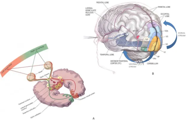
The Visual System... 33
Anatomical and physiological basis... 34
Measures, procedures, and concepts... 39
Eye movements and behaviour... 40
HARD-WIRED TO ESTABLISH AFFECTIVE BONDS ... 42
Biological Foundations of Attachment: The Attachment Behavioural System... 43
Individual differences in the attachment system’s functioning... 45
The Internal Working Models... 47
Attachment Patterns... 48
The emerging neurobiological underpinnings of attachment... 50
BRIDGING EMOTION, COGNITION, AND ATTACHMENT THROUGH THE PHYSIOLOGICAL ROUTE ... 54
E EXXPPEERRIIMMEENNTTAALLSSEECCTTIIOON ... 61N STUDY I - Attachment insecurity and strategies for regulation: When emotion triggers attention... 63
STUDY II - Hidden emotions under the skin: Autonomic reactivity on attentional regulation in insecure attachment... 85
STUDY III - Looking at the dark side of life? Eyes caught by attachment anxiety ... 111
G GEENNEERRAALLDDIISSCCUUSSSSIIOON... 137N Study I... 139 Study II ... 140 Study III... 141 Limitations... 142 Interpretation ... 143 Perspectives ... 154 R REEFFEERREENNCCEES ... 157S
xix
List of Figures
Figure 1. Overview of the sympathetic and parasympathetic branches of the autonomic nervous system ... 15
Figure 2. Anatomy of the eccrine sweat gland in various layers of skin ... 18
Figure 3. Recommended placement of electrode sites for the measurement of skin resistance and skin potentials (Redrawn from Venables & Christie, 1980)... 21
Figure 4. Graphical representation of the main EDA components ... 22
Figure 5. The pulmonary and systemic circulation systems (A); Anatomy of the human heart (B) ... 26
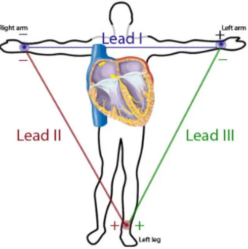
Figure 6. Representation of the Einthoven triangle with the standard limb leads I, II and III... 30
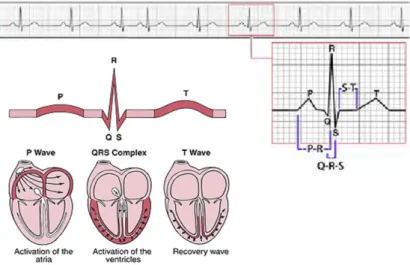
Figure 7. Electrocardiogram wave components... 31
Figure 8. Anatomy of the human eye (A); Structure of the retina (B), and Retinal layers (C). ... 35
Figure 9. The optic nerve (A) and the visual pathways (B). ... 36
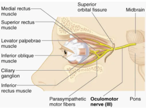
Figure 10. Extrinsic ocular muscles ... 38
Figure 11. Scan path showing the number, location, and duration of gaze fixation on both neutral and emotional pictures... 40
1
I
3
Overview
Human beings are inherently driven to establish and maintain affective bonds with conspecifics (Bowlby, 1969, 1973, 1980). This need finds its roots in humans’ intrinsic evolutionary history. More precisely, this need lies in a fundamental motivation in which survival has since long depended, as these bonds represent the source of cooperation, protection, and acquisition of knowledge essential for environmental adaptation (Caporael & Brewer, 1995; Wilson, 1978). The importance of attachment ties is obvious, both in the strong emotional reactions they elicit (Scherer, Summerfield, & Wallbott, 1983), as well as in the consequent behavioural responses, such as to approach or avoid. This is of particular relevance, considering that individuals navigate in a socio-emotional world.
Deeply embedded in human nature, approach and avoidance tendencies are fundamental building blocks underlying the complexity of social behaviours (Carver & Harmon-Jones, 2009). In the social context, stimuli readily captured are thought to define these specific responses to social demands in a protective manner. Eye movements play here a key role, which continuously scanning the outside world, allow individuals to learn, interpret, and adaptively guide behaviour. Indeed, the way people perceive and attend social and emotional experiences are critical in launching those behavioural tendencies (Harmon-Jones, 2003; Marsh, Ambady, & Kleck, 2005), in the subsequent information processing (Neumann & Strack, 2000), as well as in physiological reactions, such as sweat or increasing heart beat (e.g., Lang, Greenwald, Bradley, & Hamm, 1993). This clearly entails an evolutionary advantage for humans who can efficiently recognize and detect threatening stimuli or events in their environment, since this ability prompts them to trigger defensive behaviours (Öhman, Lundqvist, & Esteves, 2001; Öhman & Mineka, 2001, 2003). Importantly, this puts in evidence individual differences in the deployment of specific adaptive and regulation strategies people use in such encounters.
Within this scope, the evolutionary-based attachment theory (Bowlby, 1969, 1973, 1980) offers an enriching framework to further investigate such individual variations. Speculating on the underlying psychobiological mechanisms by which attachment may critically shape development and behaviour, Bowlby (1969) conceived the existence of a species-universal program aiming at providing species fitness: The attachment behavioural system, which has a core function in governing the transactions between the individual and the environment. In agreement, the system is thought to evolve along the interaction between unique biological features and a particular socio-emotional history. Therefore, the singular
4
outcome of both factors allegedly raises individual differences in attachment styles, and in subsequent regulation strategies. Interestingly, its inherent protective function confers to the system a fear-wariness facet, as it promptly detects potential threats in addition to specific plans of actions towards preservation. Ultimately then, the attachment behavioural system is assumed to shape emotions, cognitions and behaviours.
Regardless of the vast amount of research that has indubitably added valuable knowledge to the field (e.g., Main, 1990; Mikulincer & Shaver, 2007; Schore, 2003; Soares, Dias, Machado, Klein, 2002a), the patchwork nature of attachment styles and its impact on biobehavioural aspects is yet not clearly understood, remaining therefore a focus of debate.
Under this scenario, the goal underlying this thesis is to contribute to the current understanding on how the strategies endowed to insecure attachment styles operate and influence basic abilities critical for everyday life in a social world. These include regulation strategies at emotional, cognitive and physiological levels. Thus, applying an integrative outlook, the studies presented here attempt at bridging emotion, cognition, and psychophysiology towards a broader comprehension of insecure attachment.
This thesis is organized in three major sections. The Introduction section provides a description of the evolutionary-based attachment theory framework as a privileged model to investigate approach-avoidance tendencies in the influence of emotion on cognitive processes, such as threat detection, and its underlying regulation strategies. Also, this section briefly reviews prior research on the portrayal of the insecure attachment styles as described in terms of emotional and cognitive profiles towards adaptive functioning. The Experimental section includes three studies conducted, using different techniques: The first focusing on regulation abilities in attentional bias towards emotion information; the second investigating the psychophysiological aspects underlying such biases; and the third, examining these effects with the newly eye tracking method. Finally, the Discussion section will summarize the key findings within a multidisciplinary and comprehensive approach.
5
HARD-WIRED TO ATTEND EMOTION
I feel, therefore I am...
Emotion...
Since immemorial times, emotions have been an intriguing and central part in understanding human life, captivating thinkers and scientists, leading to theories, questions, some answers, and further questions... Yet, its fascination still holds today. Whilst emotions are thought to be inherently part of human nature, its complexity has precluded a definite and unambiguous definition, acknowledging that several conceptions prevail.
Tracing back to a few thinkers among many, it is possible to find valuable insights towards a comprehension of emotions since the pre-Socratic era. By then, and perhaps for some still today, emotions were a menace to the virtues of reason. Aristotle (384–322 B.C.) soundly proclaimed that emotions are connected to actions. Whenever an action takes place, the outcome may be of contentedness, disenchantment, or shame. Emotional experiences are therefore shaped by the subjective evaluation of the individual as a result of his own engagement in the social world. Also Descartes (1649/1989) agreed with Aristotle, stressing that emotions do not only involve the sensations caused by the physical agitation, but also perceptions and desires. Accordingly, an emotion that arises from the perception of a critical event, potentially harmful, involves the desire to avoid it. Nonetheless, emotions are conceived as a disruptive type of passion that contaminates reason. Conversely, David Hume (1739/1888) insisted that emotions were the source that enthused behaviour, and such source could not be identified with the impression or sensation alone, but only by more complex ideas, that is, reason.
In the late 19th and early 20th centuries major contributions arouse. Charles Darwin (1872/1965) pondered on emotions, emphasizing its adaptive function. Linking emotions to the past history of human species, emotional expressions such as tears when upset, are conceived as vestigial patterns of action. Therefore, fundamental emotions including fear or anger derive from habits that in our evolutionary past were vital for survival, and so are intrinsic across species and cultures. Later and also influenced by Darwin, William James (1890, 1884) highlighted the physiological grounds: Emotions are coined as behavioural and physiological adaptive response tendencies triggered in a straightforward fashion by evolutionarily relevant circumstances. In his own words, “…bodily changes follow directly
6
the perception of the existing fact… and feeling of the same changes as they occur, is the emotion” (1890, p. 449). That is, when we perceive the exciting fact, the emotion becomes the perception of body changes as we react to it. Lange (1882), joined James on the embodied nature of emotion, explicitly stating that “We owe . . . the emotional side of our mental life, our joys and sorrows, our happy and unhappy hours, to our vasomotor system.” (p. 80). While Lange clearly emphasized the importance of the viscera (e.g., stomach, heart), James further included other bodily responses (e.g., skin, peripheral muscles). Afar from the physiological account, Arnold and Gasson (1954) suggested that the appraising of critical events was the core feature of emotions. This involved either an attraction or repulsion from an event, which indicated whether the emotion was positive or negative. That is, specific emotions take place according to those appraisals. This standpoint thereby accentuates a large focus on input or bottom up perceptual processes. Integrating these internal and external dimensions, Schachter and Singer (1962) proposed that emotions were shaped both by physiological arousal and appraisal.
So far, this brief overview offered historical milestones to the understanding of emotions. But what are emotions? How can they be conceptualized to further pursue its scientific study?
But what are emotions? The nature of emotions and its evolutionary
grounds
“Like all primates, humans are intensively social species.
Indeed, we probably owe our success as species to our sociality“.
(Robin Dunbar, 2001)
In the 18th century, David Hume (1739/1888) notoriously declared: “reason is, and ought to be, the slave of the passions.” As Solomon (2008) agreeably puts it, this long debate between reason and passion; or between cognition and emotion is nicely illustrated by the metaphor of “master and servant”. This dissociation places cognition safely in a bright and in control path, whereas emotions, as a source of suspicion from tremulous grounds, are believed to be uncontrollable and more primitive. And so, the question “What is an emotion?” has proved to be a hard task to resolve.
Mutual features on the foregoing considerations sketch on the behavioural, cognitive and the physiological substrates of emotions. It is clear that emotions have a complex constellation of components (Lang et al., 1993; Levenson, 1999). Inherently part of the social life, emotions are embedded in human communication through facial expressions, bodily
7
posture, gestures, touch, and voice, reflecting the involvement of physiological responses in the body and the brain. Beyond and intertwined with social purposes, emotions are goal-directed, boosting regulatory behaviours to surpass actual contingencies. Emotions enable selection and swift orientation towards relevant events, driving attention to significant threats and opportunities, thereby facilitating adaptive actions and reactions (e.g., Levensson, 1999). Stressing the subjective experience of emotion, appraisals are strikingly marked by parallel psychophysiological changes in both the central and autonomic nervous system’s activity (e.g., Ekman, Levenson, & Friesen, 1983; Levenson, Ekman, & Friesen, 1990). Emotions trigger bodily actions: the heart pounds; palms sweat; muscles tense and relax; blood boils; faces blush, frown, and smile. In everyday life people recognize the strong organic reverberation to crude emotions such as fear, rage, grief, and love (James, 1890). Accordingly, emotions are thought to be organized in psychophysiological reactions about ongoing relationships with the environment (Lazarus, 1991). Unquestionably, emotions are biologically rooted, as its own etymological origin portrays - the Latin word movere, meaning to move - emotions can therefore be conceived as action dispositions, mobilizing the body for behaviour (Lang, 1995; Lang, Bradley, & Cuthbert, 1990). Stimuli prompt affective responses. When emotions are intense, people move: they freeze, flee, or fight, depending on the environmental context (Bradley & Lang, 2007). Furthermore, these affective responses appear to be in concert with inner motivations (Oatley & Jenkins, 1996).
In spite of the intricacy in defining what emotions are, and embracing all these aspects, Lang (1968) cogently argued that every emotional state consists of three basic components: A behavioural response, a subjective feeling, and a physiological correlate. Specifically, whenever an emotion is experienced, overt adaptive behaviours are triggered, such as approach or avoidance. These are further accompanied by subjective verbal reports, such as “I’m afraid”, and its inherent rating, implying the knowledge or the cognitive evaluation about the intensity of the reported description. While pleasant emotions are held to be associated with appetitive tendencies, unpleasant ones are roughly indexed to defensive trends (Cacioppo & Berntson, 1994; Lang et al., 1990). Moreover, hedonically valenced events or stimuli differ in the degree to which they engage a behaviour, which is also related to intensity arousal. Valence and arousal features are in turn critical in organizing the pattern of physiological responses in emotional reactions, thereby providing explanations to the emotional experience and to its subjective evaluation (Cuthbert, Schupp, Bradley, Birbaumer, & Lang, 2000; Russell, 1980). Finally, the physiological reactions are those bodily events that, out of the individual’s awareness, respond to specific demands: the heartbeat increases,
8
hands sweat, reflexes become sharper (Lang et al., 1983; Levensson, 1999). This multi-component model offers a more detailed framework to the scientific inquiry of emotions. Additionally, recent research has provided knowledge to the inquest of how emotions are embodied in the brain, demonstrating the contributions of key brain regions, including the prefrontal cortex (PFC), amygdala, hypothalamus and the anterior cingulate cortex (ACC), to the processing of emotions (e.g., Dalgleish, 2004; LeDoux, 1996).
All this, definitely bestows emotion with an evolutionarily advantage for the human involvement in the social world. Indeed, for the human species, the physical and social environment has functioned as selection pressures in which evolution has occurred, and the extraordinary capacity of cooperation appears to have been a powerful determinant to grant reproduction and survival (e.g., Buck, 1999, 2002). Within the evolutionary ground, this great task was accomplished in relation with other individuals and the establishment of relationships and communities. Underlying this gathering force is emotion, a species-characteristic trait, profoundly social, supporting affective attachments between conspecifics (e.g., Barret & Campos, 1987; Bowlby, 1969). In the same vein, it has been argued that emotions have evolved to become the basis of human social bonding (Oatley, Keltner, & Jenkins, 2006). This approach has gathered wide support (e.g., Bowlby, 1969; Ekman, 1992; Izard, 1971, 1977), converging to the idea that emotions, provided with biological script-like behaviours shaped by cultural factors, may be the language of human social life, thereby settling human attachment at the heart of the debate.
...and Cognition
"My experience is what I agree to attend to." (William James, 1890)
A hallmark feature in adaptive cognitive functioning is the capacity to selectively focus and process relevant information about ongoing behaviour, while other interfering sources are ignored (Lavie, 2005). Crucial to accomplish this are attentional mechanisms, in particular visual attention, which guides human behaviour while navigating in the cluttered mosaic environment (e.g., Driver, 2001).
As one of the most striking human abilities, attention has been since long a central theme in psychology research. At the turn of the 20th century Titchener (1909) referred to it as
9
emphasized: “Everyone knows what attention is. It is the taking possession of the mind in clear and vivid form of one out of what seem several simultaneously possible objects or trains of thought” (p. 403). Attention therefore refers to selectivity of processing: The ability to shift focus between countless stimuli in the environment through direct or avert strategies for further scrutiny (Lavie, Hirst, Fockert, & Viding, 2004). Importantly, attention can modulate or enhance the selected information according to specific goals of the perceiver. This implies that individuals are not merely passive receivers of information. But how does the brain bewares with potential overload? In the late 1950’s Broadbent (1958) advocated that filtering of irrelevant sensory information occurred at initial stages of processing. According to this early selection framework, further developed by Treisman and Geffen (1967), unattended information is not processed beyond its initial physical properties, such as spatial location or colour. It is only at later stages that attentional processes are called upon to further integrate the selected information. Alternatively, a late selection explanation was also offered, positing that selection takes place only after categorization and semantic analysis of all inputs has occurred (Deutsch & Deutsch, 1963; Duncan, 1980). Later, Pashler (1998) suggested that unattended information was not completely filtered; however, it was not processed to the same degree as attended information either.
A conciliation between these two accounts was provided by Lavie (1995, 2005), who proposed that the early/late selection debate could be resolved by considering the overall perceptual load. In order for attention to remain focused on a specific target, the overall perceptual load must be sufficiently high to ensure that no capacity remains to process distracter events; otherwise attention spreads to irrelevant cues (Lavie, 1995; Lavie & Tsal, 1994). That is, in contexts of high perceptual load early selection processes take place, whereas in the opposite situation late selection is likely to occur. This suggests that different mechanisms of selection exist and may operate simultaneously during perceptual processing. In agreement, more recent research acknowledges that visual selective mechanisms operate in a hierarchical fashion along non-selective and selective pathways. The non-selective pathway involves preattentive processes that operate independent of attentional focus, recognizing basic features of the visual surroundings very quickly. Importantly, these processes occur automatically, and quite efficiently. Conversely, the selective pathway entails attentive processes, which deploy full attention to a specific object, binding its features together into a cohesive image. However, the proficiency of attentive processes is of limited capacity. While these employ a serial processing mode, the preattentive ones, encompassing a monitoring system that constantly scans the environment, processes information in parallel (Wolfe 2000;
10
2007). Thus, this preattentive ability is crucial to direct attention toward or away from relevant targets. The combined action of both pathways provides a powerful device to guide humans through the visual world.
Beyond selectivity of processing, and following James (1890), researchers further distinguished between two modes of attention deployment: a goal-driven mode that based on top-down processing is controlled by the individual’s intentions; and a stimulus-driven mode, which based on bottom-up processing, can steer attention automatically. The goal-driven mode typically requires voluntary and processing effort while selecting relevant incoming sensory information for further analysis. In addition, top-down mechanisms employ longer-term cognitive strategies, involving the individual’s expectations, knowledge, and current goals. On the contrary, the stimulus-driven mode, which depends on the nature of the stimuli such as saliency and novelty, is thought to be faster and more potent. This is because bottom-up mechanisms are thought to operate on raw sensory input, quickly and involuntarily shifting attention to prominent visual features of potential relevance that pop-out (e.g., Jonides, 1981; Yantis, 1998; Yantis & Jonides, 1984). Thus, bottom-up attention may alert to salient stimuli in the environment, such as a red spot against green trees that could be a delightful fruit, or a sudden movement that could be a predator. In contrast, top-down attention may modulate bottom-up signals when focus on a specific task is needed, biasing attention toward colour spots when the observer is ravenous or toward sudden movements if the observer is frightening (e.g., Yantis & Egeth, 1999). This implies that although top-down and bottom-up systems emphasise distinct routes to information processing, the guidance of attention is governed by dynamic interactions between both systems (Corbetta & Shulman, 2002; Posner & Petersen, 1990). This is especially true when manifold stimuli compete for neural representation in the visual cortex (e.g., Torralbo & Beck, 2008). It has been suggested that this competition may be resolved through prefrontal and parietal sources of top-down attentional control, which can enhance the representation for a particular visual stimulus at the expense of others (Beck & Kastner, 2009; Scalf & Beck, 2010).
Nevertheless, the stimulus itself or relevant contextual cues may prompt pop-out effects in the visual cortex, independent of top-down control (Beck & Kastner, 2005). Additionally, this dominance of stimuli-specific favouring bottom-up processing may also gain neural representation via connections with the amygdala (Beck & Kastner, 2009). This is the particular case of emotionally relevant stimuli (e.g., LeDoux, 2000; Sander, Grafman, & Zalla, 2003). In agreement, these processes plainly convey an adaptive advantage either to swift and effortless attend unexpected events, such as potential threats, as well as to redirect
11
processing resources and promote shifts of attention to new focus of interest. Thus, while emotion drives attention, other attention control processes may in turn take over such interfere.
The two-way link between emotion and cognition
The interplay between these aspects involving emotion and cognition entails the evolutionary value of attention mechanisms. For adaptive purposes, the perceptual system is biased to efficiently recognize and detect threat (e.g., Lang, Bradley, & Cuthbert, 1997), as well as pleasure (Lang et al., 1990). Indeed, within the evolutionary framework (Öhman, Carlsson, Lundqvist, & Ingvar, 2007; Öhman et al., 2001), automatic processing of such stimuli is assumed to be functional in readily recruiting resources to either cope with potentially harmful events, or to take advantage of beneficial situations (e.g., Lang et al., 1997; LeDoux, 1996; Öhman, 1993). Accordingly, this capacity serves an important function in governing behaviour, prompting rapid preservative tendencies such as avoidance or approach, thereby minimizing aversive outcomes and increasing the likelihood of survival (Öhman et al., 2001; Öhman & Mineka, 2001; 2003). This seemingly pop-out effect, thought to occur at a preattentive perceptual stage, is especially notorious towards fearing cues (for reviews see Mathews & MacLeod, 2005; Mogg & Bradley, 1998). Evidence showing that threatening stimuli capture and hold attention supports this view (Öhman, Flykt, & Esteves, 2001; Pratto & John, 1991). Stimuli with a positive value have also been found to yield a capture effect, though not to the same degree (Anderson, 2005; Arnell, Killman, & Fijavz, 2007; Blair et al., 2007; Schimmack, 2005). The less prominent influence of positively valenced stimuli or activities, such as feeding and procreation, may be related to its less pressing function. Although these are of crucial importance, pleasure may be simply less urgent than pain, which carries imperative signals implying that an action should be quickly taken (e.g., Pratto & John, 1991). Interestingly, it has been recently argued that positive stimuli should have a similar effect as negative ones, as the prompt detection of food may be as essential for survival as the detection of threat (Blair et al., 2007). That is, the emotional content of external cues informs about how these are related to the individual’s needs and well-being.
Of note, this emotional modulation of attention is also known to impair processing of concurrent information (Blair et al., 2007; Pratto & John, 1991). Yet, emotional stimuli tend to facilitate encoding, wherein the information is recalled with higher accuracy and vividness
12
than neutral ones (e.g., Kensinger & Corkin, 2003; Ochsner, 2000; Vuilleumier, 2005). The cognitive system must therefore, be prepared to carry out efficient appraisals, which are critical for the success of goal-directed behaviour.
It has been suggested that the increased perceptual processing of emotional stimuli, in particular threat-related, might result from direct feedback signals imposed by the amygdala on cortical pathways, potentially with other top-down influences induced by attentional systems in the frontal and the parietal cortex (Vuilleumier, 2005). Indisputably, emotion readily biases the selection of sensory inputs. However, other higher-level influences beyond perception, including memories, thoughts and actions possibly coerce emotion biases on top-down control (Miller & Cohen, 2001). This means that the flow of sensory processing and response selection may be accomplished by various functional pathways. Behaviourally, this twofold influence can be found in individual differences, in which a modulatory effect on the magnitude of emotional biases in attention is observed, in both clinical and non-clinical populations (e.g., Fox, Russo, Bowles, & Dutton, 2001; Mogg & Bradley, 1999; Yiend & Mathews, 2001). Then, this two-way link may underlie functional interactions between emotional and cognitive factors that regulate the allocation of processing resources and determine goals in behaviour (Vuilleumier, 2005).
Although the impact of emotion on cognition is not yet completely understood (Blair et al., 2007), this prospect is especially interesting when considering emotion regulation: Control processes are called upon, so the individual is not overridden by emotion (Gross, 1998; Ochsner & Gross, 2004; 2005). Similarly then, emotion regulation, involving the initiation or alteration of ongoing behavioural responses is too at the core of human adaptation. Yet, this dual interplay on emotion information processing is not only found on cognitive functions such as attention. As a reminder, whenever an emotion is experienced, physiological changes are likely to occur (e.g., James, 1890; Lang, 1968). Under this scope, a door for a deep understanding is unlocked by the psychophysiological science, which provides the assessment of responses from specific biological systems related to emotion information processing.
13
THE WAY BACK ALONG PSYCHOPHYSIOLOGICAL
PATHWAYS
“…the principal function of the nervous system is the coordinated innervation of the musculature. Its fundamental anatomical plan and working principles are understandable only on these terms.”
(Sperry, 1952)
Foundations of the Psychophysiological Science
The interest regarding the physiological substrates of behaviour has accompanied the course of intellectual history, and such wonderings have ultimately placed psychophysiology at the core of the mind-body debate (Greenfield & Sternbach, 1972). Indeed, as its literal definition asserts, psychophysiology studies the interactions between the mind and the body. In agreement, the psychophysiological science is based on the assumption that psychological processes are an embodied phenomena, and that measures of physical processes can therefore shed light on the human mind (Cacioppo & Petty, 1981). It is currently acknowledged that social, cultural, and interpersonal contexts are powerful determinants of brain and behaviour. As a natural consequence, monism has replaced any remnants of dualism, as psychological states are more reliably to be conceived as represented in, and acting through cortical, limbic, and brainstem regions. These states in turn influence the activity at autonomic and neuroendocrine levels, which subsequently modulate crucial cellular and molecular processes (Cacioppo, Tassinary, & Berntson, 2007). The human brain is highly complex, with several processes occurring simultaneously, however, only a few are relevant to any particular peripheral organ or effect.
Traditional approaches to the investigation of human behaviour typically manipulate psychological states to observe consequent changes in the brain. Considering this perspective as a form of conversion of the natural flow of neuronal impulses in a representation and interpretation of the world, Sperry (1952) urged science to view the brain as an organ that transforms patterns of sensory experience into patterns of motor response. Focusing on the afferent information that travels from the periphery to the central nervous system (CNS), one can observe its influence on the brain and behaviour. There is a vast amount of research on the integration of autonomic and somatic responses, showing that such blending is primarily accomplished within the CNS, while the coordinated autonomic-behavioural activity is reflected at the periphery. That is, this autonomic-behavioural activity is mirrored in anticipatory and preparatory phases of a given output (Germana, 1969). Sperry (1952)
14
therefore invited researchers to pursue the way back. Indeed, this is the path followed in psychophysiological science: Psychological processes are reflected in efferent patterns, as evinced by the modulatory effect of muscles and glands. The subject matter of psychophysiology is, after all, an embodied phenomenon. Thus, emphasizing specific relations between psychological and physiological underpinnings of human behaviour, psychophysiology can be defined as the scientific inquiry of cognitive, emotional, and behavioural phenomena as related to, and revealed through, physiological principles and events in functional organisms interacting with the environment (Cacioppo & Tassinary, 1990; Cacioppo, Tassinary, & Berntson, 2007). This implies that a hallmark feature of psychophysiology is a multi-component analysis framework; brain and body processes occurring on environmental transactions. The human brain plays here a crucial role, as the chief regulator. Having evolved to refine behavioural adaptive capacities, its structural and functional organization is oriented towards a continuous efferent path, which characterizes its own architecture.
The structural and functional architecture of the human brain is divided into two major subsystems: the central nervous system (CNS) and the peripheral nervous system. The CNS consists of the spinal cord and the brain. As the key governor and coordinating system, the CNS detects, interprets, and responds to changes to internal and external conditions. Specifically, it integrates information and generates appropriate feedback by sending electrochemical impulses through nerves to effector organs such as muscles and glands. The nerves connecting effectors and receptors to the CNS compose the peripheral nervous system. This system includes sensory receptors, and both sensory and motor neurons. Sensory receptors are activated by stimuli in the internal or external environment, which are in turn forwarded to sensory neurons in the CNS. The CNS then processes the signal, and transmits a message back to an effector organ through a motor neuron.The spinal cord, as an extension of the brainstem, carries messages between the CNS and the rest of the body. The peripheral system is further divided into the somatic nervous system and the autonomic nervous system (ANS), also called visceral system. Receiving sensory information from peripheral and external sources, the somatic nervous system enables humans to react consciously to environmental changes. It includes 31 pairs of spinal nerves containing both sensory and motor neurons, with which it voluntarily controls the movements of the skeletal muscles. It also comprises 12 pairs of cranial nerves involved in sensory functions related to balance, sight, olfaction, taste and touch. On the other hand, the ANS mainly receives neural signs from the limbic system and the hypothalamus, and has the core function of maintaining
15
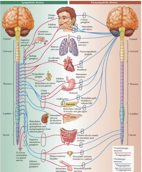
internal and external homeostasis. As its own designation asserts, the ANS operates in an automatic fashion, without voluntary input. The ANS has an afferent pathway, which consists of postganglionic neurons within the viscera that transmit the information to the CNS, and an efferent pathway comprising preganglionic neurons arising from the CNS. By releasing specific neurotransmitters, these preganglionic neurons trigger the action back to the effectors: The smooth and cardiac muscles and glands. The ANS further divides into the sympathetic and parasympathetic nervous systems (see Figure 1).
Figure 1. Overview of the sympathetic and parasympathetic branches of the autonomic nervous system
Briefly, the sympathetic division adaptively mobilizes bodily resources under challenging situations, thereby inducing fight-or-flight responses. The neurons that drive such effects are primary motor neurons arising from the thoracic and lumbar regions of the spinal cord. The postganglionic sympathetic neurons typically release norepinephrine, which
16
activates adrenergic receptors on the peripheral target tissues. Others, however, release acetylcholine that activates muscarinic receptors, such as the ones involved in sweat glands innervation. Thus, heightened levels of sympathetic activity increase alertness, and allow the body to maximize the use of its own resources: The pupils dilate, the blood vessels of the skin and gut constrict, the bronchi dilate increasing oxygenation, the heart rate accelerates, the force of cardiac contraction is enhanced, blood is shunted towards skeletal muscles, while digestive and other vegetative functions become quiescent. In contrast, the parasympathetic division has the functional role of restoring the organism’s equilibrium that were suspended during intense activity, increasing metabolic and other energetic resources when the body is at rest. The parasympathetic neurons rise from distributed areas in the brainstem: the cranial and vagus nerves, and the sacral part of the spinal cord. Through the release of acetylcholine its activity constricts the pupils, as well as the coronary and blood vessels, slows the heart rate, and increases the peristaltic activity of the gut (for details see Kandel, Schwartz & Jessell, 2000).
To accomplish its endeavours, psychophysiology has developed a number of non-invasive recording procedures capable of capturing the nervous system activity. Typically then, these techniques are traditionally organized in terms of the registered physiological activity and the underlying neurophysiological mechanism (e.g., Andreassi, 2000). To name a few among many, psychophysiological techniques focused on the CNS activity include electroencephalography (EEG), event-related brain potentials (EPRs), and neuroimaging methods such as positron emission tomography (PET) and functional magnetic resonance imaging (fMRI). Of note however, is the fact that the PET technique has the inconvenient of using contrast agents. Physiological measures of the peripheral, somatic nervous system’s activity comprise electromyography, electrooculography, and eye tracking. Finally, those involving the peripheral, ANS include the recording of the electrodermal activity (EDA), electrocardiography (ECG), electrogastrography, and also eye tracking. Indisputably, these methods and procedures allowed psychophysiological research to flourish. The demonstration that autonomic events are highly correlated with behavioral responses highlights that the core function of the CNS is to provide adaptive behaviour (Germana, 1969), thereby offering insights into almost every facet of human nature.
17
The Electrodermal System
Highly sensitive to psychological states and processes, the electrodermal activity (EDA) is a widely used psychophysiological measure. Research involving EDA include investigation on cognitive functioning, such as attention and information processing, as well as emotional processes on both typical and atypical behaviour.
The discovery of electrodermal activity is ascribed to the pioneering studies of Féré (1888) and Tarchanoff (1890). The inquest of the psychological effects on the electrical changes in human skin began over 100 years ago with Jean Charcot and his collaborators, and their known work with patients suffering of hysteria. While Vigouroux (1879, 1888) measured electrical activity as a mean of diagnosis for the disorder, Féré (1888) found that by passing a small electrical current across two electrodes placed on the surface of the skin, it was possible to momentarily measure skin conductance changes in response to stimuli. Importantly, these findings launched the first attempts to establishing a link between the electrical phenomena in skin changes and psychology, that is, in establishing its psychological significance. This type of measurement is today referred to as exosomatic method, since the recording of the skin resistance response (or its reciprocal, the skin conductance response) relies on the passage of an external current across the skin. Shortly after, Tarchanoff (1890) noted that the electrical potential between two electrodes placed on the skin could occur without applying an external current, further suggesting that such variations in skin potential were the result of sweat glands secretion. Tarchanoff’s assumption was later supported (Darrow, 1927), and it is currently accepted that the activity of the sweat glands, not sweat on the skin per se, is crucial for EDA. This procedure is termed as endosomatic method, as the recording of the skin potential response does not involve an external current. However, its interpretation is less well understood. Contemporary research uses preferably the exosomatic method for the recording of skin conductance levels (SCLs) and skin conductance responses (SCRs) (Fowles et al., 1981). Tracking these early findings, the modern era on EDA research began in the 1970s when Lykken and Venables (1971) proposed standardized techniques for the recording skin conductance, as well as units of measurement.
Anatomical and physiological basis
Conveyed with a major adaptive significance, the human skin is a selective barrier that prevents the entry of foreign organisms into the body, selectively facilitating passage of
18
materials from the bloodstream to the exterior of the body. Additionally, it regulates water balance and body temperature, mainly through vasoconstriction and dilation, and variations in the production of sweat. The skin consists of three major layers (see Figure 2): the epidermis, the dermis, and the hypodermis. The epidermis comprises three strata: (1) the outermost stratum is the corneum or horny layer, composed of a coating of dead cells that serves to protect the internal organs; (2) beneath lays the lucidum, containing tough connective tissue and blood vessels, and (3) the Malpighii or panniculus adiposus, the deeper stratum made of fat and connective tissue. The eccrine sweat glands are found on the layer bellow, the dermis. These are simple tubular structures, consisting of a coiled compact body, where the secretory segment is located, and by a sweat duct, a long tube whereby the gland excretes sweat. The secretory portion has a spherical structure of about 0.3 to 0.4 mm in diameter. The sweat duct remains relatively straight in its path through the stratum Malpighii and stratum lucidum, it then spirals through the stratum corneum and opens on the surface of the skin as a small pore (Edelberg, 1972).
Figure 2. Anatomy of the eccrine sweat gland in various layers of skin
There are, however, two types of secretory glands. Besides eccrine glands, which are located all through the body, but in dense concentrations on surface of hands and feet, there are apocrine glands, which are found under armpits and genital areas. These are relatively large, open into hair follicles that continuously secrete fatty sweat. Apocrines are scent glands stimulated by epinephrine hormones that to date, have been less attractive for
19
psychophysiology. Conversely, the eccrine glands are of major interest. It is estimated that a square centimetre on the palm contains 2.000 sweat glands (Jacob & Francone, 1970; Vila, 2006). Cholinergic stimulation via fibres from the sympathetic nervous system (SNS) constitutes the foremost influence on the production of sweat by eccrine glands (e.g., Shields, MacDowell, Fairchild, & Campbell, 1987; Wallin, 1981). Specifically, the sweat glands are innervated by postganglionic sympathetic nerve fibres, while the sweat ducts act as a set of variable resistors wired. The column of sweat rise in the ducts in varying amounts and in varying number of sweat glands, depending on SNS activation. The higher the sweat rises, the lower the resistance in the resistors. As sweat fills the ducts, the path through stratum corneum becomes increasingly conductive. Therefore, examining meaningful changes in the skin conductance amplitude entirely based on the sweat glands constitutes a reliable method (Edelberg, 1993).
Notabily, the SNS has both excitatory and inhibitory influences distributed in various parts of the brain and therefore the neural mechanisms and pathways implicated in the CNS control of EDA are many and complex. The hypothalamus, which has a core function in maintaining homeostasis, such as thermoregulation, is the head ganglion of the autonomic nervous system, acting as the major subcortical center regulating its activities (Truex & Carpenter, 1964). Yet, other cortical areas are also thought to be involved. It has been suggested that EDA is influenced by emotion and arousal (e.g., Boucsein, 1992). Accordingly, additional limbic structures, such as the cingulate gyrus, and the hippocampus, contribute for electrodermal responses to temperature and emotional cues. The reticular formation, besides informing the hypothalamus about skin temperature, also plays a part on EDA following arousal states. More recent research on the neural substrates underlying the production of SCRs found that the ventromedial prefrontal cortex, the right inferior parietal region, and the anterior cingulate were associated with elicitation of SCRs while participants evaluated stimulus significance. Moreover, when the stimulus were emotionally relevant, the amygdala and the orbitofrontal cortex, in addition to the areas mentioned above, were also involved (e.g., Bechara, Damasio, & Lee, 1999; Critchley, Elliot, Mathias, & Dolan, 2000; Tranel & Damasio, 1994). Importantly, these areas are at the core of the so-called social brain, being recruited for emotional and social information processing (e.g., Baron-Cohen et al., 2000; Johnson, 2005).
20
Measures, procedures, and concepts
Following the exosomatic method, EDA is measured by passing a small current through a pair of electrodes placed on the surface of the skin. The principle invoked in the measurement of skin resistance or conductance is that of Ohm’s law, which states that skin resistance (R) is equal to the voltage (V) applied between two electrodes placed on the skin surface, divided by the current (I) being passed through the skin. This law can be expressed as R = V/I. The circuits used to measure skin conductance are of two basic types: constant current or constant voltage. When using the constant current it is possible to measure the voltage between the electrodes, which varies directly with skin conductance changes. Conversely, when the voltage is held constant across the electrodes the current flow is measured, varying directly with the reciprocal skin conductance changes. Lykken and Venables (1971) strongly argued for a constant voltage amplification system, as it allows a direct measurement of skin conductance. Conductance is expressed in units of Siemens whereas skin conductance changes are expressed in units of microSiemens (µS) (e.g., Dawson, Schell, & Filion, 2007; Mendes, 2009; Vila, 2006).
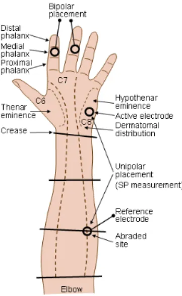
There are, however, important methodological considerations underlying an optimal EDA recording. Silver-silver chloride (Ag/AgCl) cup electrodes are generally recommended because they significantly reduce bias potentials and polarization (Venables & Martin, 1967). These electrodes, usually with 1.0 to 1.5 cm in diameter, can be easily attached to the recording site through the use of double-sided adhesive collars, which also serve to control the size of the skin area that comes in contact with the electrode paste. This electrode paste is a central parameter, because it is the contact area, not the size of the electrode that affects the conductance values. Thus, it serves as a conductive medium between the electrodes and the skin. In order to preserve the electrical properties of the skin, the paste should closely resemble sweat in its salinity, or sodium chloride (NaCl), near 2.9gr/100ml water (Venables & Christie, 1980). Skin conductance is recorded using two electrodes (bipolar recording), both placed on active sites. The recordings are typically taken from specific locations on the palms of the hands, with several acceptable placements. The most common is the electrode bipolar placement in the thenar eminences of the palms, and the volar surface of the medial or distal phalanges of the fingers (see Figure 3). Although EDA can be measured from any of these sites, the values obtained are not necessarily comparable. It is frequent to place the electrodes in the non dominant hand because it is less likely to have lesions, and because it leaves the dominant hand free to simultaneously perform a manual task. Adequate hygiene of
21
the recording site is also important. Hands should not be cleaned with alcohol or others potential abrasive agents that may diminish the natural conductive properties of the skin. Nevertheless, individuals should be asked to wash their hands with a nonabrasive soap prior to the electrodes attachment. The skin should be kept clean and dry (Venables & Christie, 1973). Finally, environmental factors, such as temperature and humidity levels are significant as well, as they may constraint an optimal hydration of the stratum corneum. A room temperature of 23◦
C is recommended (Boucsein, 1992).
Figure 3. Recommended placement of electrode sites for the measurement of skin resistance and skin
potentials (Redrawn from Venables & Christie, 1980)
Changes occurring in the skin conductance reflect different types of EDA: tonic levels, phasic responses, and spontaneous fluctuations, that in spite of closely related, reveal different psychological processes (Venables & Martin, 1967). The tonic level refers to relatively stable EDA. These fluctuations, which occur relative to the baseline activity at any given time and in the absence of discrete stimulation, are designated as skin conductance levels (SCL). Tonic SCLs usually ranges between 2 and 50 µS (e.g., Vila, 2006). Conversely, momentary fluctuations occurring in the presence of specific stimuli are termed as phasic responses and correspond to skin conductance responses (SCR; Venables & Martin, 1967). The SCRs represent the number of conductance changes (enlarged or reduced) that reach a maximum value of change, tending to recover to baseline levels afterwards. To determine such changes, minimum values between .01 and .05 µS are frequently used. Phasic SCRs are characterized by high frequency rates and its amplitude typically ranges between .05 and 5.0 µS (e.g., Mendes, 2009). In addition, the scoring of specific SCRs depends on the time latency
22
window during which a response is assumed to be elicited by the stimulus. Latency windows of a 1–3 or 1–4 seconds are generally accepted. The presentation of novel, unexpected, emotional stimuli are likely to elicit SCRs referred to as “specific” SCRs. Indeed, studies have demonstrated that such fluctuations may mirror attentional and orienting responses (e.g., Öhman, Hamm, & Hugdahl, 2000), as well as motivational and emotional dimensions of the given stimuli (Gomez, & Danuser, 2004). Finally, the spontaneous fluctuations are a particular type of phasic response, in which the elicit stimulus is not identifiable. In such cases, the occurring SCR is referred to as a spontaneous or nonspecific SCR (NS-SCR). These responses may be elicited by means of deep breaths, body movements, or cognitive and emotional automatic processing (e.g., phobic reactions). However, if these occurrences are also recorded, a tangible explanation may be offered (e.g., Hugdahl, Fredrikson, & Öhman, 1977). The measurement of NS-SCR activity is their rate per minute, which classically takes place between 1 and 3 min while subjects are resting.
Specific stimulus-elicited SCR are most commonly analyzed in terms of the amplitude or size of the SCR waveform. The amplitude of the SCR is quantified as the amount of increase in conductance measured from the onset of the response to its peak. The mean value computed across trials during which a measurable (nonzero) response occurred will provide the amplitude of SCR (Humphreys, 1943). Because skin conductance amplitude is frequently found to be positively skewed, several correction methods are advised, as for instance the use of logarithmic transformations (Venables & Christie, 1980).
In addition to the SCR amplitude, there are other EDA components related to temporal characteristics of the SCR waveform (see Figure 4). These include the onset latency (time between a stimulus and the onset of SCR); rise time (time between the onset and peak of the SCR); and recovery halftime (time between peak SCR and 50% of recovery to prestimulus baseline). However, these temporal characteristics are not yet fully understood, and therefore are less commonly reported (e.g., Dawson et al., 2007).
23
The electrodermal system and behaviour
Research on the physiological underpinnings of human behaviour through EDA encompasses distinct experimental approaches; those involving the presentation of either discrete or long-lasting stimuli or situations, and those involving correlates of individual differences. Paradigms involving discrete stimulus are the most often used (e.g., Dawson et al., 2007; Vila, 2006). Extensive research has demonstrated the effectiveness of examining SCRs to brief or discrete stimuli, underscoring the relevance of this physiological measure. Determining the psychological meaning of a specific SCR is dependent on a well-controlled experimental paradigm set, requiring knowledge of both the stimulus condition, as well as the response system. The SCR is believed to be extremely responsive to certain stimuli properties such as novelty, surprise, arousal value, and significance (e.g., Shakhar, Gati, Ben-Bassat, & Sniper, 2000; Lang et al., 1990). Additionally, it may be the case that SCR occurs without the individual’s awareness. That is, SCRs may be elicited by stimuli properties that were not consciously processed, but that grabbed preattentive mechanisms instead (Öhman, 1979). Lang, Bradley and Cuthbert (1998) developed a set of widely used pictures (International Affective Picture System, IAPS), rated for both their arousal and valence quality. The SCRs elicited by these pictures have reliably been found to be related to the arousal dimension, with larger SCR amplitude, as arousal rating increased for both positively (e.g., erotic) and negatively valenced pictures (e.g., snakes; Lang et al., 1993; Cuthbert, Bradley, & Lang, 1996).
Conversely, paradigms involving long-lasting or continuous stimulus are best conceptualized in terms of modulating increases and decreases in tonic arousal. Therefore, the SCL, which represents reactions over relatively long periods of time, is the electrodermal measure commonly preferred. Experimental conditions that reliably produce increases in levels of skin conductance are the ones requiring the performance of a stress inducting task (e.g., arithmetic tasks). Typically, SCLs are thought to increase about 1 µS above resting levels during anticipation, followed by another increase of 1 or 2 µS during task performance (e.g., Lacey, Kagan, Lacey, & Moss, 1963). The finding that the SCL is consistently elevated in such conditions suggests that tonic EDA reveals an effortful allocation of attentional resources on task performance, associated with heightened autonomic activation (Jennings, 1986). Other types of long-lasting stimulus likely to spur electrodermal arousal include the presentation of films eliciting emotional states (Gross & Levenson, 1993; Gross, 1998), and