Universidade de Lisboa
Faculdade de Farmácia
Mestrado Integrado em Ciências Farmacêuticas
2019
Classification of dermatophytes by a multilocus phylogenetic approach
based on Tef-1α, beta tubulin and ITS genes
Universidade de Lisboa
Faculdade de Farmácia
Classification of dermatophytes by a multilocus phylogenetic approach
based on Tef-1α, beta tubulin and ITS genes
Carolina Pedro Gomes Tomé
2019
Orientador: Doutora Maria Manuel Pereira Lopes, Professora Auxiliar
Co Orientador: Doutora Rosalie Sacheli, Investigadora
Monografia de Mestrado Integrado em Ciências Farmacêuticas apresentada à
Universidade de Lisboa através da Faculdade de Farmácia
3
Resumo
Os dermatófitos são fungos que têm a capacidade de invadir tecidos queratinizados de humanos e de outros animais. As infecções causadas por dermatófitos são comumente conhecidas como tinhas, e podem afetar a pele, cabelos e unhas.
Este estudo diz respeito a Trichophyton mentagrophytes, Trichophyton interdigitale, Trichophyton violaceum e Trichophyton soudanense, quatro espécies conhecidas por causar Tinea capitis, uma infecção que afeta os folículos capilares sendo predominantemente uma doença que afecta crianças pré-adolescentes.
Embora as dermatofitoses não sejam consideradas de natureza urgente, são um problema de Saúde Pública. O aumento dramático da incidência das dermatofitoses e a necessidade de aumentar o nosso conhecimento acerca da ecologia e epidemiologia dos dermatófitos têm implicado uma investigação mais intensa em métodos de diagnóstico para identificação rápida e precisa destes patogéneos fúngicos.
A filogenia dos dermatófitos, no entanto, permanece incerta, porque os seus membros são filogeneticamente e taxonomicamente muito próximos e as suas características fenotípicas às vezes são precárias.
Este trabalho descreve uma tentativa de um método de identificação de dermatófitos, usando a sequenciação de DNA fúngico de três genes específicos, ITS, BT2 e Tef-1α para identificar filogeneticamente e distinguir T. mentagrophytes de T. interdigitale e T. violaceum de T. soudanense, uma vez que foram identificados erroneamente ao longo dos anos, a fim de fornecer informações sobre a sua taxonomia e promover a possibilidade de efetuar uma terapia direcionada.
Palavras-chave: Tinea capitis; Trichophyton interdigitale; Trichophyton mentagrophytes; Trichophyton violaceum, Trichophyton soudanense
4
Abstract
Dermatophytes have the capacity to invade human keratinized tissues and other animals to produce infections, dermatophytosis. Dermatophyte infections are commonly known as ringworm or Tinea, and can affect the skin, hair and nails.
This study concerns Trichophyton mentagrophytes, Trichophyton interdigitale, Trichophyton violaceum and Trichophyton soudanense, four species known to cause Tinea capitis, an infection that affects the hair follicles. It is predominantly a disease of preadolescent children. Although a dermatophyte infection is not an emergency, the dramatic increase in the incidence of opportunistic fungal infections and the need to increase our knowledge of the dermatophytes ecology and epidemiology has led to a critical need for diagnostic methods that can rapidly and accurately identify fungal pathogens.
The phylogeny of dermatophytes, however, remains unclear because their members are phylogenetically and taxonomically very closely related, and their phenotypic features are sometimes poor.
This project describes an attempt to establish a method of identification of dermatophytes by sequencing the fungus DNA, Internal transcribed spacer (ITS) , Beta tubulin gene (BT2) and the nucleotide sequence of translation elongation factor 1-α (Tef-1α) were used as tools to identify phylogenetically these four species, and distinguish T. mentagrophytes from T. interdigitale and T. violaceum from T. soudanense, since they have been misidentified over the years, in order to provide information about their taxonomy and promote the possibility for a targeted therapy.
Key words: Tinea capitis; Trichophyton interdigitale; Trichophyton mentagrophytes; Trichophyton violaceum, Trichophyton soudanense
5
Agradecimentos
Em primeiro lugar, gostaria de demonstrar a minha enorme gratidão à Professora Maria Manuel da Faculdade de Farmácia da Universidade de Lisboa pela sua ajuda ao longo da elaboração deste projeto, pela disponibilidade e apoio que sempre demonstrou, e pela enorme paciência.
Seguidamente gostaria de agradecer à professora Marie-Pierre Hayette do Centre Hospitalier Universitaire de Liège, que me proporcionou a oportunidade de trabalhar e conhecer um pouco melhor esta área e tornou possível o desenvolvimento deste trabalho. Teço um grande agradecimento à professora Rosalie Sacheli, Centre Hospitalier Universitaire de Liège, pelo constante apoio e dedicação, e especialmente pelo acompanhamento que me deu durante os três meses que estive na Bélgica.
Gostaria também de agradecer ao Fabrice, pela sua boa disposição constante e pela forma sempre calorosa como me acolheu no laboratório.
Aos amigos que conheci em Liège, um agradecimento parece-me pouco, vocês tornaram o meu Erasmus numa das minhas melhores recordações.
Aos meus amigos da FFUL, que me acompanharam durante 5 anos que passaram a correr, um obrigada especial, vocês ensinaram-me que não importa quanto tempo passou, mas sim a intensidade como vivemos cada momento.
Aos meus amigos da AMSC, que no fundo são e serão sempre a família que eu escolhi. Aos meus pais, que estão sempre presentes em todas as minhas conquistas e derrotas, pelo seu apoio incondicional, ao meu irmão Herberto por ser um exemplo para mim, à Mariana e à Sara, que foram e sempre serão uma constante na minha vida, e ao Miguel por ser o meu porto seguro.
6
Acknowledgments
Firstly, I would like to show my deep gratitude to Professor Maria Manuel from Faculty of Lisbon of University of Lisbon for her help throughout the writing of this project, for her constant availability, support and for always being patient with me.
Next, I would like to thank Professor Marie-Pierre Hayette from Centre Hospitalier Universitaire de Liège,who granted me the opportunity to work and to learn more about this area, and who made possible the development of my thesis.
I am very grateful to Professor Rosalie Sacheli, from Centre Hospitalier Universitaire de Liège, for her constant help and dedication, and especially for the support she gave me during the three months I spent in Liège.
I would also want to thank Fabrice for his constant joy and kindness, and for always making me feel welcome in the laboratory.
To the friends I met in Liège, a thank you is not enough. You made my Erasmus in one of my favourite memories.
To my FFUL friends, who accompanied me for 5 years who taught me that time is not important, only the intensity as we live each moment.
To my AMSC friends, who have been and will be, always, the family I chose.
To my parents, who are always present in all my achievements and defeats, for their unconditional support, to my brother Herberto, who is an example for me, to Mariana and to Sara, who have been and always will be a constant in my life and to Miguel, for being my safe harbour.
7 List of Contents Resumo………. 3 Abstract………. 4 Agradecimentos………... 5 Acknowledgements………. 6 List of Contents……… 7 List of Figures……….. 8 List of Tables……… 8 List of Abbreviations……… 10 1. General Introduction………... 11 1.1 Dermatophytes……….. 11 1.2 Tinea Capitis……… 12 1.3 Importance of Diagnosis……… 15 1.4 Diagnostic Methods……… 16 1.4.1 Traditional Methods……….. 16 1.4.2 Molecular Methods……… 17 2. Objectives………. 22
3. Materials and Methods………... 23
3.1 Strains……….. 23 3.2 Culture Conditions……….. 23 3.3 DNA Extraction……… 23 3.3.1 Maxwell Procedure………... 24 3.4 DNA Amplification……….. 25 3.4.1 PCR………. 25 3.5 Sequence Method……….. 27
3.5.1 Purification of the PCR products……… 27
3.5.2 Sequencing Reaction………... 27
3.5.3 Purification of the sequencing reaction………. 28
3.6 Phylogenetic Analysis……… 29
4. Results……….. 30
4.1 Classification of the strains by sequencing method and later analysis in CBS and BLAST database……… 30 4.2 Phylogenetic Analysis……… 32 5. Discussion……… 35 5.1 Strains Identification ……….. 35 5.1.1 ITS………... 35 5.1.2 β- Tubulin Gene……… 36 5.1.3 Tef-1α………. 36
5.2 Amplification and sequencing of DNA………. 36
5.3 Phylogenetic analysis……… 37
6. Conclusion……… 38
7. Future Perspectives……… 38
8. Bibliography………. 39
8
List of Tables
Table 1: Primers of ITS, BT2 and Tef-1α used to amplify dermatophytes DNA... 25
Table 2: ITS program of PCR technique………. 25
Table 3: Tef-1α program of PCR technique……… 25
Table 4: BT2 program of PCR technique……… 26
Table 5: Reagents and their amounts used in sequecing reaction………. 27
Table 6: Description of the Program applied to sequence the dermatophytes DNA……… 27
Table 7: Discrimination of Species of T. interdigitale sequenced and analysed by CBS and BLAST databases………. 29
Table 8: Discrimination of Species of T. mentagrophytes sequenced and analysed by CBS and BLAST databases……… 30
Table 9: Discrimination of Species of T. violaceum sequenced and analysed by CBS and BLAST databases……….. 30
Table 10: Discrimination of Species of T. soudanense sequenced and analysed by CBS and BLAST databases………. 31
Table 11: Results of ITS sequencing in T. interdigitale and T. mentagrophytes. 44 Table 12: Results of Tef-1α sequencing in T. interdigitale and T. mentagrophytes……… 56
Table 13: Results of BT2 sequencing in T. interdigitale and T. mentagrophytes.. 65
Table 14: Results of ITS sequencing in T. soudanense and T. violaceum……... 71
Table 15: Results of Tef-1α sequencing in T. soudanense and T. violaceum….. 82
Table 16: Results of ITS sequencing in T. soudanense and T. violaceum……... 91
List of Figures Fig 1: Tinea capitis kerion-type………. 11
Fig 2: Diagram of the DNA IQ Reference Sample Kit/Maxwell 16 Instrument….. 24
Fig 3: Dendrogram representing the phylogenetic differentiation of T. mentagrophytes, T. interdigitale, T. soudanense, and T. violaceum……….. 32
Fig 4: Representation of the rDNA gene complex in fungi denoting gene order and position of the ITS……… 35
9
List of Abbreviations
aLRT: Approximate likelihood ratio test
BLAST: Basic Local Alignment Search Tool database BCP: Bromcresol purple
BT2: β Tubulin Gene
CBS: Centraalbureau voor Schimmelcultures database DMSO: Dimethyl sulfoxide
DNA: Deoxyribonucleic acid
dNTP: Deoxyribonucleotide triphosphate GTR: Generalised time reversible
HPLC: High performance liquid cromatography
IHEM: Belgian Coordinated Collections of Microorganisms
ITS: Internal transcribed spacer mtDNA: Mitochondrial DNA
MUSCLE: Multiple Sequence Comparison by Log-Expectation PCR: Polimerase chain reaction
PCR-RFLP: Polimerase chain reaction - Restriction fragment length polymorphism PhyML: Phylogeny software
rDNA: Ribosomal DNA
RT PCR: Real time Polimerase chain reaction Tef-1α: Translation elongation factor 1-α gene T. inter: Trichophyton interdigitale
T. menta: Trichophyton mentagrophytes T. soud: Trichophyton soudanense T. viol: Trichophyton violaceum UV: Ultraviolet radiation
10
1. General Introduction
1.1 Dermatophytes
Dermatophytes are the most common, superficial, filamentous fungi that cause skin, hair and nail infections (1). They are a unique, highly specialized, and a closely interrelated group of filamentous fungi that share the peculiar ability to digest and grow on keratinized tissue of the host (2), and usually colonize the non-living, cornified layer of the epidermis, as they are unable to penetrate the deeper tissues of an immunocompetent host (3) (4).
They are classified in three anamorphic genera, based on the conidial morphology and the accessory structures: Trichophyton, Microsporum and Epidermophyton (1).
Based on their ecology, dermatophytes have been divided into three groups, as anthropophilic, zoophilic, and geophilic species (4). Anthropophilic dermatophytes are primarily associated with humans, causing mycoses, and rarely infect animals. Zoophilic species are common pathogens of animals, and occasionally infect humans, whereas geophilic dermatophytes are primarily associated with keratinous materials (i.e., hair, feathers and horns) present in the environment, and they might be transmitted to humans and animals through contact with soil (5).
The distribution of dermatophytes in a given geographical area depends on climate, like a highly humid weather, environmental or socioeconomic factors which tend to change over time, whereby immigration, tourism, poor hygienic conditions and over population (6) (7).
The infection which is caused by these fungi is termed as dermatophytosis and it is the most common fungal infection and one of the most common infectious diseases. The majority of superficial cutaneous fungal infections are caused by 10 of the approximately 40 species of dermatophytes (8).
It is a contagious disease and the severity of the infection depends on the pathogenic attributes of the invading species, and factors associated to host and local environmental conditions (9).
The infections affect different parts of the body and can be divided by those who infect the glabrous skin (Tinea corporis, Tinea cruris, Tinea faciei), the highly keratinised skin
11 (palms, soles), skin rich in terminal hair follicles (Tinea capitis, Tinea barbae) and nail infections (10).
1.2 Tinea capitis
The one that we will be focusing on is Tinea capitis, predominantly a disease of preadolescent children.
When the hair is invaded by dermatophytes, the infections are classified as favus, endothrix, or ectothrix.
Favus is inflammatory, characterized by yellow, cupshaped crusts around the hair shafts.
Endothrix hair invasion where hyphae grow down the hair follicle and then penetrate the hair shaft. The fungus grows completely within the hair shaft, and the cuticle surface of the hair remains intact. The hyphae within the hair are converted to arthroconidia (spores);
Ectothrix hair invasion where the hyphae invade the hair shaft at mid follicle, then grow out of the follicle and cover the hair surface. Arthroconidia may develop both within and without the hair shaft. The arthroconidia that surround the hair have the appearance of a sheath. Ectothrix hair invasion develops in a manner similar to endothrix except that the hyphae destroy the hair cuticle and grow around the exterior of the hair shaft. The hyphae are subsequently converted into infectious arthroconidial spores (11).
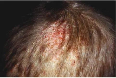
In very severe forms, a frank kerion may be formed which is a swollen mass, discharging pus, as we can see in Figure 1 (10).
These infections frequently spread among family members and classmates. Certain hairdressing practices such as shaving of the scalp, plaiting or the use of hair oils may promote disease transmission, but their precise role remains the subject of study (12).
12 Until recently, Tinea capitis was thought to have a high prevalence mainly in the developing world because of poor hygiene, overcrowding and low socio-economic standards. During the last few decades, a substantial increase in the prevalence of mycotic scalp infection and a remarkable change in the pattern of the causative dermatophytes among different European countries have been observed. Moreover, in most countries differences have been found between rural and urban areas (13). Over the last few years there have been some significant changes in the reported incidences with an overall increase in the number of cases of anthropophilic infections (14).
Evidence continues to accumulate indicating that Tinea capitis poses an increasing public health concern since it is the most common paediatric dermatophyte infection. (11).
There are approximately 30 species that are responsible by Tinea capitis. We will focus on four, Trichophyton mentagrophytes, Trichophyton Interdigitale, Trichophyton soudanense and Trichophyton violaceum (15).
T. mentagrophytes is the second most important pathogen in most countries. It is a prevalent pathogen of human and animal dermatophytosis. It started of being a species complex containing many species and varieties with different morphological characters in the past. With the aid of modern molecular taxonomic analysis, species and varieties of T. mentagrophytes species complex have been reduced to four species only, T. mentagrophytes sensu stricto, T. interdigitale, T. erinacei, and Arthroderma benhamiae.
Some members of the species complex are zoophilic and have different host predilections: T. mentagrophytes sensu stricto infects voles, T. erinacei infects hedgehogs, and A. benhamiae infect the rodents such as mice and rabbits. Humans sometimes contract infections through close contact with diseased rabbits, dogs, cats and hedgehogs and although some zoophilic varieties of T. mentagrophytes produce only minor or subclinical infections in animals, they can initiate a severe inflammatory response in man (7)(16).
T. interdigitale is the only dermatophyte taxon that unifies strains of both human and animal-associated lineages (17), but It is mostly anthropophilic and frequently found in man (18).
Microscopically, both varieties possessed septate hyphae with single-celled hyaline microconidia, of which T. interdigitale were subspherical to pyriform and T.
13 mentagrophytes were spherical to subspherical. Nevertheless, T. mentagrophytes isolates generally had smoother, thin walled, clavate multiseptated macroconidia than T. interdigitale. For some isolates, the cultural characteristics were inconsistent, and further testing was required to confirm their identity (18).
However, some authors refer to T. interdigitale as a separated species from T. mentagrophytes because most T. interdigitale and T. mentagrophytes isolates could be distinguished on the basis of their gross colonial and microscopic morphology, as the former tended to have powdery to downy colonies and the latter powdery to granular colonies (18) (19) and also, these two species have been distinguished by molecular methods before (20).
Moreover, the phylogeny of this complex remains unclear because the phenotypic features of members of T. mentagrophytes complex are poor and many isolates from medical and veterinary samples have lost their sexual activity (21).
From a clinical point of view, because T. mentagrophytes complex includes both anthrophilic and zoophilic species, it is important to have a reliable method to identify the species of the complex (22) and also from an epidemiological point of view, it is necessary to determine the variety of T. mentagrophytes complex and its origin in order to prevent spread of infection (20) (16).
Trichophyton violaceum and Trichophyton soudanense are common causes of Tinea capitis in parts of Africa and West Asia, respectively, and are a lot of times misidentified and misdiagnosed (23) (24).
Regarding colony morphology and microscopic characteristics, T. violaceum isolates were slow-growing, with waxy, glabrous, wrinkled colonies. Colonies were a distinctive, deep, purple-red and sometimes had a white, waxy fringe. Over time, some cultures became fluffy and lost their characteristic colony morphology. Microscopically, there was lack of sporulation.
T. soudanense isolates were also slow-growing. Colonies were flat, with a suede-like texture, a spidery edge, and a distinctive yellow or orange-yellow color. Over time, the colonies became folded and pleomorphic. Microscopically, no conidia were seen. Reflexive branching, in which the hypha bends back toward the inoculum site, was observed (23).
T. soudanense was reduced to synonymy with T. violaceum based on molecular techniques but the unification of T. soudanense and T. violaceum may conceal
14 possible evolutionary diversification, since both species have their unique geographical distributions (25)(26).
1.3 Importance of diagnosis
Over $500,000,000 per year are spent globally on medications that target dermatophytosis (26) (17). Additionally, dermatophytosis is considered a public health problem in developed and developing countries, as it is already a problem for immunocompromised patients (27) (28), and its incidence has been increasing (17)(6). However, nowadays, nothing is done in terms of public health measures to control the spread of dermatophyte infection (29).
The dramatic increase in the incidence of opportunistic fungal infections along with the development of new antifungal agents with various spectra of activity and the emergence of antifungal resistance has led to a critical need for diagnostic methods that can rapidly and accurately identify fungal pathogens (6). Also, identification of the dermatophyte species is essential to verify if the infection is due to relapse or due to a newly acquired organism (30) (17) and, most importantly, to start the appropriate treatment at the earliest (9) (31)(32).
Adding to this, there are countless scientific queries that benefit from the ability to discriminate pathogen strains within the species, to apply the preventive strategies for infection, as well as to extend our knowledge in the realm of dermatophytes ecology and epidemiology (2)(33)(34), and also to establish an accurate taxonomy of individual species, because it has changed fundamentally. During the last decade, progress has been made in the modern systematics of dermatophytes, nevertheless some pending issues remain (17) (35).
However, identification of dermatophytes sometimes remains difficult or uncertain due to their overlapping phenotypic characteristics, variability and pleomorphism (3) (4). Accurate and rapid diagnosis of the infection and reliable species identification of the isolates in clinical laboratory are noteworthy issues in clinical studies of dermatophytes and dermatophytosis (36) (1) (29) because if the diagnosis is based on clinical symptoms alone, about 50 % of patients are presumably misdiagnosed (36).
Without the tools necessary to reliably distinguish strains belonging to a singular species of microorganism, investigators would be unable to examine the origins of phylogenetic diversity within a species; track the introduction, maintenance, and disappearance of strains within a community or broaden their understanding of the unique relationship that exists between a pathogen and its host (33).
15 1.4 Diagnostic Methods
1.4.1 Traditional Methods
Traditional diagnosis of cutaneous tinea infections have been based on the colony morphology (pigmentation of the surface and reverse sides, topography, texture and rate of growth) and microscopic characteristics (size and shape of macroconidia and microconidia, spirals, nodular organs) in combination with biochemical and physiological tests (nutritional requirements, temperature tolerance, enzyme production, hair perforation and others) (9).
Gross microscopic morphology is the basis of classification and identification of dermatophytes. However, it often varies with culture conditions (3). It involves scraping of the scaling skin, and application of a dilute KOH 10% to 20% solution to look for hyphal elements by light microscopy. Either heat or addition of DMSO 20% to 40% solution can be used to break up the keratinocytes to aid in visualization of the hyphae (27).
In Tinea capitis, a UV-A light (Wood’s lamp) can be used to identify fluorescence of fungal infected hair shafts. Short, broken, easily removed hairs can also be examined with KOH and heat or DMSO (37).
Moreover, despite a positive KOH preparation, pathogen cultivation may not be possible, especially if antifungal treatment has already been initiated.
Culture-based diagnostics should therefore be conducted (38).
Identifying characters include colony pigmentation, texture, growth rate and distinctive morphological structures, such as micro conidia, macro conidia, spirals, pectinate branches, pedicels and nodular organs. Some usable media are as follows:
1. Urea agar or broth is used to assist gratitude of urease-negative species of Trichophyton genera.
2. Bromcresol purple (BCP)-milk solids-glucose agar is used to distinguish dermatophytes as T. rubrum, T. mentagrophytes, T. soudanense, T. megninii, M. persicolor and M. equinum, on the divergence of releasing ammonium ion from casein and the catabolite domination by glucose.
3. Potato flake agar or Cycloheximide amended potato glucose use of isolation, identification of T. rubrum by quick red pigmentation in germfree, usual isolates and with relatively antibiotic-susceptible contaminants.
16 4. Casamino acids-erythritol- albumin medium. It is an extremely useable medium for isolating dermatophytes from heavily contaminated by bacteria or cycloheximide-tolerant such as C. albicans.
5. Another isolation medium is (BCP)-casein-yeast extract agar which grows all dermatophytes(39).
Even successful cultivation does not guarantee correct pathogen identification as it may not be sufficiently distinctive in the first culture (38). Also, culture of fungal organisms is usually slow (1-4 weeks), requires considerable skill in identification of the fungus, and has a relatively low sensitivity and specificity (26) because it does not enable differentiation between dermatophytic and non-dermatophytic infections, especially in nail samples, thus has a high false-negative rate: up to 40% of microscopy-positive samples fail to become positive in culture (40)(28)(35).
Furthermore, the results are often misleading due to the pleomorphic appearance and change of colony morphology resulting from subculturing of the strains (41).
Also, the colonies evaluated on specific media, along with analysis of some nutritional requirements or biochemical characters, remain imprecisely identified due to the high similarity and variability in morphology, the pleomorphism phenomenon, and a long time required for emergence of phenotypic characteristics (2).
In addition, some strains cannot be cultivated and hence the pathogen cannot be identified at the species level (31).
Epidemiological studies of dermatophyte infection have now entered a new era with the ability to differentiate strains of fungi by molecular strain typing methods. The epidemiology of many fungal infections can now be studied in more detail (29).
1.4.2 Molecular methods
Molecular techniques appear to offer the promise of faster, more accurate diagnosis of superficial dermatophyte infections, with potential turnaround times of 3 days or less. (36). Molecular techniques are attractive because of their reproducibility, simple handling and degree of skill required in comparison with traditional methods (1). Molecular diagnosis requires extracting fungal DNA from a clinical sample and then the use of various methods of polymerase chain reaction (PCR) to amplify the fungal DNA (42)(4) which enables rapid detection and determination of various dermatophytes from minute amounts of starting materials (18). Several groups have successfully applied PCR technology to the phylogeny and diagnosis of pathogenic fungi, including
17 dermatophytes (43). PCR technique consists in three steps being the first the denaturation of the double stranded DNA to form a single stranded that can then hybridize with specific primers that will allow the elongation of the chain in order to create several copies.
Several studies have applied molecular diagnostics to fungal infections. During the last decade, a wide variety of DNA-based techniques have become available for studies on species identification, such as species-specific PCR, PCR- Restriction fragment length polymorphism (RFLP), real-time PCR (RT PCR), nested PCR and various targets such as ribosomal-DNA (rDNA), β tubulin gene (BT2), mitochondrial DNA (mtDNA), and others (6). A common argument in favour of these molecular techniques in the scientific reports is that genotypic differences are considered more stable and more precise than phenotypic characteristics (9)(44).
RT PCR is a potentially more sensitive approach that is based on detection with cyber green or with specific fluorophore-labelled oligonucleotide probes.
The RT PCR has several advantages like the omission of post-PCR analysis, which diminish the risk of false-positivity (45) and the increase of sensitivity assay which could markedly improve the investigation of scalp dermatophytoses, where conventional mycology often suffers from low sensitivity due to a combination of low-level or patchy fungal load, heavy encrustation of debris blocking microscopy, and the contamination of samples by clinically ineffective topical antifungals such as ketoconazole from shampoos. In addition, Tinea capitis of the elderly, an entity often remaining unrecognised could be further elucidated by RT PCR (46)(47).
However, it is not practical enough for a large number of laboratories that are either small scale or very tightly budget (48).
PCR-RFLP is a technique based on detection of genomic restriction fragments by PCR amplification, which can be used with DNA of any organism and has been proven to be useful for rapid and correct identification of dermatophyte species and able to generate species-specific DNA polymorphisms with many dermatophyte species on the basis of characteristic band patterns detected by agarose gel electrophoresis (49). (50).
Unfortunately, it is a complex technique with poor discriminative power to make an easy and specific diagnosis (48) (47) .
Nested PCR involves the use of two primer sets and two successive PCR reactions and can be used to identify dermatophytes in clinical samples, which is particularly useful for Tinea capitis in which adequate treatment depends on the incriminated
18 dermatophyte and should be initiated as soon as possible, being that it depends on the identification of the source of the infection (48).
Furthermore, it can be used when only a small amount of material is collected for mycological analysis and the number of species that can be identified by sequencing a nested PCR product is unlimited (51).
It was observed to be more sensitive for the detection of dermatophytes than single-round PCR, it is faster than traditional methods and it is helpful for diagnosis of cases with dermatophytoses which were recently treated with antifungal agents, or showed uncultivable filaments (48).
Also, It has been possible to discriminate without ambiguity closely related species such as T. violaceum and T. soudanense by this method (51).
An inconvenience with nested PCR is the high risk of contamination and the need of two negative controls (51).
Nevertheless, the main disadvantage of these methods is their higher cost relative to other detection methods (27).
With the introduction of molecular methods it has become clear that the phenotypes and genotypes of this group of fungi are not always congruent (26).
To help distinguish this and other species that are usually confused, a lot of methods have been applied, as it was described earlier. One of them, relies on genome sequencing and was developed to study the future outbreaks on the biology, virulence, pathogenicity and the host specificity of the clinically important dermatophytes (52)(31).
1.4.2.1 ITS gene
The ribosomal DNA (rDNA) genes are found in all microorganisms and known to accumulate mutations at a slow constant rate over time. Nucleotide sequence heterogeneity within this region can be used to phylogenetically classify microorganisms. Interspaced among the highly conserved sequences of the rDNA genes are regions of variable sequences called spacer regions historically referred to as spacers since they separate the functional DNA sequences of the various rDNA genes.
Since mutations within the spacer regions of the rDNA gene complex occur with greater frequency than with the rDNA genes, the sequence heterogeneity within this
19 area has been useful for the separation of both genera and species. Similarly, it is also recognized that eukaryotic cells such as fungi have a rDNA gene complex region with comparable characteristics (53). The intervening ITS regions have become important molecular targets for taxonomy and identification (54).
Due to greater sequence variation and polymorphism (44), the ITSllITS2 domains are more suited for species and strain identification than the 18s region (small subunit), the 5.8s region and the 28s region (large subunit). For example the 5.8S region is only about 160 bp long and highly conserved within major organism groups.
Several groups have developed methods utilizing the ITS regions to identify species and strains of a range dermatophytes, yeasts and moulds (55)(21).
Most studies have shown that sufficient variation exists within the ITS regions to allow species identification. The ITS regions have been used as targets to investigate the validity of dermatophyte taxonomy, and to determine the phylogenetic relationships among other fungal species by comparing the base pair sequences of the ITS1 regions (22).
Some of the cited studies (and others) have provided ITS sequence information for a variety of fungi and are available in GenBank for analysis using a single representative isolate of each taxon (44)(40).
This ITS based identification system saves time (it takes 2 to 3 days) and is accurate and applicable even to strains with atypical morphological features (22).
Currently, ITS-rDNA sequencing is the golden standard for species delineation of dermatophytes. However, the method is expensive and may be impracticable for largescale analysis (55).
1.4.2.2 Beta tubulin gene
Beta tubulin is a monomeric globular protein involved in the generation of microfilaments. The locus includes some introns which are known to be good estimators for distinction of closely related species (17). BT2 protein-encoding gene has been broadly used in fungal phylogenetic analyses because it contains both variable and highly conserved regions and it can provide high resolution and support for fungal systematics. It is known that most Trichophyton spp., especially the human-adapted species, are similar to each other in ITS (17)(2).
20 1.4.2.3 Tef-1α
The nucleotide sequence of translation elongation factor 1-α (Tef-1α) gene encoding a part of the protein translation machinery was first used in Fusarium. The gene appears to be consistently single-copy and shows a high level of sequence polymorphism among related species (56)(57).
Due to this, this study aims to investigate if it is possible to identify and distinguish these species phylogenetically, T. mentagrophytes and T. interdigitale, T. violaceum and T. soudanense, by sequencing and comparing three parts of the fungal genome such as internal transcribed spacer (ITS), beta-tubulin gene (BT2) and the translation elongation factor 1-α (Tef-1α) gene.
21
2. Objectives
This investigation work was developed in the CHU de Liège, Le Centre hospitalier universitaire, in the microbiology department, as it was an Erasmus+ Placement project.
The search of methods to identify dermatophytes has been studied for many years, and although some researchers have found some reliable techniques, some species of dermatophytes are continuingly misdiagnosed and misidentified.
The use of ITS, BT2 and Tef-1α have been described by several authors as promising genes that are capable of identifying fungus to the species level, although they are not always successful.
The aim of this project is to combine these three genes in order to establish a method that would be able to improve the identification of these species as well as to improve the knowledge in their taxonomy and phylogeny.
To pursue this goal some objectives were established: Extraction and purification of fungus DNA, amplification of DNA by PCR technique for the three genes and sequence of the amplified DNA.
22
3. Materials and Methods
3.1 Strains
We worked with 65 different strains that had been isolated in culture at the CHU of Liège from samples collected of infected hair from patients (15 had been previously classified as T. interdigitale, 14 as T. mentagrophytes, 13 as T. violaceum and 23 as T. soudanense). These strains were conserved frozen in the laboratory at -78ºC.
Reference strains (IHEM) were also included in this study, IHEM 620 and IHEM 584 as reference strains of T. interdigitale, IHEM 10342, IHEM 4268, IHEM 4203 as reference strains of T. mentagrophytes and IHEM 13492 as reference strain of T. soudanense.
3.2 Culture Conditions
The dermatophytes were cultured in liquid medium and the medium of choice was Sabouraud, prepared in the laboratory with 30 g of Sabouraud powder (Sigma) and 1 litre of distilled water.
Sabouraud’s agar containing antibiotic(s) (chloramphenicol ± gentamicine) and cycloheximide (Actidione) is commonly used as primary isolation medium, and it is the reference medium to culture dermatophytes (58).
Cultures are usually incubated at 20–25ºC, but higher incubation temperatures of 30– 32ºC can be used when lesions are suspected to be caused by some species (37). The procedure consists in 15 mL of medium applied inside a falcon tube and one or two drops of milk containing the strain put after in the tube. Then the tubes are left in a stove at 30ºC for approximately a week.
We also prepared solid cultures, and the medium used was Sabouraud plus chloramphenicol in a glass tube. The strains were applied with a inoculation loop and left in the stove at 30ºC for approximately a week. (58).
It is important to mention that the tubes were not completely closed because the dermatophytes need oxygen to grow.
23 A common protocol for DNA extraction involves two principal steps: first, the breakdown of cell walls that is generally carried out by mechanical disruption with bead-beating material, enzymatic digestion or homogenization with liquid nitrogen, and second, the isolation and purification of DNA. Frequently, filamentous fungi DNA is extracted by conventional methods (i.e., disrupt the cell wall and nuclear membrane, removing contaminant proteins, and resuspension in buffered solution), nevertheless, the DNA integrity and purity are regularly compromised, even more, loss of DNA yield on purification step can occur (59).
It was used bead beating material and several buffers, one of them being proteinase K, an enzyme that catalyse fungal cell lysis and removes the proteins (60).
The DNA extraction from the culture was performed with liquid medium where the material to extract was introduced in a 2 mL Eppendorf tube with small glass beads and 1 mL of distilled water.
Every time that an extraction is performed, is mandatory that a negative control is introduced (an Eppendorf with distilled water) to guarantee the absence of contamination.
The tubes are vortexed for 30 minutes, centrifuged for 10 minutes at 16,100 rpm. The supernatant is withdrawn, and it is added of 200 µL of AL buffer (Qiagen), 180 µL of ATL buffer (Qiagen), and 20 µL of proteinase K (Qiagen).
Finally, the mixture is incubated in an inoculation plate for 2h at the temperature of 56ºC.
This procedure joins a mechanical lysis with an enzymatic lysis, which is very important to guarantee that the cell wall is broken so that we can access the genetic material.
3.3.1 Maxwell Procedure
The Maxwell 16 Instrument makes use of paramagnetic particles to capture DNA following cell lysis. After cell lysis occurs, the particles are introduced to the samples to bind the DNA.
Following a series of sequential washes, the target DNA is eluted into a storage buffer suitable for downstream amplification applications.
Because the paramagnetic particles exhibit a maximum binding capacity, by standardizing the number of particles employed for extraction of the various samples
24 one can expect the amount of DNA eluted during the final step to be fairly consistent between samples. The instrument bases sample transfer on magnetic handling, problems normally associated with clogged tips, partial liquid transfers, rotational mixing, and maintenance of consumables are not an issue when using this system. Magnetic handlers are inserted into disposable plunger mechanisms that are used for a single sample throughout the purification process, which minimizes the risk of sample-to-sample contamination (61).
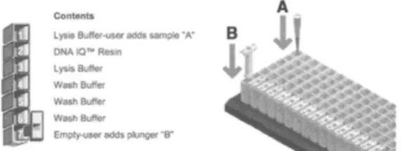
We used Maxwell (Promega) machine and the Cell DNA Purification Kit (Promega). Magnetic handlers are inserted into disposable plunger mechanisms that are used for a single sample throughout the purification process, which minimizes the risk of sample-to-sample contamination. The DNA IQ Reference Sample Kit comes with cartridges that are sealed to contain pre-loaded aliquots of purification solutions (Figure 2). It contains a lysis buffer, DNA IQ Resin, and a wash buffer.
We applied:
1. 400 µL of the samples in the first well; 2. A plunger in the last well which serves to
sheath the magnetic poles during processing;
3. 300 µL of elution buffer in the elution tubes.
The procedure takes 35 minutes and then the DNA withdrawn (approximately 200 µL) is ready to amplify.
3.4 DNA Amplification
3.4.1 PCR
The PCR of ITS, Tef-1 and BT2 were performed in a Thermo Cycler Hybaid using: 1. 10 µL of the DNA sample
2. 40 µL of a mix that contains: a. 5 µL of buffer II 10X (AB) b. 7 µL of MgCl2 (25mM, from AB)
c. 0.4 µL of dNTP (25 mM, from Promega) d. 0.5 µL of each primer
Figure 2: Diagram of the DNA IQ Reference Sample Kit/Maxwell 16 Instrument(61).
25 e. 0.25 µL of Taq polymerase (5U/ µL, from AB)
f. 26.35 µL of distilled water (Braun).
A positive control is used to check if there the procedure occurs normally and a negative control to guarantee that there are not any contaminants in the samples. The primers used for ITS were ITS1 5’ TCCGTAGGTGAACCTGCGG 3’ and ITS4 5’ TCCTCCGCTTATTGATATGC 3’, for BT2 gene were Bt2a 5’
GGTAACCAAATCGGTGCTGCTTTC 3’ and Bt2b 5’
ACCCTCAGTGTAGTGACCCTTGGC 3’ and for Tef 1-αF 5’
CACATTAACTTGGTCGTTATCG 3‘ and Tef 1-αR 5’ CATCCTTGGAGATACCAGC 3’, as it is shown in Table 1 and the programs of PCR applied differed between genes, as it can be seen in Table 2, Table 3 and Table 4.
Primers Foward Reverse
ITS (IDT) ITS 1: 5’
TCCGTAGGTGAACCTGCGG 3’ ITS 4 5’ TCCTCCGCTTATTGATATGC 3’ BT2 (IDT) Bt2a: 5’ GGTAACCAAATCGGTGCTGCTTTC 3’ Bt2b: 5’ ACCCTCAGTGTAGTGACCCTTGGC 3’ Tef 1-α (IDT) 5’ CACATTAACTTGGTCGTTATCG 3 ‘ 5’ CATCCTTGGAGATACCAGC 3’ Table 1: Primers of ITS, BT2 and Tef-1α used to amplify the dermatophytes DNA.
The following programs are applied:
ITS Program
Temperature Time Cycles
94º C 5 min x1 94ºC 15 sec x35 55ºC 5 sec 68ºC 40 sec 68 ºC 7 min x1 4ºC infinit
Table 2: ITS program of PCR technique.
Tef -1α Program
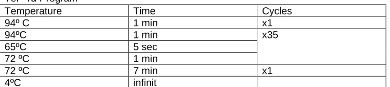
Temperature Time Cycles
94º C 1 min x1 94ºC 1 min x35 65ºC 5 sec 72 ºC 1 min 72 ºC 7 min x1 4ºC infinit
26 BT2 Program
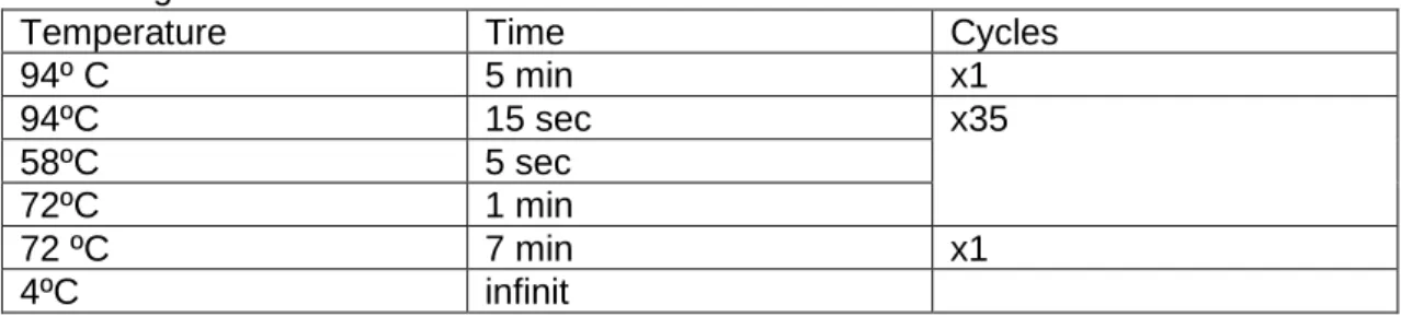
Temperature Time Cycles
94º C 5 min x1 94ºC 15 sec x35 58ºC 5 sec 72ºC 1 min 72 ºC 7 min x1 4ºC infinit
Table 4:BT2 program of PCR technique.
After PCR is done, the amplified products are revealed in an agarose 2% gel, in a E- Gel power Snap (Invitrogen) and the procedure consists in applying 10 µL of distilled water in each well, 10 µL of the 100 bp marker in the first well, 10 µL of the samples on the other wells and 10 µL of distilled water in every well. After 10 minutes, while the gel is running, the bands will get separated by size and will appear by applying an UV light because the gel is pigmented and it will bind with DNA.
3.5 Sequence Method
To obtain the sequence of each strain the following steps have to be accomplished: 1. Purification of the PCR products by ExoSap-IT technique
2. Sequence of the DNA fragments previously purified 3. Purification of the sequencing reaction
3.5.1 Purification of the PCR products
A mix of 1 µL of ExoSapIT pure solution and 5 µL of Nuclease Free Promega water is done and 6 µL of it is put in the 96 well plate along with 6 µL of the samples.
The ExoSap programme is applied for 15 minutes at 37ºC and then 15 minutes at 80ºC.
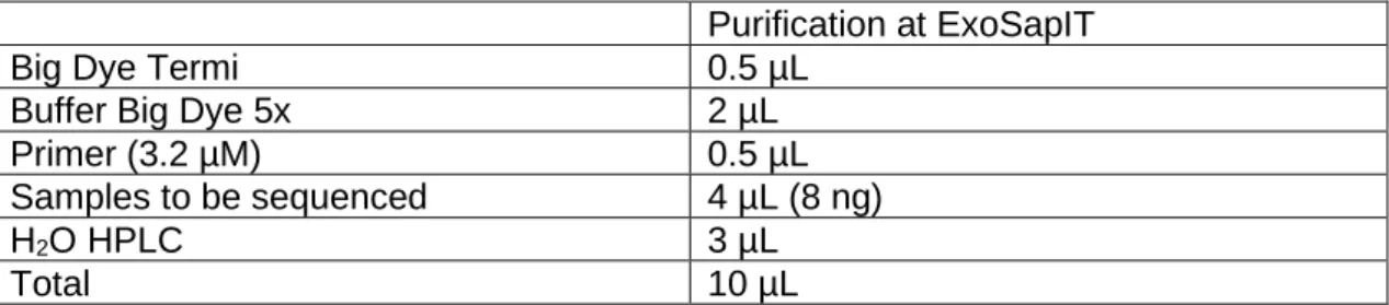
3.5.2 Sequencing Reaction
To initiate the sequencing procedure the samples were added to the mixture seen in Table 5, and then the tubes are centrifuged and the sequence program, shown in table 6 is applied and the DNA fragments are sequenced.
27 Purification at ExoSapIT
Big Dye Termi 0.5 µL
Buffer Big Dye 5x 2 µL
Primer (3.2 µM) 0.5 µL
Samples to be sequenced 4 µL (8 ng)
H2O HPLC 3 µL
Total 10 µL
Table 5: Reagents and their amounts used in the sequencing reaction.
Temperature Time Cycles
96ºC 3 min x1
96ºC 15 sec x25
50ºC 15 sec
60ºC 4 min
10ºC infinity
Table 6: Description of the Program applied to sequence the dermatophytes DNA.
3.5.3 Purification of the sequencing reaction
The purification of the sequencing reaction is done by the CleanSeq Agencourt Technique, with the Kit CleanSeq Agentcourt Ref. A29151 (800 rnx) A29154 (5000 rnx).
Finally the sequence is analysed by CBS database or BLAST database in Tef case (annexes).
28 3.6 Phylogenetic Analysis
Samples of interest were sequenced for the following three genes: ITS, Tef-1α and BT2.
The following two species were chosen as outgroups: Purpureocilum lilacinum and Aspergillus fumigatus.
The sequences for the three genes of interest for each one of the two outgroups species were retrieved from Genbank.
Namely the accession numbers for Purpureocilum lilacinum sequences are: KJ476413.1 (Tef-1α), MH613756.1 (BT2) and LC413751.1 (ITS); the ones for Aspergillus fumigatus are: KJ476410.1 (Tef-1α), KU714964.1 (BT2) and AJ853744.1 (ITS).
For each one of the three genes, sequences from all samples and from the two outgroups were grouped.
Sequences were complemented when needed to correct the Sanger sequence and allow for alignment.
Alignments were performed using MUSCLE in Seaview (https://www.ncbi.nlm.nih.gov/pubmed/19854763) for each gene. The alignments for each of the three genes were then concatenated together based on the name of the sample and gaps were added between genes.
Phylogenies were then inferred on the concatenated alignment using PhyML (https://www.ncbi.nlm.nih.gov/pubmed/20525638).
PhyML was launched with a GTR model with aLRT branch support, optimized nucleotide equilibrium frequencies, optimized invariable sites and optimized across site rate variation. Tree search operations were performed using the best of NNI and SPR.
29
4. Results
4.1 Classification of the strains Classification of Strains by Sequencing method and later analysis in CBS and BLAST database
All the strains were grown in Sabouraud medium and DNA was amplified from all strains using PCR technique for all the three genes, Tef-1α, ITS and BT2, with the exception of 5 strains that the PCR was negative when examined in the electrophoresis gel, perhaps because of an error during in the extraction.
The DNA was sequenced, and the results were analysed by CBS database, concerning ITS and BT2 genes, and BLAST database concerning Tef-1α.
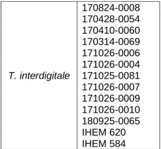
As we can see in Table 7, 13 of the species analysed were classified as T. interdigitale, where two of them were reference strains, IHEM 620 and IHEM 584.
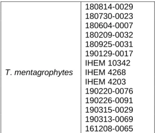
The strains identified as T. mentagrophytes (Table 8) were 14 in total in which 3 were reference strains, IHEM 10342, IHEM 4268 and IHEM 4203.
T. interdigitale 170824-0008 170428-0054 170410-0060 170314-0069 171026-0006 171026-0004 171025-0081 171026-0007 171026-0009 171026-0010 180925-0065 IHEM 620 IHEM 584
Table 7: Discrimination of species of T. interdigitale sequenced and analysed by CBS and BLAST databases.
30 Ten strains were identified as T. violaceum and are presented in Table 9. For each species, at least one reference strain was included in the study except for T. violaceum, because although this strain was a part of the research, it was not possible to extract the DNA, probably due to not enough time of growth in culture.
Besides this strain, other 3 were contaminated so after a second analysis we excluded them from the study.
T. mentagrophytes 180814-0029 180730-0023 180604-0007 180209-0032 180925-0031 190129-0017 IHEM 10342 IHEM 4268 IHEM 4203 190220-0076 190226-0091 190315-0029 190313-0069 161208-0065 T. violaceum 180227-0044 180227-0045 180227-0072 180410-0017 180410-0021 180420-0056 180611-0016 180611-0049 180621-0047 180625-0012
Table 8: Discrimination of species T. mentagrophytes sequenced and analysed by CBS and BLAST databases.
Table 9: Discrimination of species of T.
violaceum sequenced and analysed by CBS
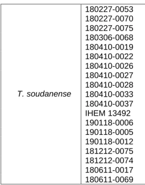
31 Table 10: Discrimination of species of T. soudanense sequenced
and analysed by CBS and BLAST databases.
Of all the 65 strains analysed, 19 were classified as T. soudanense (Table 10), where one is the reference strain, IHEM 13492.
There was concordance on the results between the three segments analysed, which means that when sequencing every segment, ITS, BT2 and Tef-1α, they all classified each strain as one species.
4.2 Phylogenetic Analysis
After sequencing, the dendrograms were elaborated concerning T. mentagrophytes, T. interdigitale, T. violaceum and T. soudanense (Figure 3).
T. soudanense 180227-0053 180227-0070 180227-0075 180306-0068 180410-0019 180410-0022 180410-0026 180410-0027 180410-0028 180410-0033 180410-0037 IHEM 13492 190118-0006 190118-0005 190118-0012 181212-0075 181212-0074 180611-0017 180611-0069
32
Figure 3: Dendrogram representing the phylogenetic differentiation of T. mentagrophytes, T. interdigitale, T. soudanense, and T. violaceum.
33 Firstly, Purpureocilum lilacinum from the class Sordariomycetes, and Aspergillus fumigatus from the class Eurotiomycetidae were used as outgroups, and they were chosen due to the lack of homology between them and the species that are being investigated.
It is necessary to include at least one distantly related sequence from the sequences being analysed in order to elaborate the dendrogram.
It can be seen in Fig. 4, there are cleary seen two clusters that separate the species of T. violaceum and T. soudanense, from T. mentagrophytes and T. interdigitale.
In the first cluster it is present a separation between T. violaceum and T. soudanense, and this cluster can be divided in two, of the two species, but in the T. mentagrophytes and T. interdigitale cluster the separation between species is more tenuous, being that we cannot determine the existence of two clusters that separate both species.
As it can be observed, some strains were not classified as they had been by CBS and BLAST platform. In the T. mentagrophytes dendrogram we can see that all the strains that were classified as this species are classified correctly but 2 strains that were well classified as T. mentagrophytes, appear in the dendrogram area of T. interdigitale (IHEM 4268 and 190313-0069), one of them being a reference strain.
On the other hand, all species that were previously classified as T. interdigitale were well evaluated and maintained their identification.
Analysing the T. violaceum dendrogram, all the strains were identified as they were by sequencing, although 2 strains were in T. soudanense dendrogram (180410-0017 and 180410-0021), and in the T. soudanense dendrogram the same happened, as they were all identified as previously but one strain (180306-0068) was in T. violaceum dendrogram.
34
5. Discussion
The overall increase of Tinea capitis during the last decades is due to the emerging anthropophilic infections in Europe probably as a result of increasing numbers of immigrants of different ethnic origin. (13)
Whatever the reasons behind the current outbreak, the procedures for surveillance are clearly not able to detect cases in order to control the spread of infection (14).
Tinea capitis does not respond to topical therapy alone, and therefore requires oral antifungal agents, which implies the need of the exact treatment regimen dependent on the species (38).
Worldwide Tinea capitis is most commonly caused by several dermatophytes, being that the aim of this study was to identify and distinguish some of them, T. interdigitale from T. mentagrophytes and T. violaceum from T. soudanense.
We used only a molecular method to identify the strains because the morphological proprieties of the strains are not always accurate to identify them with certainty, as they have been misidentified before (62), also it is hard to differentiate some based on conventional methods (26) and due to the time traditional methods take to develop, since that factor was limited.
5.1 Strains Identification
To identify each strain this study focused on ITS, BT2 gene and Tef-1α which have been used before to discriminate fungal species (35)(63).
5.1.1 ITS
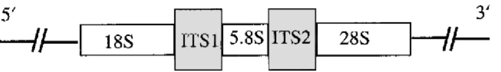
The Internal transcribed spacer (Figure 4) is located in the rDNA gene complex of all eukaryotic cells between the 18S and 5.8S rDNA genes (ITS1) and the 5.8S and 28S rDNA genes (ITS2) (Figure 3) and has areas of high conservation and areas of high variability. It is an ideal starter for the development of specific PCR primers for identification of fungal species (68)(55).
35
Figure 4: Representation of the rDNA gene complex in fungi denoting gene order and position of the ITS regions (Iwen et al., 2002).
Once isolated, the ITS regions can be compared to the wealth of other sequences in the sequence database.
Although the ITS region is the main target, other genes are becoming more widely studied, in particular the β-tubulin gene.
5.1.2 β-Tubulin gene
Microtubules are found in all eukaryotic cells and are involved in maintenance of cell shape, mitosis, and a variety of other morphogenic events. At the core of these multimeric structures, lie heterodimeric tubulin filaments made up of two very similar α- and β-tubulin proteins. Despite the highly conserved nature of α-and β-tubulins, sufficient differences exist between cell types and taxa to provide opportunities for identification based on these tubulin proteins (64).
The database of β-tubulin sequences is not as large as that for ribosomal ITS sequences but can be particularly useful when ITS sequence variation or conservation is not suitable for production of a taxon-specific diagnostic (65)(66).
5.1.3 Tef-1a
Finally, Tef-1a gene, which encodes part of the protein translation machinery, has shown a high level of sequence polymorphism among related species and is recommended for the discrimination of fungal species (67).
5.2 Amplification and sequencing of DNA
PCR is the enzymatic exponential amplification of a specific target region using short primers, leading to detectable amounts of amplified DNA from one or a few original sequences (68).
36 The Sanger sequencing method uses 2’,3’ dNTPs which lack a hydroxyl group on the 3’ carbon of the sugar ring. In a regular nucleotide, the 3’ hydroxyl group acts as a “hook," allowing a new nucleotide to be added to an existing chain.
Once a dideoxy nucleotide has been added to the chain, there is no hydroxyl available and no further nucleotides can be added. The chain ends with the dideoxy nucleotide, which is marked with a particular colour of dye depending on the base (A, T, C or G) it carries.
Dermatophyte species identification by sequence comparison is often problematic using the publicly available NCBI database. It is plethoric with identical sequences under different names (especially ITS and 28S sequences).
A way to circumvent this problem is to directly use the publicly available ITS sequence database at the CBS dermatophyte database website. Identification of fungi in dermatological samples using PCR is reliable and provides significantly improved results in comparison with cultures (32).
As our results show, 65 strains were analysed and with the combination of the three genes it was achieved an identification by DNA sequencing and comparison with CBS database and BLAST database.
5.3 Phylogenetic analysis
It could be could inferred this method as good approach to evaluate and distinguish dermatophytes, however, after making a phylogenetic study with these three genes the results were not satisfactory due to lack of separation between species.
This can be explained because they are very closed related, and they have been misidentified before, or perhaps because the targeted genes were not ideal to separate these specific species. Some authors wrote that the variations found in ITS analysis were very slight suggesting that these differences are not useful markers for strain identification (32) (3) and there are some studies that still consider T. interdigitale a subtype of T. mentagrophytes, and see them as only one specie.
It has also been written that T. soudanense and T. violaceum had also been recognised as a synonym before, but the unification of these species may conceal a possible evolutionary diversification since both species have their unique geographical distribution (3)(1) (70)(23).
37
6. Conclusion
In conclusion, it was possible to discriminate the different species of dermatophytes by the sequencing method but the phylogenetic analysis performed did not have the expected results, because the species that we were trying to distinguish are very close related, and are a lot of times misidentified.
To sum up, this project helped understand the differences and difficulties in discriminating dermatophytes and also the importance of its identification being that more research has to be done to achieve a more reliable method.
7. Future perspectives
Firstly, since this project lacked the morphologic evaluation and research, this analysis should be performed in order to sustain the identification reached with the molecular studies, since although the results with the traditional methods take longer, and have some disadvantages as it was said before, they still are very important to discriminate dermatophytes.
Secondly, being that the genes amplified have been investigated before, and were successful on identifying several species our focus should be placed on different types of techniques.
There are several types of molecular techniques and procedures that have been used before to identify dermatophytes, among them, RT PCR, nested PCR and PCR- RFLP, which could all be used as an alternative to the technique performed in this study. Given the advantages and performance, a nested PCR could be performed to the samples analysed in this study, to search if the results obtained would be more satisfactory.
38
8. Bibliography
1. Mirhendi H, Motamedi M, Makimura K, Satoh K. Development a diagnostic pan-dermatophyte TaqMan probe real-time PCR assay based on beta tubulin gene. 2016;520–7.
2. Abastabar M, Mirhendi H, Rezaei-matehkolaei A, Shidfar MR, Kordbacheh P, Makimura K. Restriction Analysis of β -Tubulin Gene for Differentiation of the Common Pathogenic Dermatophytes. 2014;96(December 2012):91–6. 3. Li HC, Bouchara J, Hsu MM, Barton R, Su S, Chang TC. Identification of
dermatophytes by sequence analysis of the rRNA gene internal transcribed spacer regions. 2019;(2008):592–600.
4. Med J, Li R, Wang D, Academy C, Hospital TF. Typing of Common Amplification Dermatophytes of Polymorphic by Random. 1997;38:239–46.
5. Cafarchia C, Iatta R, Stefania M, Gräser Y, Otranto D. Infection , Genetics and Evolution Molecular epidemiology , phylogeny and evolution of dermatophytes. Infect Genet Evol [Internet]. 2013;20:336–51. Available from:
http://dx.doi.org/10.1016/j.meegid.2013.09.005
6. Ahmadi B, Mirhendi H, Shidfar MR, Jalalizand N, Geramishoar M, Shokoohi GR. ORIGINAL ARTICLE / ARTICLE ORIGINAL A comparative study on
morphological versus molecular identification of dermatophyte isolates. J Mycol Med [Internet]. 2015;25(1):29–35. Available from:
http://dx.doi.org/10.1016/j.mycmed.2014.10.022 7. Hay RA. Letter to the Editor. 2016;83:247–50.
8. Laniosz V, Wetter DA. What’s new in the treatment and diagnosis of dermatophytosis? 2014;33(September).
9. Tro A, Zie P, Gnat S, Nowakiewicz A. Evaluation of growth conditions and DNA extraction techniques used in the molecular analysis of dermatophytes. 2017; 10. Degreef H. Clinical Forms of Dermatophytosis ( Ringworm Infection ).
2008;(May):257–65.
11. Elewski BE. Tinea capitis: A current perspective. 2000;1–20.
12. Higgins EM, Fuller LC. Guidelines for the management of tinea capitis. 2001;(January):53–8.
39 in Europe : current state and changing patterns. 2007;50:6–13.
14. Robles W, Midgley G. Tinea capitis in Europe : new perspective on an old problem. 2001;229–33.
15. Gupta AK, Summerbell RC. Tinea capitis. 2000;(March):255–87.
16. Makni F, Ayadi A. Polymorphisms in the ITS rDNA regions for differentiating strains of the Trichophyton mentagrophytes complex in Sfax-Tunisia. 2014;453– 9.
17. Rezaei-matehkolaei A, Mirhendi H, Makimura K, Hoog GS De, Satoh K, Najafzadeh MJ, et al. Nucleotide sequence analysis of beta tubulin gene in a wide range of dermatophytes. 2014;(July):674–88.
18. Liu D, Coloe S, Baird R, Pedersen J. MYCOLOGY PCR identification of
Trichoph yton men tagroph ytes mentagrophytes dermatophytes with a random primer. 2019;46(1997):1043–6.
19. Fathallah A. Relationship Between Phenotypic and Genotypic Characteristics of Trichophyton mentagrophytes Strains Isolated from Patients with
Dermatophytosis. 2017;487–93.
20. Chauvin MFDE. Genetic diversity among Trichophyton mentagrophytes isolates using random ampli ® ed polymorphic DNA method. 1999;839–44.
21. Michizuki, Takashi; Kawasaki, Masako; Ishizaki, Hiroshi; Makimura K. Identification of Several Clinical Isolates of Dermatophytes Based on the Nucleotide Sequence of Internal Transcribed Spacer 1 (ITS 1) in Nuclear Ribossomal DNA. 1999. p. Vol. 26: 276-281.
22. Makimura K, Mochizuki T, Hasegawa A, Uchida K, Saito H, Yamaguchi H. Phylogenetic Classification of Trichophyton mentagrophytes Complex Strains Based on DNA Sequences of Nuclear Ribosomal Internal Transcribed Spacer 1 Regions. 1998;36(9):2629–33.
23. Magill SS, Manfredi L, Swiderski A, Cohen B, Merz WG. in Baltimore , Maryland ᰔ. 2007;45(2):461–5.
24. Summerbell RC, Haugland RA, Li A. rRNA Gene Internal Transcribed Spacer 1 and 2 Sequences of Asexual , Anthropophilic Dermatophytes Related to
Trichophyton rubrum. 1999;37(12):4005–11.
25. Zomorodian K, Uthman U, Tarazooie B. The effect of griseofulvin on the gene regulation of b -tubulin in the dermatophyte pathogen Trichophyton rubrum. J
40 Infect Chemother [Internet]. 2007;13(6):373–9. Available from:
http://dx.doi.org/10.1007/s10156-007-0552-5
26. Ilkit M, Durdu M. Critical Reviews in Microbiology Tinea pedis : The etiology and global epidemiology of a common fungal infection Tinea pedis : The etiology and global epidemiology of a common fungal infection. 2015;7828.
27. Didehdar M, Khansarinejad B, Amirrajab N, Shokohi T. Development of a high-resolution melting analysis assay for rapid and high-throughput identification of clinically important dermatophyte species. 2016;442–9.
28. Motamedi M, Mirhendi H, Zomorodian K, Khodadadi H, Kharazi M, Ghasemi Z, et al. Clinical evaluation of β - - time PCR for rapid diagnosis of dermatophytosis , a comparison with mycological methods. 2017;(May):692–6.
29. Yazdanparast A, Jackson CJ, Barton RC, Evans EG V. Clinical and Laboratory Investigations Molecular strain typing of Trichophyton rubrum indicates multiple strain involvement in onychomycosis. 2003;51–4.
30. Howell SA, Barnard RJ, Humphreyst F. Application of molecular typing methods to dermatophyte species that cause skin and nail infections. 2019;48(April 1997):0–7.
31. Elavarashi E, Kindo AJ, Kalyani J. Optimization of PCR – RFLP Directly from the Skin and Nails in Cases of Dermatophy- tosis , Targeting the ITS and the 18S Ribosomal DNA Regions. 2013;7(4):646–51.
32. Verrier J, Monod M. Diagnosis of Dermatophytosis Using Molecular Biology. Mycopathologia. 2017;182(1):193–202.
33. Abdel-rahman SM. Strain Differentiation of Dermatophytes. 2008;(October 2007):319–33.
34. Article O. Use of Single-enzyme PCR-restriction Digestion Barcode Target- ing the Internal Transcribed Spacers ( ITS rDNA ) to Identify Dermatophyte Species. 2012;41(3):82–94.
35. Rezaei-matehkolaei ALI, Makimura K, Hoog GSDE, Shidfar MR. Discrimination of Trichophyton tonsurans and Trichophyton equinum by PCRRFLP and by β -tubulin and Translation Elongation Factor 1- α sequencing. 2012;(October):760– 4.
36. Gräser Y, Czaika V, Ohst T. Diagnostic PCR of dermatophytes – an overview. 2012;2012(Band 10):721–5.