Detection of
Sclerotinia sclerotiorum
in soybean seeds by
conventional and quantitative PCR techniques
1Luana da Silva Botelho
2, Ellen Noly Barrocas
3*,
José da Cruz Machado
4, Rayana de Sá Martins
5ABSTRACT -Sclerotinia sclerotiorum, the etiological agent of the “white mould” in soybean, is responsible for severe losses in this crop and soil contamination. The introduction and dissemination of the disease can made through the use of seed lots contaminated with sclerotia and by seeds infected by mycelium. Therefore, seed health quality is one aspect to be monitored by means of health testing before to sowing time. In this study conventional and quantitative PCR techniques were used to assess their viability to detect S. sclerotiorum in artificially and naturally infected soybean seed samples. For that, seeds were inoculated by osmotic conditioning technique for 0, 24, 48 and 72 hours of contact of the seed with the fungal colony and mixed with healthy seeds generating incidence levels of 1, 2, 10, 20 and 100% for each incubation time. The cPCR was sensitive to detect S. sclerotiorum in samples with at least incidence 1% inoculated for 72 hours while the qPCR detected the pathogen in all incidence/inoculum potential combinations. The conventional PCR was able to detect 0.25% of the incidence of S. sclerotiorum in soybean seed lots naturally infected added a preincubation step.
Index terms: seed healthy, molecular detection, seed borne pathogen, white mould.
Detecção de Sclerotinia sclerotiorum em sementes de soja pelas
técnicas de PCR convencional e quantitativo
RESUMO - Sclerotinia sclerotiorum causador do “mofo branco” na cultura da soja, é responsável por redução da produção e contaminação do solo. A introdução e disseminação da doença podem se dar pelo uso de sementes contaminadas com escleródios e por sementes infectadas pelo micélio. A qualidade das sementes deve ser monitorada por testes de sanidade antes da semeadura. Neste estudo foram utilizadas técnicas de PCR convencional e quantitativo para avaliar a viabilidade de uso das mesmas para a detecção de S. sclerotiorum em amostras de sementes de soja artificialmente e naturalmente infectadas. Para isso, as sementes foram inoculadas usando a técnica do condicionamento osmótico por 0, 24, 48 e 72 horas de contato das sementes com a colônia do fungo e misturadas à sementes sadias gerando incidência de 1, 2, 10, 20 e 100% para cada tempo de incubação. A técnica de cPCR foi sensível para detectar S. sclerotiorum em amostras com, no mínimo, incidência de 1% inoculadas por 72 horas, enquanto que com o qPCR foi possível detectar o patógeno em todas as combinações incidência/potencial de inóculo. A PCR convencional foi capaz de detectar 0,25% de incidência de S. sclerotiorum em lotes de sementes de soja naturalmente infectados adicionando-se uma etapa de pré-incubação das sementes.
Termos para indexação: sanidade de sementes, detecção molecular, patógeno de sementes, mofo branco.
1Submitted on 10/08/2014. Accepted for publication on 12/17/2014. 2Departamento de Microbiologia Agrícola, Instituto Federal do Norte de Minas Gerais, Caixa Postal 3037, 38680000 - Arinos, MG, Brasil.
3Department of Plant and Environmental Sciences, Copenhagen University, 2360 - Taastrup, Denmark.
4Departamento de Fitopatologia, Universidade Federal de Lavras, Caixa Postal 3037, 37200-000 - Lavras, MG, Brasil.
5Departamento de Ciências Florestais, Universidade Federal de Lavras, Caixa Postal 3037, 37200-000 - Lavras, MG, Brasil.
*Corresponding author < [email protected]>
Introduction
White mold of soybean caused by Sclerotinia sclerotiorum
is a highly destructive disease that is capable of infecting species of great economic importance to Brazil as it is a cosmopolitan occurrence. The importance of this disease becomes even more impressive considering the participation of
soybeans in the socio-economic development in agribusiness.
Concerns with this pathogen are also justified because it is a
necrotrophic fungus with a wide host range and could prevent
the planting of crops in areas for periods until 10 years (Boland
that requires special attention. Because of its nature and wide spread, this pathogen has been considered denominated as a non- quarantine pest regulated with a zero tolerance level in seed
certification programs in Brazil (Machado and Pozza, 2005).
Despite the control of white mold being a serious challenge for the nation’s agriculture, since it requires must involve different management measures, the use of seeds
with certified health quality appears as the starting point to minimize the problems caused by this disease. It is known
that the seeds carrying the inoculum of this pathogen, in the form of dormant mycelium or as sclerotia mixed with seeds, may be an important factor in introduction and re-introduction
in the same fields.
The improvement of diagnostic methods for pathogens in seeds is still a challenge for quality control programs all over
the world. Conventional methods for detecting S. sclerotiorum
include the incubation of seeds on filter paper (blotter and roll
tests) and incubation of seeds in semi-selective medium of
agar bromophenol blue (NEON). Both methods have some disadvantages such as long incubation periods and difficulties
in distinguishing between S. sclerotiorum and other organisms
also present in the seeds.
Molecular methods based on the technique of
Polymerase Chain Reaction (PCR) have been investigated
for the detection of different pathogens in seeds whose
morphological characteristics are very similar (Mbofung and Pryor, 2010; Glynn Edwards, 2010; Ioos et al., 2012). These
techniques show promise for this purpose, since they have
high sensitivity, specificity, and they have the advantage of
evaluating a large number of samples in a short period time
and may be used in certification programs.
In this work the objective was to evaluate the viability of using conventional PCR (cPCR) and quantitative PCR (qPCR) for detection of S. sclerotiorum in artificially and
naturally infected soybean seeds.
Material and Methods
Origin, isolation of isolates and specificity of the primers
For the development of this work the fungal isolates were
obtained from infected seeds as indicated in Table 1. The fungi
were grown on potato dextrose agar medium, Merck®, (PDA)
following the manufacturer’s protocol. After confirmation of
their identities and purity were transferred to Petri dishes with 15 cm on the same medium, maintained in incubation at a
temperature of 20 ± 2 °C and a photoperiod of 12 h for seven
days for S. sclerotiorum and at a temperature of 25 ± 2 °C and
a photoperiod of 12 h for seven days for the other fungi chosen for this study. After growth of each isolate, the mycelium
produced on the surface was scraped, washed with sterile water and the DNA was extracted with the Wizard Genomic
DNA Purification kit® following the manufacturer’s protocol
(Promega).The quality of each DNA 10 µL was checked in 1.0% agarose gel in TBE buffer at 100V for approximately
1 h. The gel was stained with Red Gel® (Biotium) and the
PCR products were observed in a Transiluminator UV L-PIX - Transiluminator (Loccus-Biotecnologia). All DNAs samples were quantified in a Nano Drop spectrophotometer 3300 (Thermo Scientific).
The specificity was assessed through cPCR technique
using all isolates described in Table 1. For that it was used the primer SSFWD-5’GCTGCTCTTTCGGGGCCTTGTA3’ and SSREV-5’TGACATGCACTCAATACCAAGCTG3’, by Freeman et al. (2002) wich amplify a fragment with 278 base pairs.
The total reaction volume of 25 µL of Top Taq Master
Mix® (Qiagen) containing 0.625 M of each primer and 2 µL
of DNA template (50 ng). The amplifications were performed in the thermocycler Multigen (Labnet, NJ, USA). The conditions of the cycle for amplification were 95 °C for 10 min. followed by a total of 30 cycles of denaturation 94 °C for 30 s., carrying out the “touchdown” technique at annealing temperatures (decreasing 1 °C per cycle of 72 ºC to 65 ºC) for 1 min. and an extension at 72 °C for 1 min. and a final extension at 72 °C for 10 min. (Freeman et al., 2002).Was evaluated 6 µL for each PCR product in 1.0% agarose gel in TBE buffer at 100V for approximately 2 h. The gel was
stained with Red Gel® (Biotium) and the PCR products were
observed in a Transiluminator UV L-PIX - Transiluminator (Loccus-Biotecnologia).
Inoculation of S. sclerotiorum in soybean seeds and sensitivity assay
Soybean seeds, from the Conquista cultivar, were
sterilized with 1% sodium hypochlorite for 30 s, rinsed in distilled water and dried on germitest paper in laminar flow hood for 48 h. The inoculation of seeds was performed with
the isolate S. sclerotiorum code CMLAPS 242, through the
physiological priming technique as described in literature
(Machado et al., 2004). Soybean seed were distributed, in
single layers, on pure colonies of S. sclerotiorum with 5
days old developed on PDA medium plus mannitol, adjusted
to -1.0 MPa, according to MPPS software (Michel and
Radcliffe, 1995). The seeds were incubated at temperature
of 20 ± 2 °C and photoperiod of 12 h for periods of 0, 24,
48 and 72 h, here named inoculum potential (Ψ). After each incubation time, the seeds were dried in a laminar flow hood
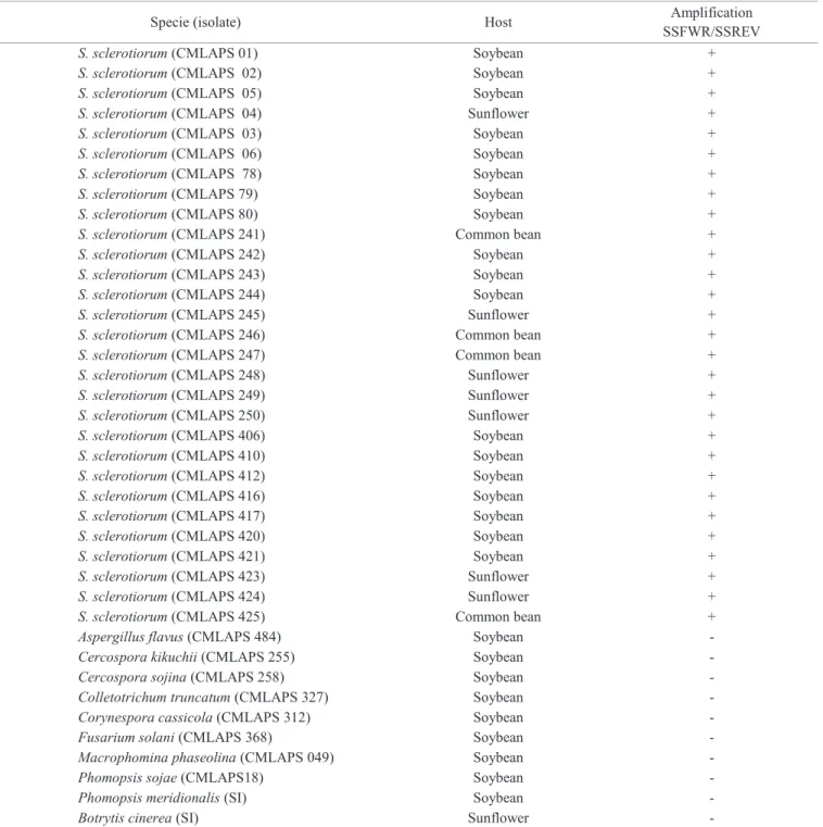
Table 1. Specificity of primers SSFWR SSREV in pure cultures of fungi from different cultures.
Specie (isolate) Host Amplification
SSFWR/SSREV
S. sclerotiorum (CMLAPS 01) Soybean +
S. sclerotiorum (CMLAPS 02) Soybean +
S. sclerotiorum (CMLAPS 05) Soybean +
S. sclerotiorum (CMLAPS 04) Sunflower +
S. sclerotiorum (CMLAPS 03) Soybean +
S. sclerotiorum (CMLAPS 06) Soybean +
S. sclerotiorum (CMLAPS 78) Soybean +
S. sclerotiorum (CMLAPS 79) Soybean +
S. sclerotiorum (CMLAPS 80) Soybean +
S. sclerotiorum (CMLAPS 241) Common bean +
S. sclerotiorum (CMLAPS 242) Soybean +
S. sclerotiorum (CMLAPS 243) Soybean +
S. sclerotiorum (CMLAPS 244) Soybean +
S. sclerotiorum (CMLAPS 245) Sunflower +
S. sclerotiorum (CMLAPS 246) Common bean +
S. sclerotiorum (CMLAPS 247) Common bean +
S. sclerotiorum (CMLAPS 248) Sunflower +
S. sclerotiorum (CMLAPS 249) Sunflower +
S. sclerotiorum (CMLAPS 250) Sunflower +
S. sclerotiorum (CMLAPS 406) Soybean +
S. sclerotiorum (CMLAPS 410) Soybean +
S. sclerotiorum (CMLAPS 412) Soybean +
S. sclerotiorum (CMLAPS 416) Soybean +
S. sclerotiorum (CMLAPS 417) Soybean +
S. sclerotiorum (CMLAPS 420) Soybean +
S. sclerotiorum (CMLAPS 421) Soybean +
S. sclerotiorum (CMLAPS 423) Sunflower +
S. sclerotiorum (CMLAPS 424) Sunflower +
S. sclerotiorum (CMLAPS 425) Common bean +
Aspergillus flavus (CMLAPS 484) Soybean -
Cercospora kikuchii(CMLAPS 255) Soybean -
Cercospora sojina (CMLAPS 258) Soybean -
Colletotrichum truncatum(CMLAPS 327) Soybean -
Corynespora cassicola (CMLAPS 312) Soybean -
Fusarium solani (CMLAPS 368) Soybean -
Macrophomina phaseolina (CMLAPS 049) Soybean -
Phomopsis sojae(CMLAPS18) Soybean -
Phomopsis meridionalis (SI) Soybean -
Botrytis cinerea (SI) Sunflower -
Abreviations: CMLAPS = Mycological Collection from Seed Pathology Laboratory, Lavras-MG, Brazil, + = positive reaction, -= negative reaction; SI = without identification.
The sensitivity was evaluated by cPCR and qPCR. The DNA extractions, primers used and conditions of cycle were the same for both.
To determine the sensitivity of detection of S. sclerotiorum
when associated to soybean seeds, portions of 100 seeds were prepared by mixing artificially infected seeds with healthy seeds, generating incidence levels of 1, 2, 10, 20 and
100% for each incubation time described above. Each seed
sample, was ground separately in a Basic IKA A11 grinder containing liquid nitrogen. The DNA for each sample was
extracted, checked their quality and quantity as described
the mill was systematically cleaned and disinfected with 2%
sodium hypochlorite solution after each sample changed.
For the cPCR the reactions were described previously. For qPCR the reaction was processed using a total volume of 20 µL of reagent “SYBR Green PCR kit” (Qiagen) with 0.625 µM of each primer and 2 µL of DNA template (50 ng). The Cq
values of each reaction were determined using the Rotor-Gene
(Corbett) software version 1.7.75. by cycler Rotor-Gene 6500 (Corbett Research, Mortlake, Australia) with optimization of gain for each tube before the first acquisition of fluorescence.
The qPCR experiment was conducted also in quintuplicate.
The specificity and sensitivity were evaluated in accordance
with the guidelines: Minimum Information for Publication of
Quantitative Real-Time PCR Experiments (Bustin, 2010).
Incidence and detection of S. sclerotiorum in naturally infected seeds using semi-seletive agar bromophenol blue
medium (NEON) and cPCR
The S. sclerotiorum presence was analyzed in thirteen
seed lots from different soybean production fields by NEON test and cPCR. Four hundred seeds/lot were distributed
in eight Petri dishes with 15 cm diameter containing agar
bromophenol blue medium (Brasil, 2009), which were then incubated in chambers at temperature of 20± 2 °C and photoperiod of 12 hours for 5 days. The seeds were distributed maintaining equidistance between them.
After the incubation period, the incidence was assessed by the formation of a yellow halo around each seed. Then, all seeds from each lot were removed separately, ground and extracted their DNAs. The experiment was done with 4 replicates. The grind, DNA extraction and PCR protocol were followed as described above. In NEON test were considered the yellow halo around each seed as positive. In PCR test, were considered positive those in which at least showed band in one replication.
The NEON test in this experiment was performed to
evaluate the incidence of S. sclerotiorum in seed lots naturally
infected, as an incubation step and as reference to detection using cPCR technique.
Results and Discussion
Specificity of the primers
The evaluation of the PCR products through
electrophoretic analysis in 1,0% agarose gel, obtained from the amplification of DNA fragments of isolates with specific
primers designed for S. sclerotiorum, generated bands of 278
base pairs, indicating specificity for the species in question. As
for the other fungi found in soybean seeds such as Aspergillus
flavus, Cercospora kikuchii, Cercospora sojina, Colletotrichum
truncatum, Corynespora cassicola, Fusarium solani,
Macrophomina phaseolina, Phomopsis sojae, Phomopsis
meridionalis and Botrytis cinerea from sunflower seeds, the
results were negative, confirming that the pair of primers was specific for the detection of S. sclerotiorum (Table 1).
As reported in literature, other primers have been described as successful for the detection of S. sclerotiorum for ascospores
and in plant tissues infected by the pathogen (Yanni et al., 2009; Kim and Knudsen, 2008; Rogers et al., 2009). In this work
these primers were tested in soybeans seeds inoculated with S.
sclerotiorum but without success. Only the primers described
by Freeman et al. (2002) showed good results when tested in
soybeans seeds inoculated with S. sclerotiorum, in both types
of PCR using the “touchdown” method. The touchdown PCR
has been used to avoid amplification of non-specific sequences
caused by phenolic compounds or other inhibitors, in PCR´s reactions. This is important in seed pathogen’s detection which some interference may reduce the sensitivity of detection and can
generate false negative results (Barrocas et al., 2009). In addition to this one of the difficulties highlighted in the sanitary quality
control of seeds has been the occurrence of pathogens in low incidence and low inoculum in the seeds.
Furthermore, for the detection of fungi, specifically in
seeds, it is necessary that, whatever method adopted, the primer
pair be tested for specificity and optimized for maximum
sensitivity for pure fungal colony and pathogen in association with seeds. It is also important to test the primers published by
other authors in different isolates of the region where the work is being developed. Variability naturally found among fungal
isolates from the same species but from different regions or hosts can lead to contradictory results of detection. The primers
used had already been tested by Freeman et al. (2002) for S.
sclerotiorum isolates from other countries, as well as similar species, for that only brasilian isolates were used.
Inoculation of S. sclerotiorum in soybean seeds and sensitivity assay
According to results in Table 2 the cPCR wasn’t enough
sensitive to detect S. sclerotiorum in samples with low
concentrations of this pathogen. In combinations with lower
incidences (1 and 2%) and lower inoculum potential (24 and 48 hours) the cPCR wasn’t able to detect the fungi in samples evaluated. For the samples with the same incidence but 72 hours of
the inoculum potential, only 2 from the 5 replicates were detected. The cPCR was sensitive only to samples with incidences
higher than 10%, considering at least 24 hours of the inoculum
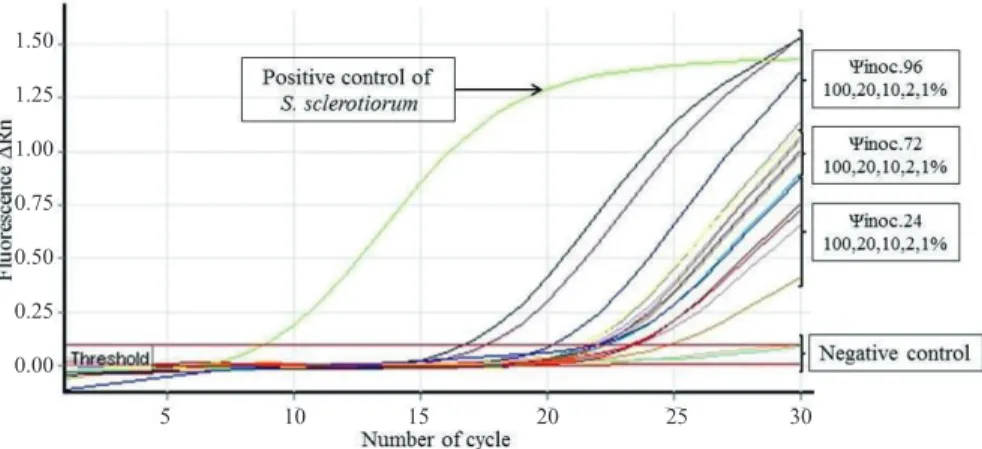
In assessing the qPCR (Table 2, Figure 1) it was possible
to detect the pathogen in all replications of incidence/inoculum potential combination even in samples that were exposed to
the pathogen for only 24 h and 1% of incidence.
Table 2. Amplification of genomic DNA from S. sclerotiorum by cPCR and qPCR at different levels of incidence in soybean seeds.
Incidence (%) potential Inoculum (hrs) Results cPCR
Results qPCR
*Negative control 0 0/5 5/5
24 0/5 5/5
1 48 0/5 5/5
72 2/5 5/5
24 0/5 5/5
2 48 0/5 5/5
72 2/5 5/5
24 5/5 5/5
10 48 5/5 5/5
72 5/5 5/5
24 5/5 5/5
20 48 5/5 5/5
72 5/5 5/5
24 5/5 5/5
100 48 5/5 5/5
72 5/5 5/5
**Positive control - 5/5 5/5
*Negative control – seeds without inoculation. **Pure culture from S. sclerotiorum CMLAPS 241.
In all extractions of DNA in seeds with different combinations of incidence/inoculum potential, the Cq values
were between 16.77 and 29.09 and confirmed the presence of the pathogen in all inoculated samples (Figure 1). The
eficiency (E) was 1.32 and R2 was 0.924 (Figure 2).The
increase of fluorescence values (Δ Rn) was higher as the
increased time of exposition the colony of the fungus S.
sclerotiorum in artificial inoculation and wasn´t observed increase in fluorescence in healthy seeds.
The osmotic conditioning technique used to inoculate seed, in this paper, ensured its infection at varying levels,
as has been demonstrated for some pathosystems (Barrocas et al., 2014; Siqueira et al., 2014; Machado et al., 2004) and
can be used as a tool that may help to validate techniques used for detecting pathogens in seeds. Therefore, it´s recommended to use the seeds inoculated with that
technique, as a first study on the detection sensitivity seed
borne pathogens.
In this study, an intercalating dye SYBR Green® that,
although less sensitive than probes showed satisfactory
results in the detection of seed infected by S. sclerotiorum,
even with low levels of infection, 1 and 2% in qPCR since
the objective was to detect S. sclerotiorum in association with
soybean seeds. The detection of this fungus was obtained in seeds with low incubation time and low incidence of infection in the seed samples tested. The qPCR technique showed good
repeatability, unlike cPCR.
Figure 1. Amplification curves on the detection of S. sclerotiorum through quantitative polymerase chain reaction technique
(qPCR) using SYBR® Green, indicating the increase of the fluorescence signal. Soybeans seeds healthy (Negative
control) and soybeans seeds inoculated with S. sclerotiorum in different inoculum potential (Ψinoc.) 24, 48, and 72
with incidences of 1, 2, 10, 20 and 100% respectively in each potential.
It is important to consider that the cPCR technique only indicates the presence or absence of the pathogen in
a sample, which may be a limiting factor for the healthy tests in seed pathology. The detection and quantification 1.50
1.25
1.00
0.75
0.50
0.25
0.00
of the pathogen in a sample of seeds have been important for some epidemiological studies of view have been used to examine diverse seedborne pathogens such as bacteria
(Becker et al., 2011; Cottyn et al., 2011), fungi (Chen et al.,2013;
Duressa et al., 2012; Montes-Borrego et al., 2012; Ioos et al., 2012; Kuan et al., 2011) and viruses (Ling et al., 2011),
including in detection for many seed borne pathogens in
the same time (Feng et al., 2014).
Figure 2. Quantitative polymerase chain reaction (qPCR) standard curve of the log of amount of S. sclerotiorum DNA, ranging
from 250 ng µL-1 to 0,5 ng µL-1 per PCR versus the corresponding cycle quantitative (Cq) values.
For molecular detection of S. sclerotiorum in soybean seeds in Brazil, where the presence of this fungus in seeds lots isn´t accepted, the test may be recommended as a screening procedure, which aims to inform if the lot is infected or not.
For lots that are considered infected other complementary
techniques should be used to indicate the incidence and viability of the pathogen in the samples. On the other hand, the qPCR technique may represent another form of information that can be used in the sanitary control of seeds. The
quantification of pathogens in the seed lot, isn´t considered in
routine testing as for the recommendation of fungicide to be used in seed treatment. This information could be important,
since the treatment of seeds with high inoculum quantity of pathogens may not produce desired results.
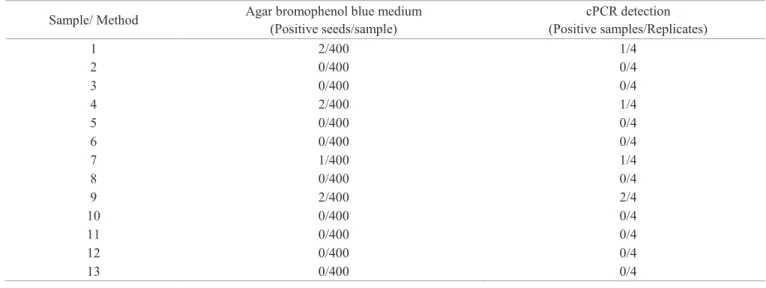
Incidence and detection of S. sclerotiorum in naturally
infected seeds
According Table 3 the lots 1, 4, 7 and 9 showed S. sclerotiorum incidence of 0.5%, 0.5%, 0.25% and 0.5%, respectively when
were evaluated by agar bromophenol blue medium. The same lots showed positive results when analyzed by cPCR. The lots 1.4 and 7 showed bands in one replication and lot 9 in two replications. The other seed lots were negative when were evaluated by agar
bromophenol blue mediumand also in cPCR test.
Table 3. Detection of S. sclerotiorum in thirteen samples of soybean seed assessed by agar bromophenol blue medium and cPCR.
Sample/ Method Agar bromophenol blue medium (Positive seeds/sample)
cPCR detection (Positive samples/Replicates)
1 2/400 1/4
2 0/400 0/4
3 0/400 0/4
4 2/400 1/4
5 0/400 0/4
6 0/400 0/4
7 1/400 1/4
8 0/400 0/4
9 2/400 2/4
10 0/400 0/4
11 0/400 0/4
12 0/400 0/4
13 0/400 0/4
Quantification cycle (Cq)
Log of DNA concentration (ng/µL) 15
14
13
12
11 10
9 8
Specifically for the detection of S. sclerotiorum in soybean seeds naturally infected or inoculated, Hennenberg
et al. (2012), concluded that traditional tests described for
the detection of that pathogen in soybean seeds, as paper roll, agar bromophenol blue medium and the blotter test aren´t sensitive for the detection of the pathogen in seeds
from areas with incidence the disease, but are efficient for detecting seed artificially inoculated. This study showed that the agar bromophenol blue medium and cPCR were efficient
in detecting seeds infected by S. sclerotiorum in the same lots.
For detection by cPCR an additional step as incubation time
could be useful.
This extra incubation period before PCR may represent a viable alternative to increase the biomass of the fungus in the
seed, diluting the influence of potential inhibitors during the incubation period and ensuring detection and quantification of viable organisms (Blanco-Meneses and Ristaino, 2013; Barrocas et al., 2009).
As mentioned PCR technique isn’t be able to give information about incidence of pathogen in each sample. But since in this experiment the preincubation time used was the semi-seletive agar bromophenol blue medium, then there was extra information about incidence/lot and we could compare both techniques.
While the cPCR detected S. sclerotiorum at incidence
level of 1% in seeds wich were incubated for 72 hours of the
exposition time of the pathogen’s colony, the same technique
could detect 0,25% of the pathogen in seeds naturally infected
using a preincubation time. This process has been adopted successfully by some authors for other pathosystems with
success (Mbofung and Pryor, 2010; Ioos et al., 2012) and
should be better investigated to reduce this period to be used in routine tests.
The use of the combined methods can improve the sensitivity of the pathogen detection. The molecular tests are the new alternative to solve it when intends detect small quantities of pathogen, but the choice of the most appropriated methodology to detect seed borne pathogen to represent the healthy conditions of a seed lot is still a challenge and must be investigated for each pathosystem.
Conclusions
The primers described by Freeman et al. (2002) are
specific and suitable to detect S. sclerotiorum in soybean seeds inoculated and naturally infected.
The cPCR is suitable to detect S. sclerotiorum in soybean
seeds inoculated with at least 1% of incidence and 72 hours of
inoculum potential and in seeds naturally infected can detect
0.25% when combined with incubation period.
The qPCR is suitable to detect S. sclerotiorum in soybean
seeds inoculated with at least 1% of incidence and 24 hours of
inoculum potential.
Acknowledgements
Thanks are due to the National Council for the Improvement of Higher Education (CAPES), to State Research Foundation of Minas Gerais (Fapemig) and to National Council for Scientific Research (CNPq) for their support to this research work.
References
BARROCAS, E.N.; MACHADO, J. C.; ALVES, M C.; CORRÊA, C.L. Desempenho de sementes de algodão submetidas à deficiência hídrica e presença de Colletotrichum gossypii var cephalosporioides. Bioscience Journal, v.30, n.2, p. 421-428, 2014. http://www.seer.ufu.br/index.php/
biosciencejournal/article/viewFile/17993/13757
BARROCAS, E.N.; MACHADO, J.C.; FIGUEIRA, A.R.; SOUZA, R.M.; ISHIDA, A.K.N.; ZACARONI, A.B.; ROCHA, H.S. Uso de técnicas moleculares para a diagnose de patógenos em sementes (Use of molecular techniques to pathogens seeds diagnose). Informe Agropecuário, v.30, n.253,
p.24-32, 2009.
BECKER, J.; HACKL, M.; RUPP, O.; JAKOBI T.; SCHNEIDER, J.; SZCZEPANOWSK, R.; BEKEL, T.; BORTH, N.; GOESMANN, A.; GRILLARI, J.; KALTSCHMIDT, C.; NOLL, T.; PÜHLER, A.; TAUCH, A.; BRINKROLF, K. Unraveling the Chinese hamster ovary cell line transcriptome by next-generation sequencing. Journal Biotechnology, v.156, p.227-235, 2011. http://www.sciencedirect.com/science/article/pii/ S0168165611005517
BLANCO-MENESES, M.; RISTAINO, J.B. Detection and quantification of
Peronospora tabacina using a Real-Time Polymerase. Plant Disease, v.95, n.6, p.673- 682, 2013. http://apsjournals.apsnet.org/doi/pdf/10.1094/PDIS-05-10-0333
BOLAND, G. J.; HALL, R. Index of plant hosts to Sclerotinia sclerotiorum.
Canadian Journal of Plant Pathology, v.16, n.2, p.93-108, 1994.
BRASIL. Ministério da Agricultura Pecuária e Abastecimento. Manual de Análise Sanitária de Sementes (Handbook on Seed Health Testing) Ministério
da Agricultura, Pecuária e Abastecimento. Brasília: MAPA-ACS, 2009. 200p. http://www.agricultura.gov.br/arq_editor/file/12261_sementes_-web.pdf
BUSTIN, S.A. Why the need for qPCR publication guidelines? The case for MIQE. Methods, v.50, n.4, p.217-226, 2010.http://www-sciencedirect-com. ez26.periodicos.capes.gov.br/science/article/pii/S1046202309002618
CHEN, Y. Y.; CONNERA, R. L.; GILLARDC, C. L.; MCLAREND, D. L.; BOLANDE, D. L.; BALASUBRAMANIANF, P. M.; STASOLLAG, C.; ZHOUH, Q. X.; HWANGH, Q. X.; CHANGH, Q. X.; BABCOCKI, C. A quantitative real-time PCR assay for detection of Colletotrichum
COTTYN, B.; BAEYEN, S.; PAUWELYN, E.; VERBAENDERT, I.; VOS, P.; BLEYAERT,P.; HOFTE, M.; MAES, M. Development of a real-time PCR assay for Pseudomonas cichorii, the causal agent of midrib rot in greenhouse-grown lettuce, and its detection in irrigating water. Plant Pathology, v.60, p.453–61, 2011. http://onlinelibrary.wiley.com/doi/10.1111/j.1365-3059.2010.02388.x/pdf
DURESSA, D.; RAUSCHER, G.; KOIKE, S.T.; MOU, B.; HAYES, R.J.; MARUTHACHALAM, K.; SUBBARAO, K.V.; KLOSTERMAN, S.J. A real-time PCR assay for detection and quantification of Verticillium dahliae
in spinach seed. Phytopathology, v.102, p.443–451, 2012. http://apsjournals. apsnet.org/doi/pdf/10.1094/PHYTO-10-11-0280
FENG, C.; MANSOURI, S.; BLUHM, B.H.; DU TOIT, L.J.; CORRELL, J.C. Multiplex real-time PCR assays for detection of four seedborne spinach pathogens. Journal of Applied Microbiology, v.117, p.472-484, 2014. http:// onlinelibrary.wiley.com/doi/10.1111/jam.12541/pdf
FREEMAN, J.; WARD, E.; CALDERON, C.; MCCARTNEY. A polimerase chain reaction (PCR) assay for the detection of inoculums of Sclerotinia sclerotiorum. European Journal of Plant Pathology, v.108, n.9, p.877-886, 2002. http://download.springer.com/static/ p d f / 2 9 3 / a r t % 2 5 3 A 1 0 . 1 0 2 3 % 2 5 2 FA % 2 5 3 A 1 0 2 1 2 1 6 7 2 0 0 2 4 . pdf?auth66=1412838790_f078f0ae28911d99dbf8e1d6528645ba&ext=.pdf
GLYNN, N.C.; EDWARDS, S.G. Evaluation of PCR assay for quantifying seed-borne infection by Fusarium and Microdochium seedling blight pathogens. Applied Microbiology, v.108, p.81-87, 2010. http://onlinelibrary. wiley.com/doi/10.1111/j.1365-2672.2009.04410.x/full
HENNENBERG, L.; GRABICOSKI, E.M.G.; JACCOUD-FILHO, D.S.; PANOBIANCO, M. Incidência de Sclerotinia sclerotiorum em sementes de soja e sensibilidade dos testes de detecção. Pesquisa Agropecuária Brasileira, v.47, n.6, p.763-768, 2012. http://www.scielo.br/pdf/pab/v47n6/47n06a05.pdf
IOOS, R.; FOURRIER, C.; WILSON, V.; WEBB, K.; SCHEREFFER, J.L. DE LABROUHE, D.T. An optimized duplex realtime PCR tool for sensitive detection of the quarantine Oomycete Plasmopara halstedii in sunflower seeds. Phytopathology, v.102, p.908–917, 2012. http://apsjournals.apsnet. org/doi/pdfplus/10.1094/PHYTO-04-12-0068-R
KIM, T, G.; KNUDSEN, G.R. Quantitative real-time PCR effectively detects and quantifies colonization of sclerotia of Sclerotinia sclerotiorum
by Trichoderma spp. Applied Soil Ecology, v.40, n.1, p.100-108, 2008. http:// www-sciencedirect-com.ez26.periodicos.capes.gov.br/science/article/pii/ S0929139308000565
KUAN, C.P., WU, M.T., HUANG, H.C., CHANG, H. Rapid detection of
Colletotrichum lagenarium, causal agent of anthracnose of cucurbitaceous crops, by PCR and real-time PCR. Journal of Phytopathology, v.159, p.276-282, 2011. http://onlinelibrary.wiley.com/doi/10.1111/j.1439-0434.2010.01765.x/pdf
LING, K.S.; WECHTER, W.P.; WALCOTT, R.R.; KEINATH, A.P. Development of a real-time RT-PCR assay for squash mosaic virus useful for broad spectrum detection of various serotypes and its incorporation into a multiplex seed health assay. Journal of Phytopathology, v.159, p.649–656, 2011. http://onlinelibrary. wiley.com/doi/10.1111/j.1439-0434.2011.01814.x/pdf
MACHADO, J.C.; GUIMARAES, R.M.; VIEIRA, M.G.G.C.; SOUZA, R.M.; POZZA, E.A. Use of water restriction technique in seed pathology.
SeedTesting International.ISTA News Bulletin, v.128, p. 14-18, 2004.
MACHADO, J.C.; POZZA, E.A. Razões e procedimentos para o estabelecimento de padrões de tolerância a patógenos em sementes (Reasons and procedures to establish tolerance standards of pathogens in seed). In: ZAMBOLIM, L.
Sementes qualidade fitossanitária, Viçosa, MG: UFV, 2005. p.375-398.
MBOFUNG, G.C.; PRYOR, B.M. A PCR-Based assay for detection of Fusarium oxysporum f. sp. lactucae in lettuce seed. Plant Disease, v.94, n.7, p.860-866,
2010. http://apsjournals.apsnet.org/doi/pdf/10.1094/PDIS-94-7-0860
MICHEL, B. E.; RADCLIFFE, D. A computer program relating solute potential to solution composition for five solutes. Agronomy Journal, v.87, n.1, p.126-130, 1995.
MONTES-BORREGO, M.; MUNOZ-LEDESMA, F.J.; JIMENEZ-DIAZ, R.M.; LANDA, B.B. Real-time PCR quantification of Peronospora arborescens, the opium poppy downy mildew pathogen, in seed stocks and symptomless infected plants. Plant Disease, v.95, p.143–152, 2012. http://
apsjournals.apsnet.org/doi/pdfplus/10.1094/PDIS-07-10-0499
ROGERS, S.L.; ATKINS, S.D.; WEST, J.S. Detection and quantification of airborne inoculums of Sclerotinia sclerotiorum using quantitative PCR.
Plant Pathology, v.58, n.2, p.324-331, 2009. http://onlinelibrary.wiley.com/ doi/10.1111/j.1365-3059.2008.01945.x/pdf
SIQUEIRA, C. S.; MACHADO, J. C.; BARROCAS, E.N.; ALMEIDA, M.F. Potential for transmission of Stenocarpella macrospora from inoculated seeds to maize plants grown under controlled conditions. Journal of Seed Science, v.36, n.2, p.154-161, 2014. http://www.scielo.br/pdf/jss/v36n2/
v36n2a03.pdf