Colonization of maize seeds by two species of
Stenocarpella
transformed with
fluorescent proteins and assessed through scanning electron microscopy
1Carolina da Silva Siqueira
2*, José da Cruz Machado
2,
Carla Lima Corrêa
2, Ellen Noly Barrocas
3ABSTRACT – Stenocarpella maydis and Stenocarpella macrospora species causing leaf spots and stem and ear rots, can be transported and disseminated between cultivating areas through seeds. The objective was to transform isolates of species of Stenocarpella with GFP and DsRed and to correlate different inoculum potentials with the effect caused by the presence of these pathogens in the tissues of maize seeds. The isolates were transformed with introduction of the genes in their nuclei, employing the technique of protoplast transformation. Seeds were inoculated by osmotic conditioning method with transformed and not transformed isolates, with different periods of exposition of seeds to those isolates, characterizing the inoculum potentials, P1 (24 h), P2 (48 h), P3 (72 h) and P4 (96 h). The seeds inoculated with isolates expressing GFP and DsRed in both species elucidated by means of the intensities of the emitted fluorescence, the ability of those organisms to cause infection and colonization in different inoculum potentials. The potentials P3 and P4 caused the highest levels of emitted fluorescence for the colonization by both pathogens. A comprehensive and abundant mycelial growth in the colonized seed structures were well visualized at potential P3 and P4 by means of SEM.
Index terms: seed pathology, genetic transformation, GFP, DsRed protein, fungus.
Colonização de sementes de milho por duas espécies de Stenocarpella
transformados com proteínas fluorescentes e avaliadas por microscopia
eletrônica de varredura
RESUMO – Stenocarpella maydis e Stenocarpella macrospora, espécies causadoras de manchas foliares, podridões em plantas e grãos ardidos de milho, podem ser transportadas e dispersas para áreas produtoras através das sementes. O objetivo foi transformar isolados de espécies de Stenocarpella com GFP e DsRed e correlacionar diferentes potenciais de inóculo com o efeito causado pela presença desses patógenos nos tecidos de sementes de milho. Os isolados foram transformados por meio da introdução dos genes nos seus núcleos, empregando a técnica de transformação de protoplastos. Sementes foram inoculadas pelo método de condicionamento osmótico com isolados transformados e não transformados, com diferentes tempos de exposição das sementes a estes isolados, caracterizando os potenciais de inóculo, P1 (24 h), P2 (48 h), P3 (72 h) e P4 (96 h). As sementes inoculadas, com isolados transformados expressando GFP e DsRed, de ambas as espécies, elucidaram por meio das intensidades das fluorescências emitidas, a capacidade desses organismos de causarem infecção e colonização em diferentes potenciais de inóculo, tendo P3 e P4, provocado as maiores intensidades de fluorescência pelas colonizações dos patógenos. Um abrangente e abundante crescimento micelial nas estruturas colonizadas das sementes foi observado nos potenciais P3 e P4, por meio da MEV.
Termos para indexação: patologia de sementes, transformação genética, proteína GFP, proteína DsRed, fungo.
1Submitted on 10/22/2013. Accepted for publication on 04/02/2014. 2Departamento de Fitopatologia, - UFLA, Caixa Postal 3037, 37200-000 - Lavras, MG, Brasil.
3Department of Plant and Environmental Sciences, 2360- Taastrup, Denmark. *Corresponding author <[email protected]>
Introduction
Many pathogenic fungal species, like those of the so called “Stenocarpella complex” may be transmitted by maize seeds. Those species are responsible for rots on the stalk and ear on maize, being also potential producers of toxins noxious
to animal health (Petatán-Sagahón et al., 2011) and causing
drop on the price of the product to destined as certificated
seeds in the market (Ribeiro et al., 2005).
fungi S. maydis and S. macrospora survive in the internal tissues of seeds (endosperm and embryo) mainly on mycelial form. Thereby, when the seed comes in contact with the soil water and receives the stimuli from the environment to start germination process, the fungal mycelium starts growing from internal tissues to seed’s surface. Thus, when growing on the seed the fungus reaches the roots and coleoptiles; and through coleoptiles fungus reach to soil’s surface and may cause death of that seedling (Casa et al., 2006).
Thereby, the study of pathogen dynamics, at the internal tissues infection moment, as well as the pathogen location within the seed, becomes of paramount importance for the understanding the host-pathogen relationship. In this sense, some molecular
markers, such as the green fluorescent protein (GFP) originating from the jellyfish Aequorea victoria, and the red fluorescent
protein, originating from the mushroom anemone Discosoma sp.
(DsRed) used on fluorescence microscopy; besides the scanning
electron microscopy (SEM), have been successfully used for the live observation of the fungal structures within the seeds, as well as to clarify the processes of infection and colonization of seed internal tissues by the pathogenic agents (Cristea et al., 2005; March et al., 2003).
Based on the previous information, the aim of this study was to obtain different isolates of the two species of
Stenocarpella, transformed by molecular markers expressing
the GFP and DsRed fluorescent proteins by adjusting already
described protocols; as well as correlating different inoculum potentials in the infection and colonization of the seed internal
tissues through the fluorescence microscopy and scanning
electron microscopy (SEM).
Material and Methods
Characterization of isolates and source of seeds: two different isolates of Stenocarpella maydis (Berkeley) Sutton [Syn. Diplodia maydis (Berkeley) Saccardo] as well as two different isolates of Stenocarpella macrospora (Earle) Sutton [Syn. Diplodia macrospora Earle] were used for this study. One of the S. maydis isolates, registered as CML698 was obtained at the mycological collection of Federal University of Lavras (UFLA), located at Lavras, state of Minas Gerais (MG); and the other isolate, registered as MY2 was obtained of Embrapa Maize and Sorghum, located at Sete Lagoas, MG. The two isolates of S. macrospora, registered as CMLAPS375 and CMLAPS10, respectively were obtained at the mycological collection of the Seed Pathology Laboratory of UFLA. The maize seeds (cv. RB 9308 YG), with 98% germination (Brasil, 2009a), were provided by the company Riber KWS Seeds, based at Patos de Minas, MG. The cv. RB
9308 YG were submitted to health testing which reveled the presence of Fusarium verticillioides at level of 28.5% and
Penicillium sp. at 13% incidence in the seeds (Brasil, 2009b).
Transformation of isolates: both of isolates of S. maydis
and both of S. macrospora were transformed by using the plasmids pSC001 and pSC002, which contain the hpr gene, for resistance to antibiotic hygromycin B; as well as the pToxA
promoter gene originated from Aspergillus nidulans (Maor et al. 1998). Following methodology described by Sambrook and Russell (2001), competent cells of bacterium Escherichia coli were used for multiplication of these plasmids; and for transformation of isolates, the protocols previously described by Maier et al. (2005) and Silva et al. (2009) for Fusarium graminearum and Sclerotinia sclerotiorum were used with
some methodological modifications.
For obtaining protoplasts of S. maydis and S. macrospora, both fungi were grown on PDA culture medium (200 g agar; 20 g dextrose; and 200 g potato) for 5 days, at 25±2 °C and photoperiod of 12/12 h (L/D). After such period, mycelium disc with 1 cm2 were cut from the growth zone of each colony and transferred to Erlenmeyer flasks with 100 mL capacity,
containing 50 mL of liquid potato-dextrose culture medium. For generation of protoplasts, immediately after inoculation the
flasks with the mycelial discs were incubated under constant
shaking, provided by a mechanized horizontal shaker, set to 75 rpm, where they were maintained, at 28 °C, for 3 h. After fungal growth, mycelial mass of each colony was aseptically
filtered and immediately dehydrated with vacuum pump.
Afterwards, 100 mg of dry mycelium, from each isolate, was added into a tube with 3 mL of the KCl osmotic stabilizer (0.7M) and 10 g of lysing enzymes [Sigma®-L1412 (lysing
enzymes from Trichoderma harzianum lyophilized powder)] at a ratio of 10 mg.mL-1 osmotic stabilizer.
Only the produced protoplasts were filtered through a
previously sterilized cheesecloth layer, and then centrifuged at 600 x g (RCF), at 4 °C, for 5 min. Immediately after, these protoplasts were re-suspended in KCl (0.7 M), at 4 °C, and again re-suspended in storage buffer composed by four parts of the solution STC [0.8 M sorbitol; 50 mM Tris HCl (pH 8.0); and 50 mM CaCl2] and one part of the solution SPTC [0.8 M sorbitol; 40% PEG 4000; 50 mM Tris HCl (pH 8.0); and 50 mM CaCl2]. Subsequently, to the suspensions
of final concentration of approximately 107 protoplasts.
mL-1, were added 10 µl of plasmid DNA, at a ratio ranging
between 0.35 and 1.66 mg.µL-1, which were then kept on ice
for 30 min. Afterwards, 1 mL SPTC was added to solution, and kept at room temperature for more 20 min. After such period, protoplast suspensions were separately poured into
[0.1% yeast extract (Sigma-Y4250 - 250 g); 0.1% hydrolyzed casein (Sigma-C8845 - 500 mg); 34.2% sucrose (Sigma-84100 – 1 kg); and 1.0% Agar granulated (Difco-1016141000 - 1 kg)]. Subsequently, this regeneration medium, containing the transformed isolates was poured into Petri dishes (9 cm Ø), and incubated, at 25±2 °C, for 72 h. After this period, to each of the plates was added 10 mL of water-agar medium containing the antibiotic hygromycin-B, at a concentration of 100 mg.mL-1, and kept at 25±2 °C, and 12/12 h photoperiod,
for 15 days, into an incubation chamber.
After protoplast transformation, mycelium of each of the two species of the fungus, containing the marker genes for proteins GFP and DsRed were separately transferred to PDA culture medium, to which was added the antibiotic hygromycin-B, at concentration of 100 µg.mL-1, as well as
to the Oatmeal-Agar (OA) culture medium (20 g agar, 30 g oatmeal, and 1 L H2O) also added with hygromycin-B at same concentration used in the PDA medium. All isolates were assessed both by their morphological characteristics as by their cultural characteristics as mycelial coloration and mode of growth of the colonies. For observing stability of the different isolates of transformed fungus, successive transfers were performed on PDA medium containing 100 µg.mL-1
of antibiotic hygromycin-B. Both for the assessments of bioassays as for stability tests, observations were performed
with the aid of a fluorescence stereomicroscope (brand Leica; model M165FC) and of a fluorescence microscope (brand
Zeiss, model AxioVision Z.1) equipped with the software
AxioVision Imaging System, which possesses special filters for observation of the marker genes for the proteins GPF (filter with a wavelength between 470 and 490 nm) and DsRed (filter
with a wavelength between 510 and 560 nm) within the cells. Subsequently, all images obtained were edited with the aid of
the software AxioVision and Microsoft Office Picture Manager.
Inoculation of seeds: before installing the experiments, all the seeds were disinfected with a sodium hypochlorite solution (1%) for 1 min., rinsed with sterile distilled water and placed to dry, at room temperature, for 48 h. Following methodology described by Machado et al. (2012) seeds were evenly distributed into Petri dishes containing PDA culture
medium, modified with mannitol solution (water potential
adjusted to -1.4 MPa, designated by Michel and Radcliffe,
1995), and fungal colonies developed for five days; as well as
each of four transformed isolates of S. maydis (MCL698-GFP, MY2-GFP and MCL698-DsRed, MY2-DsRed); and four transformed isolates of S. macrospora (CMLAPS375-GFP, GFP and CMLAPS375-DsRed, CMLAPS10-DsRed). Besides of mannitol solution addition, to the PDA medium, the antibiotic hygromycin-B was also added, at a
concentration of 100 μg.mL-1; and plates containing seeds
were then incubated at 25±2 °C, and photoperiod of 12/12 h, during different periods (inoculum potential) [P1 (24 h); P2 (48 h); P3 (72 h); and P4 (96 h)]. For the control treatment seeds inoculated with non-transformed isolates of S. maydis (isolate CML698) and S. macrospora (isolate CMLAPS375) were used; which were also used to assess behavior of the fungus in the seeds through SEM.
Preparation of inoculated seeds for analysis by SEM: the seeds inoculated with the non-transformed isolates of
S. maydis and S. macrospora were randomly chosen for observation under a scanning electron microscope. To that, seeds were transversely, longitudinally, and medially cut
for the fixation in a modified Karnovsky solution (2.5% of
glutaraldehyde; 2.5% formaldehyde diluted in 0.05 M sodium cacodylate buffer (pH 7.2) and CaCl2 0.001 M), at 4 °C, for
48 h. After fixation, the fragments of seeds of each of the
treatments were immersed in a 30% glycerol solution, for 30 min. Subsequently, these fragments were cut in smaller pieces using liquid nitrogen, and immediately immersed in distilled water. After such process, samples were immersed
in a fixative solution consisting of osmium tetroxide (1%) diluted in distilled water, for 3 h. Immediately after fixation,
the samples were again washed in distilled water and then dehydrated in a series of successive dilutions of acetone [three times in pure acetone (100%); and once in solutions containing 90%, 75%, 50%, and 25% acetone]. Then the samples were transferred to a Critical Point apparatus (Balzers;
CPD-030) for replacement of acetone by CO2 and finalization of
drying process. The specimens so obtained were mounted on
aluminum stubs, fixed with the aid of a double-sided carbon
tape, and placed on an aluminum pellicle, covered with gold in a Balzers evaporator (model SCD 050), and then observed through a scanning electron microscope (Zeiss, model LEO EVO 40 PVX). The images digitally generated by this equipment, were then recorded and registered.
Results and Discussion
Genetic transformation of S. maydis and S. macrospora
with the expression of GFP and DsRed fluorescent proteins
was effectively performed, obtaining in the end, stable transformants, as evidenced by resistance to the antibiotic
hygromycin-B and of the visualization of the fluorescent
proteins, green and red.
Stable protoplasts in sufficient quantity for transformation were obtained, observing that there are several influencing
microorganism to be used (Almeida et al., 2008; Ishikawa et al., 2010). Even for S. macrospora, which was the first attempt
to produce protoplasts, the quantities were favorable, ranging between 30-45 x 105 protoplasts/mL, and to S. maydis, of
40-45 x 105 protoplast/mL. For many fungi this is a critical point
because without obtaining intact protoplasts, the success of the transformation was questionable (Marchi et al., 2005; 2006).
With the transformation procedure, 21 transformed isolates of S. maydis containing the gene expressing for GFP and 20 containing the gene for DsRed, were obtained i.e., about 3-4 transformants of S. maydis per µg of plasmid DNA. For S. macrospora, 18 isolates were obtained with the GFP and 15 with the DsRed, resulting from 2-3 transformants per µg plasmid DNA. As the morphological characteristics, when comparing transformed and non-transformed isolates, these revealed few differences, as in color of the colony, being observed in older colonies (with 8-15 days) of these transformed, a darker color than normal and, sometimes, greenish tones. Concerning mycelial growth on PDA medium added with hygromycin-B, it was observed that the transformed
were slower, taking 2-3 days more to fill a 90 mm Petri dish than
a non-transformed isolate. On the OA culture media, added with hygromycin-B, several transformed isolates, both of S. maydis
as of S. macrospora grew normally, but did not maintain the
fluorescence. However, Xiao et al. (2013) report that the GFP
does not interfere with normal cell functions, and therefore can be used for analysis of cellular processes. In observing the stability of mycelial growth, when the transformed fungi
of both species were successively transferred for five times in
PDA medium containing hygromycin-B (100 g/mL), it was
verified a normal growth, and their respective fluorescence
maintained. About 20% have lost this ability, which may be
related to the difficulty of cellular nuclei of maintain and pass
the information.
For the transformed isolates, expressing GFP (Figure 1) and DsRed of S. maydis and S. macrospora, green and red
fluorescence were observed in the cytoplasm of hyphae and
conidia; however, the pycnidia retained their natural color, even if their conidia were positive. However, there has been
a variation in fluorescence intensity both for the green as the red. The lowest levels of fluorescence intensity were found in
the transformed isolates expressing DsRed.
Figure 1. Stereo micrographs of fluorescence of the fungi marked with green fluorescent protein, GFP. To Stenocarpella maydis: A1 (white light), A2 and C (hyphae of the isolate CML698-GFP) and B (pycnidia and hyphae of the isolate MY2-GFP). To Stenocarpella macrospora: D (hyphae of the isolate CMLAPS10-GFP), E (hyphae of CMLAPS375-GFP). Bar = 200 µm.
In the seeds inoculated with the transformed isolates,
expressing GFP, direct correlations between the fluorescence
intensity of the colonies with the inoculum potential (time of exposure of the seeds to the transformed fungus) (Figure 2)
were observed. In the seeds inoculated with P4 (96h) it was possible to observe an abundant and compact mycelial
growth, evidenced by the strong fluorescence emitted by
to monitor the development of the infection process. In a study performed with maize seeds infected by the fungus
Aspergillus flavus, Du et al. (1999) have correlated the
intensity of the fungus mycelial fluorescence with the
presence and the intensity of seed colonization by this transformed organism. They also emphasized the importance of using this tool on detecting this fungus in kernels meant to food and for selecting maize genotypes resistant to
accumulation of aflatoxins, which are toxic substances
produced by this fungus. Therefore, this study may be used as basis for further investigations correlating colonization of seeds for transformed Stenocarpella species with the presence of harmful toxins produced, such as diplodiatoxin produced by S. maydis, and toxin diplodiol produced by S. macrospora (Petatán-Sagahón et al., 2011) for which there is little information in the literature.
Figure 2. Stereo micrographs of the maize seeds inoculated with the fungi Stenocarpella maydis and Stenocarpella macrospora
marked with GFP with different inoculum potential [P1 (24 h), P2 (48 h), P3 (72 h), and P4 (96 h)]. Seeds with transformed Stenocarpella maydis, isolated CML698-GFP (B, C, D and E) and MY2-GFP (G, H, I and J). Seeds with transformed Stenocarpella macrospora, isolated CMLAPS375-GFP (L, M, N and O) and CMLAPS10-GFP (Q, R, S and T). Seeds of controls treatment, or without inoculation: A, F, K and P. Bar = 2 mm.
Stenocarpella maydis Stenocarpella macrospora
0 h
24 h
48 h
72 h
There were no major visual differences between the seeds inoculated with transformed isolates of S. maydis or S. macrospora;
however, it was possible to observe stronger fluorescence intensity
on the seeds inoculated with S. maydis. These results may be due to the fact that this species of the fungus grows easily in the maize seeds; whereas the species S. macrospora consistently associated with growth on maize leaves (Casa et al., 2006).
Seeds inoculated with transformed isolates of S. maydis
and S. macrospora expressing the DsRed protein did not
differ from seeds inoculated with isolates expressing the GFP (Figure 3). However, it was observed that seeds inoculated with the isolates expressing DsRed with higher inoculum
potential (P3 and P4) had more fluorescence; thereby
allowing to conclude that in this inoculum potential occurred a deeper infection, which have reached even the embryo and
the endosperm and that, when observed the red fluorescence
was much more evident on inoculated seeds, especially when compared to non-inoculated seeds (control) (Figure 4).
24h
48h
72h
96h
Control
A
B
C
E
D
G F
H
Stenocarpella
maydis Stenocarpella macrospora
Figure 3. Stereo micrographs of the maize seeds inoculated with the fungi Stenocarpella maydis and Stenocarpella macrospora
marked with DsRed with different inoculum potential [P1 (24 h), P2 (48 h), P3 (72 h), and P4 (96 h)]. Seeds with transformed Stenocarpella maydis, isolated CML698-DsRed (A, B, C and D). Seeds with transformed Stenocarpella macrospora, isolated CMLAPS375-DsRed (E, F, G and H). Seed of control treatment, or without inoculation (Control). Bar = 2 mm.
Besides observing that the transformed isolates, both of S. maydis as of S. macrospora, expressing the GFP and
DsRed proteins are able to show infection and colonization, seeds were also assessed by SEM; and through this tool it was 24 h
48 h
72 h
96 h
Control
Stenocarpella maydis
possible to observe in details the seed colonization process by these two pathogens.
Through SEM it was also possible to assess changes in the characteristics of the main physical structures of maize
seeds [endosperm, germ, pericarp, and tip (Paes, 2006)] and it was found that these changes in cellular structures were directly proportional to the increase of inoculum potential in the seeds inoculated with S. maydis and S. macrospora.
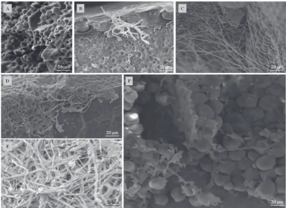
Figure 4. Electron micrographs of the epidermis of maize seeds inoculated with Stenocarpella maydis (B, C, D, E) and
Stenocarpella macrospora (G, H, I, J). A and F= Control or non-inoculated seed; G and B = inoculum potential P1 (24 h); C and H= P2 (48 h); D and I= P3 (72 h); E and J=P4 (96 h).
In the pericarp, where are the epidermis (first layer),
followed by the mesocarp, cross cells, tubular cells and testa (last layer of pericarp), in all inoculation periods, it was observed the presence of the species of Stenocarpella (Figure 4). In P1, the
hyphae of the isolates were more superficial and have caused
almost all the pericarp, with large quantities of mycelium, and this was becoming more compact and well adhered to the layers, causing drastic reduction in the volume of the protection cells, with injuries and deformation of the outer parts of the seeds. The loss of integrity of the pericarp can affect the electrical conductivity of the exudates released by the maize seeds. The same can also be observed in seeds that are submitted to a drastic drying process (José et al., 2005). No major differences were observed between the colonization of S. maydis and S. macrospora, but with S. macrospora it was
verified that mycelium was more aerial on the seed surface or
less adhered to the seeds and slightly thicker.
The combined effect of those pathogens in the inner parts of the maize seeds is of great importance because the endosperm represents about 83% of the dry weight of the maize kernels (Figures 5 and 6). The endosperm is constituted especially of starch (88%), and arranged in the form of granules and of storage proteins (8%) of prolamin type, called zeins, these proteins form the protein bodies that compose the matrix which surrounds the starch granules within the endosperm cells (Paes, 2006), i.e., the major part of the maize seed. Moreover, the species of Stenocarpella may take advantage of the inner of the maize seeds, based on the distribution of the starch granules and of matrix proteins that compose the
endosperm classified as: farinaceous and vitreous. In the first,
the starch granules are rounded and dispersed with no protein matrix surrounding these structures, which results in vacant spaces during the drying process of the seed, starting from the spaces where it previously was occupied by the water, during the development of the seed (Paes, 2006). These spaces may be occupied entirely by masses of hyphae, as observed in P3 and P4, for both species, waiting for favorable conditions to develop, besides being responsible for the damages caused to the seeds. However, in the vitreous endosperm, the protein matrix is dense, with structured protein bodies and surrounding the polygonal format starch granules, and with no spaces formation among these structures. Under such conditions it was observed hyphae very adhered in to deformed granules and developing among the protein bodies.
S. maydis and S. macrospora were able to start the infection process and to develop themselves in the maize seeds with any inoculum potential; as demonstrated in this study. In all inoculum potentials dormant structures of the fungus were found, and from there those structures may develop in the presence of water, nutrients and favorable temperature, culminating to disease development leading often to death the seeds/seedlings, as expected at higher inoculum potentials.
Figure 6. Electron micrographs of the inner parts of a maize seed inoculated with Stenocarpella macrospora during different periods. A= control (non-inoculated seed); B e C = P1 (24 h); D = P2 (48 h); E = P3 (72 h); F = P4 (96 h).
Conclusions
The protocol used for production of protoplasts and incorporation of genes that express GFP and DsRed in S. maydis and S. macrospora is efficient and satisfactory for
the transformation of those species and for its use in seed pathology studies.
Seeds inoculated with isolates of genetically transformed
Stenocarpella species elucidated, by means of the fluorescence
intensities, the varying levels of infection and colonization found in the different inoculum potentials.
Analyzes performed by SEM on maize seeds inoculated with S. maydis and S. macrospora were effective for demonstrating the damage caused to the tissues of the seeds by the infection and colonization, being the damages more severe when observed in the higher inoculum potentials.
Acknowledgments
To CNPq, CAPES and FAPEMIG for financial support for
conducting this study; to Dr. Theo van der Lee (Plant Research International, The Netherlands) who provided plasmids used for fungal transformations; to company Riber Seeds (Patos de Minas/ MG); to Plant Virology laboratory and Electron Microscopy and Ultrastructural Analysis laboratory of Department of Plant Pathology, Federal University of Lavras (Lavras/MG).
References
ALMEIDA, A. P. M. M.; DIAS, E. S.; PEREIRA, R. T. G.; TOLEDO, R.
C. C.; PFENNING, L. H. Obtenção de protoplastos do fungo filamentoso
Aspergillus ochraceus.Ciência Rural, v.38, n.5, p.1460-1462, 2008. http://
www.scielo.br/pdf/cr/v38n5/a43v38n5.pdf
BRASIL. Ministério da Agricultura, Pecuária e Abastecimento. Regras para
análise de sementes.Ministério da Agricultura, Pecuária e Abastecimento.
Secretaria de Defesa Agropecuária. Brasília: MAPA/ACS, 2009a. 395p. http://
www.agricultura.gov.br/arq_editor/file/2946_regras_analise__sementes.pdf
BRASIL. Ministério da Agricultura, Pecuária e Abastecimento. Manual de Análise
Sanitária de Sementes. Ministério da Agricultura, Pecuária e Abastecimento.
Secretaria de Defesa Agropecuária. Brasília: MAPA/ACS, 2009b. 200p. http://
www.agricultura.gov.br/arq_editor/file/12261_sementes_-web.pdf
CASA, R. T.; REIS, E. M.; ZAMBOLIM, L. Doenças do milho causadas por fungos do gênero Stenocarpella. Fitopatologia Brasileira, v.31, n.5, p.427-439, 2006. http://www.scielo.br/pdf/fb/v31n5/01.pdf
CRISTEA, I. M.; WILLIAMS, R.; CHAIT, B. T.; ROUT, M. P. Fluorescent Proteins as Proteomic Probes. Molecular & Cellular Proteomics, v.4, n.12, p.1933-1941,2005. http://www.mcponline.org/content/4/12/1933.full.pdf
DU, W.; HUANG, Z.; FLAHERTY, J. E.; WELLS, K.; PAYNE, G. A.
Green fluorescent protein as a reporter to monitor gene expression and food
colonization by Aspergillus flavus. Applied and Environmental Microbiology, v.65, n.3, p.834-836, 1999. http://www.ncbi.nlm.nih.gov/pmc/articles/ PMC91103/pdf/am000834.pdf
ISHIKAWA, F. H.; BARCELOS, Q. L.; SOUZA, E. A.; DIAS, E. S. Factors affecting the production and regeneration of protoplasts from Colletotrichum
lindemuthianum. Ciência e Agrotecnologia, v.34, n.1, p.74-79, 2010. http://
JOSÉ, S.C.B.R.; VON PINHO, E. V. R.; VON PINHO, R. G.; RAMALHO, M. A. P.; FILHO, J. L. S. Características físicas do pericarpo de sementes de milho associadas com a tolerância à alta temperatura de secagem. Revista
Brasileira de Sementes, v.27, n.1, p.125-131, 2005. http://www.scielo.br/pdf/
rbs/v27n1/25189.pdf
MACHADO, J. C.; BARROCAS, E. N.; COSTA, M. L. N.; GUIMARÃES, R. M.; MACHADO, C. F. Uso da técnica de restrição hídrica ou condicionamento osmótico em patologia de sementes. Revisão Anual de Patologia de Plantas, v.20, p.1-24, 2012.
MAIER, F. J.; MALZ, S.; LOSH, A. P.; LACOUR, T.; SCHAFER, W.
Development of a highly efficient gene targeting system for Fusarium
graminearum using the disruption of a polyketide synthase gene as a visible
marker. FEMS Yeast Research, v.5, n.6/7, p.653-662, 2005..http://dx.doi. org/10.1016/j.femsyr.2004.12.008
MAOR, R.; PUYESKY, B. A.; SHARON, A. Use of green fluorescent protein
(GFP) for studying development and fungal-plant interaction in Cochliobulus
heterostrophus. Mycology Research, v.102, n.4, p.491-496, 1998. http://
www.sciencedirect.com/science/article/pii/ S0953756208609053
MARCH, J.C.; RAO, G.; BENTLEY, W.E. Biotechnological applications
of green fluorescent protein. Applied Microbiology and Biotechnology,
v.62, p.303–315, 2003. http://download.springer.com/static/pdf/775/ art%253A10.1007%252Fs00253-003-1339y.pdf?auth66=1382536505_ d1804f14bb2deaf1300b5672930dd86b&ext=.pdf
MARCHI, C. E.; BORGES, M. F.; BROMMONSCHEKEL, S. H. Obtenção de protoplastos de Botryosphaeria sp. Ciências Biológicas e da Saúde, v.12, n.4, p.23-32, 2006. http://www.revistas2.uepg.br/index.php/biologica/article/view/441/442
MARCHI, C. E.; BROMMONSCHENKEL, S. H.; QUEIROZ, M. V.; MIZUBUTI, E. S. G. Isolation and regeneration of Magnaporthe grisea
protoplasts. Summa Phytopathologica, v.32, n.3, p.232-238, 2005. http:// www.scielo.br/pdf/sp/v32n3/a04v32n3.pdf
MICHEL, B. E.; RADCLIFFE, D. A computer program relating solute
potential to solution composition for five solutes. Agronomy Journal, v.87,
n.1, p.131-136, 1995.
PAES, M.C.D. Aspectos físicos, químicos e tecnológicos do grão de milho.
EMBRAPA/CNPMS, 2006. 14p. (Circular técnica, 75). http://www.cnpms. embrapa.br/publicacoes/publica/2006/circular/Circ_75.pdf
PETATÁN-SAGAHÓN, I.; ANDUCHO-REYES, M. A.; SILVA-ROJAS, H. V.; ARANA-CUENCA, A.; TELLEZ-JURADO, A.; CÁRDENAS-ÁLVAREZ, I. O.; MERCADO-FLORES, Y. Isolation of bacteria with antifungal activity against the phytopathogenic fungi Stenocarpella maydis and Stenocarpella
macrospora. International Journal of Molecular Sciences, v.12, p.5522-5537,
2011. http://www.ncbi.nlm.nih.gov/pmc/articles/PMC3189730/
RIBEIRO, N.A.; CASA, R. T.; BOGO, A.; SANGOI, L.; MOREIRA, E. N.; WILLE, L. A. Incidência de podridões do colmo, grãos ardidos e produtividade de grãos de genótipos de milho em diferentes sistemas de manejo. Ciência Rural, v.35, n.5, p.1003-1009, 2005. http://www.scielo.br/ pdf/cr/v35n5/a04v35n5.pdf
SAMBROOK, J.; RUSSELL, D.W. Molecular cloning: a laboratory manual, 3.ed. Cold Spring Harbor: Laboratory Press. 2001. 2344p.
SILVA, A. P.; BOLTON, M. D.; NELSON, B. D. Transformation of Sclerotinia sclerotiorum with the green fluorescent protein gene and fluorescence of
hyphae in four inoculated hosts. Plant Pathology, v.58, n.3, p.487-496, 2009. http://onlinelibrary.wiley.com/doi/10.1111/j.1365-3059.2009.02022.x/pdf
XIAO, R. F.; ZHU, Y. J.; LI, Y. D.; LIU, B. Studies on vascular infection
of Fusarium oxysporum f. sp. cubense race 4 in banana by field survey and

![Figure 2. Stereo micrographs of the maize seeds inoculated with the fungi Stenocarpella maydis and Stenocarpella macrospora marked with GFP with different inoculum potential [P1 (24 h), P2 (48 h), P3 (72 h), and P4 (96 h)]](https://thumb-eu.123doks.com/thumbv2/123dok_br/15921299.675373/5.977.198.768.289.1003/micrographs-inoculated-stenocarpella-stenocarpella-macrospora-different-inoculum-potential.webp)
![Figure 3. Stereo micrographs of the maize seeds inoculated with the fungi Stenocarpella maydis and Stenocarpella macrospora marked with DsRed with different inoculum potential [P1 (24 h), P2 (48 h), P3 (72 h), and P4 (96 h)]](https://thumb-eu.123doks.com/thumbv2/123dok_br/15921299.675373/6.977.184.724.312.972/micrographs-inoculated-stenocarpella-stenocarpella-macrospora-different-inoculum-potential.webp)