1011-1344/00/$ - see front matterq2000 Elsevier Science S.A. All rights reserved.
J. Photochem. Photobiol. B: Biol. 55(2000)164–171
A comparative study of the cellular uptake, localization and phototoxicity
of
meta
-tetra
(
hydroxyphenyl
)
chlorin encapsulated in surface-modified
submicronic oil
/
water carriers in HT29 tumor cells
O. Bourdon
a, V. Mosqueira
b, Ph. Legrand
b, J. Blais
a,*
aLPBC, UPRESA CNRS 7033 and Universite Pierre et Marie Curie, 4 place Jussieu, case 138, F-75252 Paris Cedex 05, France´
bLaboratoire de Physico-chimie, Pharmacotechnie et Biopharmacie, UMR CNRS 8612, Centre d’Etudes Pharmaceutiques, Universite Paris-Sud, F-92296´
Chatenay-Malabry, France
Received 4 November 1999; accepted 6 March 2000
Abstract
The poor selectivity of photosensitizers for tumor tissue remains a drawback in photodynamic therapy(PDT)and could be improved by adapted formulations. The cellular uptake, localization and phototoxicity of meta-tetra(hydroxyphenyl)chlorin(mTHPC)encapsulated in submicronic colloidal carriers have been studied in macrophage-like J774 cells and HT 29 human adenocarcinoma cells. Nanocapsules with an external layer made of poly(D,Llactic acid) (PLA NCs), PLA grafted with polyethylene glycol(PLA–PEG NCs), PLA coated with
poloxamer 188(polox PLA NCs)and oil/water nanoemulsion(NE)have been examined. The cellular uptake by J774, as determined by microspectroflorimetry, is reduced with mTHPC encapsulated into surface-modified NCs — PLA-PEG and polox PLA — compared with naked PLA, indicating a possible limitation of the clearance of such carriers by the reticuloendothelial system. Encapsulation also modifies the interaction between mTHPC and HT29 cells. Compared with the manufacturer’s solution(PEG, ethanol, water), the cellular uptake is strongly reduced. However, the HT29 phototoxicity is much less affected and a protecting effect against plasma proteins is observed. Fluorescence microscopy reveals a specific punctate fluorescence pattern with PLA–PEG and polox PLA NCs in contrast to a more diffuse distribution with NE and solution, indicating that photodamage targeting could be different. These findings suggest that photosensitizers encapsulated into surface-modified nanocapsules could be a promising approach for improving PDT efficacy and this has to be confirmed in vivo. q2000 Elsevier Science S.A. All rights reserved.
Keywords: PDT; mTHPC; Internalization; Fluorescence spectroscopy; Surface-modified nanocapsules
1. Introduction
Photodynamic therapy(PDT)of cancer is a non-invasive treatment of small and superficial tumors that is now devel-oped in a number of countries in several medical fields[1]. The therapy is based on the systemic administration of a tumor-localizing photosensitizer followed by illumination with light of appropriate wavelength. The resulting photo-dynamic reactions give rise to singlet oxygen (1O
2) and
active oxygen species that lead to tumor destruction (for review, see Refs.[2–4]).
Potentially active photosensitizers, chlorin, phthalocya-nines or benzoporphyrin derivatives, have been developed. Compared with hematoporphyrin derivative(HPD)and its commercial version Photofrinw
, which were first used for
* Corresponding author. Tel.:q33-1-4427-75-46; fax:q 33-1-4427-75-60; e-mail: [email protected]
clinical applications, they exhibit an improved photodynamic activity.
A good tumor affinity of the photosensitizer is a necessary condition for effective PDT. However, most of the photosen-sitizers of both first and second generations used in PDT have a limited selectivity for tumor tissue[5]. Large discrepancies are found in the evaluations reported in the literature, depend-ing on the animal model, organs and experimental assays
[6–8].
better selectivity would allow the injected photosensitizer dose to be decreased, a general challenge in pharmacology.
Most of the photosensitizers exhibiting a good affinity for neoplastic tissues are lipophilic compounds and drugs asso-ciated with lipid-based delivery vehicules(liposomes, oil/
water emulsions) (for review, see Ref. [6]). Liposomal formulations, which have been extensively used for a number of hydrophilic and lipophilic anti-cancer drugs [9], have been shown to improve the efficiency and tumoral selectivity of porphyrin derivatives incorporated in a liposome bilayer
[10,11]. The use of specific carriers of tumoral cell types, such as lipoproteins or monoclonal antibodies, has also been reported[6,12].
However, a major drawback of colloidal vehicles is their rapid uptake by the reticuloendothelial system(RES), which results from the adsorption of opsonins(plasma proteins)
onto the surface of the colloidal carriers. They are rapidly taken up by the cells of the immune system located mainly in the liver and spleen. A strategy has been proposed to decrease the adsorption of opsonins[13–15]. The interaction between the carrier and opsonins is of van der Waals and electrostatic origin and coating of liposomes, or more recently polymeric nanoparticles, with a long hydrophilic and flexible chain of polyethylene glycol(PEG)can thus prevent a close approach between opsonins and the encapsulated drug. It was shown that coating with grafted polymer confers some pro-tection to liposomes, resulting in an increased circulation time by reducing the RES uptake. According to several studies, the microvascular permeability is enhanced in the tumor and a better tumoral selectivity could thus be expected from an increased circulation time of the drug[16].
Long-circulating liposomes (stealthw
liposomes) have already been used for several photosensitizers[10,11,17]. The use of long-circulating nanoparticles of Zn phthalocya-nines, which are potential PDT photosensitizers, has also been reported[18]. It has been shown that the RES uptake of Zn phthalocyanines in poly(D,Llactic acid) (PLA) nano-sphere formulations was reduced by simple adsorption of polyethylene glycol (PEG) onto the surface of the nano-spheres. An improvement of the PDT tumor response has also been observed[19]. Nanocapsules are submicronic res-ervoirs that consist of a thin external layer made of a biode-gradable polymer, such as PLA, surrounding an inner oil compartment that is able to solubilize lipophilic compounds. The modification of the PLA nanocapsule surface was achieved in the same way as for nanospheres[20]. Polox-amer adsorbed onto PLA nanocapsules(NCs)has already been reported to reduce the liver uptake in mice and to enhance the tumor selectivity of encapsulated Zn phthalo-cyanine[21].
Recently, NCs have been prepared using PLA bearing a covalently attached PEG chain instead of an adsorbed chain. The interaction of these surface-modified NCs with J774 mouse macrophage-like cells, which have a high capacity of phagocytosis, has been studied.[22]. It was demonstrated that the attachment of a long PEG chain to NCs results in a
larger and more persistent decrease of the NC–cell association than when free drug is associated with PEG-adsorbed NCs. So far, such carriers, which appear to be well adapted to the delivery of lipophilic drugs, have never been used for pho-tosensitizers. Among the second-generation photosensitizers,
meta-tetra(hydroxyphenyl)chlorin (mTHPC) is now cur-rently under clinical trial and has stimulated a number of studies. However, the mTHPC tumor selectivity is still being debated and to date, few data obtained from patients have been reported. It may be noted that tumoral:normal tissue ratios of approximately 1 to 1.5 are currently observed by optical-fiber fluorimetry on patients undergoing PDT of eso-phagal tumors with mTHPC(J. Blais, unpublished results). In the present study, submicronic colloidal carriers( PEG-grafted PLA, polox PLA, naked PLA, and nanoemulsion
(NE) (oil without any polymer external wall))have been examined as potential delivery vehicles of mTHPC, used as a model lipophilic photosensitizer soluble in the inner core of the reservoir systems. The uptake, subcellular localization and phototoxicity of mTHPC have been compared in mac-rophage-like J774 cells and HT 29 human adenocarcinoma cells. Our final goal is to find an innovative formulation for the new photosensitizers under development at the present time.
2. Experimental
2.1. Materials
The meta-tetra(hydroxyphenyl)chlorin (mTHPC) was kindly provided by Scotia Quanta Nova, Guildford, UK. Mig-lyol 812N(caprilic/capric diglyceryl myristate), a biocom-patible oil, was obtained from Huls` (Puteaux, France), poloxamer 188 Symperonic PE/F68 from ICI (Clamart, France), poly(D,L-lactic acid) (PLA, MW 109kDa) from Boehringer(Ingelheim, Germany)and Epikuron 170 from Lucas Meyer (Hamburg, Germany). PLA–PEG 53–5 diblock(PLA MW 5 kDA and PEG MW 5kDA)was a gift from Dr R. Gref. It was synthesized and characterized as described by Quellec et al.[23]. RPMI 1640 without Phenol Red, phosphate-buffered saline (PBS), fetal calf serum
(FCS), glutamine, antibiotics and trypsin were purchased from Bio-Media(Boussens, France).
2.2. Nanocapsule preparation and characterization
2.2.1. Preparation
Table 1
Physical characteristics ofmTHPC colloidal carriers
PLA PLA–PEG Poloxamer Mean size"SD Polydispersity Zeta potential"SD
109 K 53–5 K 188 (nm) (mV)
PLA = – – 182.8"58.5 0.232 y53.6"0.5
Polox PLA = – = 186.0"57.0 0.132 y51.6"0.3
PLA–PEG – = – 191.0"50.7 0.237 y47.6"0.6
Nanoemulsion – – = 170.7"50.8 0.190 y48.8"0.8
the dark for 24 h at room temperature. After complete dis-solution, 0.25 ml of Miglyol containing m-THPC was added to the acetone solution. The resulting organic solution was poured into 20 ml of an aqueous solution containing hydro-philic surfactant(Symperonic PE/F68) only in the prepa-ration of polox PLA NCs, and magnetically stirred under moderate agitation. The colloidal suspension was concen-trated to the desired final volume (10 ml) by removal of excess solvent under reduced pressure. Oil/water nanoemul-sions(NEs)were prepared in the same way as polox PLA NCs without addition of polymer. For all these formulations, the final mTHPC concentration was 12.5 mg/ml. For for-mulation referred to hereafter as ‘mTHPC solution’, stock
mTHPC solution (1 mg/ml), was made according to the manufacturer’s recommendations(ethanol 20%, PEG 30%, H2O 50%). Further dilutions were performed in RPMI or
PBS. Respective formulation contents are summarized in Table 1.
The free drug concentration was determined in the clear supernatant following separation of nanocapsules from aque-ous medium by a combined ultrafiltration(Microcon 100, Amicon Inc, USA)and centrifugation technique. The total amount of mTHPC was measured after complete dissolution of the nanocapsules in acetonitrile. The mTHPC content of nanocapsules was calculated from the difference between the total and free estimated drug amounts in the nanocapsule suspension and supernatant, respectively. The mTHPC con-centration was determined by high-performance liquid chro-matography(HPLC) [24].
2.2.2. Physical properties of nanocapsules
The size of the NCs was measured by quasi-elastic light scattering with a Nanosizer(Coulter model N4 Plus, Coulter Electronic Inc., Hialeah, FL, USA). Zeta potential measure-ments were carried out with a Zetasizer 4(Malvern Instru-ments, UK)after dilution of the NCs by a constant factor 1:250.
The physical characteristics of the NCs are given in Table 1. They appeared as a homogeneous population of particles with a mean diameter of 180"60 nm and an mTHPC incor-poration rate greater than 99%. The negative zeta potential values resulted from the addition of Epikuron. NCs were stable over several months, as monitored by measuring the size and the encapsulation rate but, in order to avoid contam-ination, fresh formulations were prepared before each experiment.
2.3. Cell culture
Human colorectal adenocarcinoma (HT29) cells were allowed to grow to confluence in RPMI 1640 without Phenol Red and supplemented with 10% FCS, glutamine and anti-biotics. Cells were subcultured by dispersal with 0.25% tryp-sin and replated(105 cells/ml). The same procedure was
used with J774 cells. For microspectrofluorimetric and fluo-rescence imaging experiments, cells were grown in plastic Petri dishes. They were washed twice with PBS after incu-bation and prior to measurements.
2.4. Photocytotoxicity
HT29 cells were seeded into 96-well plates at 4=104cells/
ml(0.2 ml per well) and allowed to grow for 24 h in an incubator(5% CO2, 378C, humidified atmosphere) (Jouan,
France). On the day of the experiment, the culture medium was removed and 200ml of fresh RPMI without FCS con-taining mTHPC at a final concentration of 0.125–1.25mg/
ml were added to each well. Cells were incubated for 5 h and then washed twice with PBS, and fresh complete medium was added.
Irradiations were performed at 514 nm with an Arqlaser
(Spectra Physics-2020)in sterile conditions. The light was transmitted by a silica optical fiber(core diameter 200mm, N.A. 0.22)equipped with a diffuser(Medlight, Switzerland)
in order to obtain a uniform light delivery to the whole plate
(spot diameter of 130 mm). The power was calibrated with an energy meter(Spectra Physics- 407), and irradiation times were adjusted in order to obtain fluences of 5, 10 and 25 J/
cm2. Controls were as follows: wells containing cells treated
with photosensitizer but not exposed to light, wells containing cells without photosensitizer and without light, wells con-taining cells without photosensitizer and exposed to light.
Cell viability was measured 24 h later by the determination of mitochondrial activity using a colorimetric MTT assay according to the method described by Mosmann[25]. At the time of counting, 100ml of an RPMI–MTT(0.5 mg/ml)
solution were added to each well and replaced by 200ml of dimethyl sulfoxide(DMSO)4 h later. Optical densities of microplates were determined at 570 and 640 nm using a Labsystemw
Fig. 1. Typical fluorescence spectrum from a cytoplasmic area of a single HT29 living cell incubated(300 min)with mTHPC(0.25mg/ml) encap-sulated in PLA–PEG NCs. The contribution of cellular autofluorescence observed around 570 nm was negligible except for shorter incubation times and was taken into account in our intensity measurements(excitations514 nm, accumulation time 0.5 s).
Fig. 2. Uptake of mTHPC(0.25mg/ml)encapsulated in PLA NCs(d), NE(n), solution(m), polox PLA NCs(e)and PLA–PEG NCs(j)by macrophage-like J774 cells as determined by microspectrofluorimetry. The cellular fluorescence intensities were measured at 654 nm. For each exper-iment, data have been averaged from intensity values determined on 30 individual living cells. Experiments have been carried out in triplicate. Bars, SE.
2.5. Microspectrofluorimetry
Fluorescence measurements were performed with a microspectrofluorimeter previously described[26]. Excita-tion was performed with the 514 nm line of an Arqlaser and
a long-pass filter (MTO J560) was used on the emission path. The excitation beam was focused on a cell area of 1
mm2with a=63 Zeiss plan Neofluar objective(N.A. 1.25),
directly immersed in RPMI solution. A diaphragm was adjusted on the analysis path, in such a way that the analyzed cellular volume was about 10mm3, which was smaller than
the cell volume but large enough to take into account the heterogeneity of the cellular drug distribution. The fluores-cence spectra were recorded in the 550–830 nm spectral region with an accumulation time of 0.5–1 s. The values reported for the maximum fluorescence intensity were obtained after numerical filtration of the experimental spectra involving fast Fourier transformation(FFT). For each exper-iment, measurements were carried out on 30 individual cells.
2.6. Fluorescence imaging microscopy
Fluorescence microscopy was carried out on a Nikon epi-fluorescence microscope (Optiphot-2) equipped with a Nipkow wheel coaxial–confocal attachment (Technical Instruments, model K2 BIO). Living cells were in Petri dishes and viewed with a=63 objective immersed in PBS. Fluorescence images were detected by a cooled CCD camera
(RTEA 1317 K1CCD, Princeton Instruments). A standard rhodamine filter set(BP546, FT590, LP600)was used in our experiments. Specific organelle-staining probes were used. C6NBD ceramide(1mM)was used to visualize the Golgi
apparatus. Endoplasmic reticulum was stained with 0.1mM of 3,3-dihexyloxacarbocyanine(DiOC6). LysoTraker Green
(1mM)was used as a lysosome marker. Mitochondria were stained with 1mM Rhodamine 123. Cells were incubated for 1 min in all cases. The probes were purchased from Molecular Probes Inc.(USA). The rhodamine filter set was used with the rhodamine 123 probe, whereas the FITC filter set( BP450-FT510, 520–570)was used with the other probes.
3. Results and discussion
3.1. Cellular uptake by J774 and HT29 cells
The cellular uptake of encapsulated mTHPC emulsion was determined on the basis of the cellular fluorescence intensity. The cellular fluorescence of the cytoplasmic area of treated cells was typical of mTHPC emission(Fig. 1), with a main peak maximum at 654 nm and a weak band around 720 nm. It was found to be identical for all the formulations used. It must be noted that, in our experimental conditions (see Section 2), the cellular emission only results from the inter-nalized fluorophores. The contribution of cellular autoflu-orescence (maximum around 570 nm) only affected the
fluorescence intensity for short incubation times and its con-tribution was subtracted when necessary. The mTHPC uptake was evaluated from the fluorescence intensity measured at 654 nm.
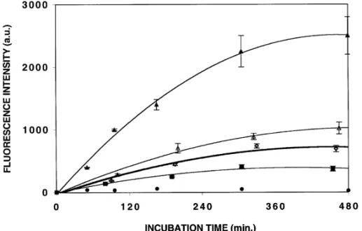
Fig. 3. Uptake of mTHPC(0.25mg/ml)encapsulated in NE(n), polox PLA NCs(e), PLA–PEG NCs(j), PLA NCs(d)and solution(m)by HT29 cells as determined by microspectrofluorimetry. The cellular fluorescence intensities were measured at 654 nm. For each experiment, data have been averaged from intensity values determined on 30 individual living cells. Experiments have been carried out in triplicate. Bars, SE.
Fig. 4. Survival fractions of HT 29 cells incubated(300 min.)with mTHPC encapsulated in PLA–PEG NCs(j), PLA NCs(d), polox PLA(e), NE
(n)and solution(m)and followed by exposure to 514 nm light(25 J/
cm2)
. Dark cytotoxicity was always negligible. Each experiment(4 wells/
experiment)has been carried out in triplicate. Bars, SE. chain attached were used. This could be considered as a first
indication that RES clearance could be limited by the use of such a carrier. It must be noted that the uptake rate determined with the solution was less than that of the naked PLA. This could be explained by a possible intermolecular interaction between mTHPC and PEG from the solvent.
Encapsulation also strongly affected the mTHPC uptake by HT29 cells(Fig. 3). Compared with the solution, the drug penetration was lowered whatever NCs were used. The uptake rate at 300 min, as determined from Fig. 3, was decreased by factors of 2.4, 3.1 and 5.4 with NE, polox PLA and PLA–PEG NCs, respectively. The time-course depend-ence of mTHPC uptake by HT29 cells was similar for all the formulations studied except for naked PLA. Surprisingly and unlike J774 cells, a specific behavior was observed in the case of PLA NCs. The drug was not internalized and only a very weak cytoplasmic fluorescence was observed.
When incubations were performed at 48C, the cellular fluorescence intensity became very weak whatever the for-mulation used and could not be determined accurately, sug-gesting an active uptake occurring via endocytosis.
3.2. Photocytotoxicity
None of the colloidal carriers used showed any dark cyto-toxicity in HT29 cells after incubation times of up to 18h. The phototoxicity of mTHPC was much less affected by encapsulation into colloidal carriers than by the drug inter-nalization process. As illustrated in Fig. 4, the phototoxicity of HT29 cells treated with mTHPC encapsulated into naked PLA, NE and polox PLA and exposed to 514 nm light with fluences of 25 J/cm2was quite similar, with a cell-survival
fraction of approximately 33% for a dose of 0.25mg/ml. It was only slightly reduced compared with that of the solution
(27%). A lower phototoxicity was found with mTHPC encapsulated into PLA–PEG NCs, with a cell-survival
frac-tion of 42%. For a dose of 1.25 mg/ml, the cell survival fractions were not significantly different with the various formulations studied(in the range 18–25%). It may be noted that the concentrations used in these experiments were lower than those generally used in the literature for phototoxicity determinations(severalmg/ml) and that the phototoxicity remained low when treated cells were exposed to fluences of 5 and 10 J/cm2.
and PLA–PEG NCs could be effective in limiting the RES uptake.
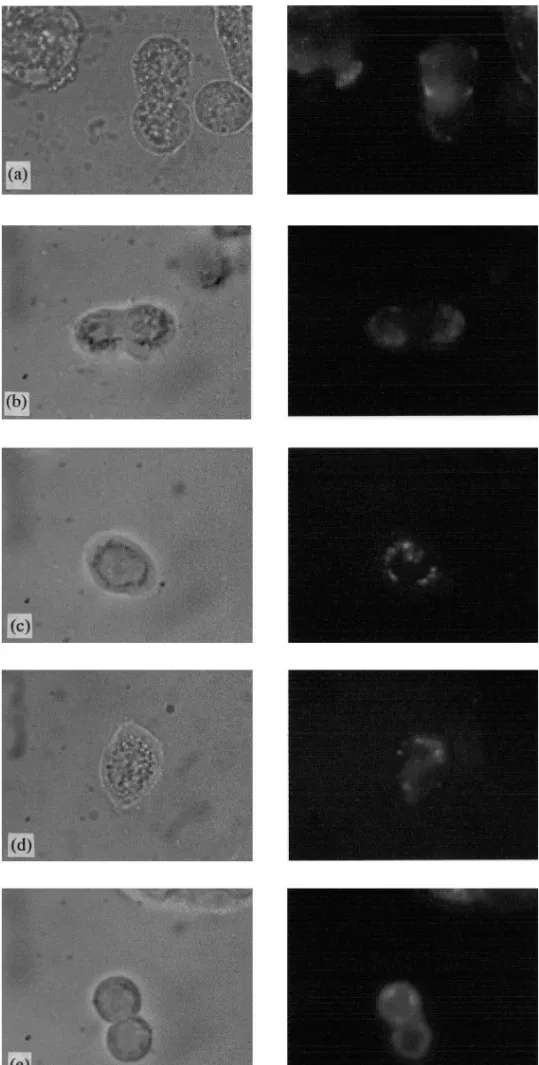
3.3. Subcellular localization in HT29 cells
The subcellullar localization of mTHPC in HT29 cells, as studied by fluorescence microscopy, was found to be for-mulation dependent(Fig. 5). The cells treated with mTHPC solution showed the fluorescence diffusively distributed throughout the cytoplasm, with a non-fluorescent nuclear area. Similar observations have been reported in V79 hamster cells[8]and in HT 29 cells[27].
The cellular distribution was markedly affected when col-loidal formulations were used. In all cases, the nuclear area remained non fluorescent. A punctate fluorescence pattern, mainly perinuclear, was seen when cells were treated with
mTHPC formulated in PLA–PEG and polox PLA NCs(Fig. 5(c),(d)). Separate incubations with specific probes give indications that these intense bright spots could arise from fluorophores localized into the Golgi system. A more accurate localization cannot be ascribed because of the specific HT29 cell morphology, which is quite thick with a small cytoplasm. Moreover, the simultaneous staining of subcellular organelles with specific probes and mTHPC was not possible in these experiments. The excitation was actually not selective enough to avoid a partial overlapping of the probe emission of high fluorescence quantum yield with that of mTHPC of low fluorescence quantum yield(0.08[28]).
In the case of PLA NCs, the fluorescence was mainly located on the external side of the plasma membrane, which is consistent with the results obtained by microspectrofluor-imetry. It may be pointed out that, surprisingly, the phototox-icity remains little affected.
As far as a specific subcellular localization of the photo-sensitizer was observed with NCs, it could be assumed that the type of photodamaged targets involved, and hence the mechanism and kinetics of cell death, are different. This could explain the absence of correlation between respective uptake rates determined with the various formulations used and the phototoxicity.
4. Conclusions
PLA–PEG and polox PLA NCs have been used for the first time as carriers for mTHPC, a photosensitizer used in PDT. First, the reduced mTHPC uptake by macrophage-like cells indicates that the NCs could limit the RES uptake. Secondly, although the encapsulation results in a lower mTHPC cellular uptake, the phototoxicity remains only slightly affected. The weak inhibition of phototoxicity by plasma proteins could be considered as a key factor for the in vivo distribution. Finally, the subcellular localization pattern observed suggests a dif-ferent drug targeting. Possibly difdif-ferent cell-death mecha-nisms could thus be involved.
These in vitro data clearly demonstrate that, as long as the colloidal carriers studied consist of an identical oily core and have a similar size, the modification of the nanocapsule sur-face is the main parameter to be considered in the interaction between encapsulated drug and cells. In vivo experiments are currently in progress to examine whether the photosensitizer biodistribution and photodynamic activity are modified by the use of PLA–PEG and polox PLA NCs as mTHPC delivery vehicles.
5. Abbreviations
PDT photodynamic therapy RES reticuloendothelial system PLA poly(D,Llactic acid)
PEG polyethylene glycol
mTHPC meta-tetra(hydroxyphenyl)chlorin PBS phosphate-buffered saline
HPLC high-performance liquid chromatography NCs nanocapsules
FCS fetal calf serum
Acknowledgements
This work was supported by the French Association for Cancer Research(Association pour la Recherche contre le Cancer, ARC). The authors want to thank Athena Kasselouri for HPLC measurements and Florence Robinet for her helpful technical assistance
References
[1] R.A. Hsi, D.I. Rosenthal, E. Glatstein, Photodynamic therapy in the treatment of cancer, Drugs 57(1999)725–734.
[2] M. Ochsner, Photophysical and photobiological processes in the pho-todynamic therapy of tumors, J. Photochem. Photobiol. B: Biol. 39
(1997)1–18.
[3] J. Schuitmaker, P. Baas, H.L.L.M. van Leengoed, F.W. van der Meulen, W.M. Star, N. van Zandwijk, Photodynamic therapy: a prom-ising new modality for the treatment of cancer, J. Photochem. Pho-tobiol. B: Biol. 34(1996)3–12.
[4] T.J. Dougherty, C.J. Gomer, B.W. Henderson, G. Jori, D. Kessel, M. Korbelik, J. Moan, Q. Peng, Photodynamic therapy, J. Natl. Cancer Inst. 90(1998)889–895.
[5] C.J. Gomer, Preclinical examination of first and second generation photosensitizers used in photodynamic therapy, Photochem. Photo-biol. 54(1991)1093–1107.
[6] E. Reddi, Role of delivery vehicles for photosensitizers in the photo-dynamic therapy of tumors, J. Photochem. Photobiol. B: Biol. 37
(1997)189–195.
[7]L. Morlet, V. Vonarx-Coinsmann, P. Lenz, M.T. Foultier, L. Xavier de Brito, C. Stewart, T. Patrice, Correlation between meta-tetra(hydroxyphenyl)chlorin(m-THPC)biodistribution and photo-dynamic effets in mice, J. Photochem. Photobiol. B: Biol. 28(1995)
25–32.
compari-son of its photobiolgical properties with those of two other photosensitizers, Int. J. Cancer 57(1994)883–888.
[9] T.M. Allen, Liposomes: opportunities in drug delivery, Drugs 54
(1997)8–14.
[10] G. Jori, E. Reddi, I. Cozzani, L. Tomio, Controlled targeting of dif-ferent subcellular sites by porphyrins in tumor-bearing mice, Br. J. Cancer 53(1986)615–621.
[11] G. Jori, Factors controlling the selectivity and efficiency of tumor damage in photodynamic therapy, Lasers Med. Sci. 5(1990)115– 120.
[12] U. Schmidt-Erfurth, H. Diddens, R. Birngruber, T. Hasan, Photody-namic targeting of human retinoblastoma cells using covalent low-density lipoprotein conjugates, Br. J. Cancer 75(1997)54–61.
[13] B. Ceh, M. Winterhalter, P.M. Frederik, J.J. Vallner, D.D. Lasic, Stealthwliposomes: from theory to product, Adv. Drug. Delivery 24 (1996)165–177.
[14] R. Gref, Y. Minamitaki, M.T. Perracchia, V. Trubetskov, V. Torchilin, R. Langer, Biodegradable long-circulating nanospheres, Science 263
(1994)1600–1603.
[15] R. Gref, A. Domb, P. Quellec, T. Blunk, R.H. Muller, J.M. Verbavatz, R. Langer, The controlled intravenous delivery of drugs using PEG-coated sterically stabilized nanospheres, Adv. Drug Delivery Rev. 16
(1995)215–233.
[16] A. Gabizon, Liposome circulation time and targeting: implication for cancer chemotherapy, Adv. Drug Delivery Rev. 16(1995)285–294.
[17] N. Oku, N. Saito, Y. Namba, H. Tsukada, D. Dolphin, S. Okada, Application of long-circulating liposomes to cancer photodynamic therapy, Biol. Pharm. Bull. 20(1997)670–673.
[18] E. Alleman, N. Brasseur, D. Benrezzak, J. Rousseau, S.V. Kudrevich,´ R.W. Boyle, J.C. Leroux, R. Gurny, J.E. Van Lier, PEG-coated poly(lactic acid)nanoparticles for the delivery of hexadecafluoro zinc phthalocyanine to EMT-6 mouse mammary tumors, J. Pharm. Phar-macol. 47(1995)382–387.
[19] E. Alleman, J. Rousseau, N. Brasseur, S.V. Kudrevich, K. Lewis, J.E.´ Van Lier, Photodynamic therapy of tumors with hexadecafluoro zinc phthalocyanine formulated in PEG-coated poly(lactic acid) nanopar-ticles, Int. J. Cancer 66(1996)821–824.
[20] H. Fessi, F. Puisieux, J. Ph. Devissaguet, N. Ammoury, S. Benita, Nanocapsule formation by interfacial polymer deposition following solvent displacement, Int. J. Pharm. 55(1989)R1–R4.
[21] V. Lenaerts, A. Labib, F. Chouinard, J. Rousseau, H. Ali, J. van Lier, Nanocapsules with reduced liver uptake: targeting of phthalocyanines to EMT-6 mouse mammary tumor in vivo, Eur. J. Biopharm. 41
(1995)38–43.
[22] V.C.F. Mosqueira, P. Legrand, R. Gref, B. Heurtault, M. Appel, G. Barrat, Interaction between macrophage cell line(J774A1)and sur-face modified poly(D,Llactide)nanocapsules bearing poly(ethylene glycol), J. Drug Targeting 7(1999)65–78.
[23] P. Quellec, R. Gref, L. Perrin, E. Dellacherie, F. Sommer, Y.M. Verbavatz, M-J. Alonso, Protein encapsulation within polyethylene glycol-coated nanospheres. I: physicochemical characterization, J. Biomed. Mater. Res. 42(1998)45–54.
[24]R.M. Jones, J.Q. Wang, J.H. Lamb, B.D. Djelal, R. Bonnett, C.K. Lim, Identification of photochemical oxidation products of 5,10,15,20-tetra(m-hydroxyphenyl)chlorin by on-line high-perform-ance liquid chromatography-electrospray ionization tandem mass spectrometry, J. Chromatogr. A722(1996)257–265.
[25] T. Mosmann, Rapid colorimetric assay for cellular growth and sur-vival: application to proliferation and cytotoxicity assays, J. Immunol. Methods 65(1983)55–63.
[26] J. Blais, C. Amirand, J.P. Ballini, P. Debey, M.T. Foultier, T. Patrice, Photofrin induced fluorescence in progressive and regressive murine colonic cancer cells: correlation with cell photosensitivity, J. Photo-chem. Photobiol. B: Biol. 27(1995)225–231.
[27] V.O. Melnikova, L.N. Bezdetnaya, C. Bour, E. Festor, M.-P. Gramain, J.-L. Merlin, A.Ya. Potapenko, F. Guillemin, Subcellular localization of in human tumor cells subjected to photodynamic treatment, J. Pho-tochem. Photobiol. B: Biol. 49(1999)96–103.
[28] R. Bonnett, P. Charlesworth, B.D. Djelal, S. Foley, D.J. Mc Garvey, T.G. Truscott, Photophysical properties of 5,10,15,20-tetrakis
(m-hydroxyphenyl)-porphyrin (m-THPP), 5,10,15,20-tetrakis
(m-hydroxyphenyl)-chlorin (m-THPC), and 5,10,15,20-tetrakis