*Correspondence: R. N. Marreto. Faculty of Pharmacy, Federal University of Goiás. Av. Universitária com 1º Avenida, s/n, Setor Universitário, 74605-220 - Goiânia - GO, Brazil. E-mail: [email protected]
A
vol. 48, n. 4, oct./dec., 2012
In vitro
skin penetration of clobetasol from lipid nanoparticles:
drug extraction and quantitation in different skin layers
Luís Antônio Dantas Silva, Stephânia Fleury Taveira, Eliana Martins Lima, Ricardo Neves Marreto
*Laboratory of Pharmaceutical Technology, Federal University of Goiás, Goiânia
Clobetasol propionate (CP) is a potent topical corticosteroid that causes several cutaneous and systemic side effects. In the present work, CP was encapsulated in nanostructured lipid carriers (NLCs) to increase drug retention in the outer skin layers and improve the safety of topical therapy. NLCs were prepared using a microemulsion technique with a mixture of lecithin, taurodeoxycholate, stearic acid, and oleic acid. In vitro penetration studies were performed in a modiied Franz-type diffusion cell, and porcine
ears were used as a model of human skin. A simple and sensitive liquid chromatographic method was developed and validated for clobetasol determination in different skin layers. NLCs presented uniform size distribution, high zeta potentialand entrapment eficiency values (> 98%). The analytical procedure was validated according to FDA guidelines. Clobetasol recoveries from skin samples were higher than 85%, with no interference of skin components and NLC ingredients. In experiments, after 6 h, a higher drug accumulation in the stratum corneum arising from NLCs compared to aqueous CP solution was observed. Thus, the NLCs demonstrated high potential for targeting CP to the skin and ensuring drug accumulation in the stratum corneum.
Uniterms: Clobetasol propionate/in vitro skin penetration. Nanostructured lipid carriers/in vitro study.
Proprionato de clobetasol (CP) é um potente corticóide tópico, que apresenta vários efeitos adversos cutâneos e sistêmicos. No presente trabalho, CP foi encapsulado em carreadores lipídicos nanoestruturados (NLCs) visando aumentar a retenção do fármaco nas camadas supericiais da pele e a segurança da terapia tópica. NLCs foram preparados usando a técnica de diluição de microemulsão com mistura de lecitina, taurodesoxicolato, ácido esteárico e ácido oléico. Estudos de penetração in vitro foram realizados em células de difusão de Franz modiicadas usando pele de orelha de porco como modelo de pele humana. Um método simples e sensível de cromatograia líquida foi desenvolvido e validado para a determinação de clobetasol nas diferentes camadas da pele. NLCs apresentaram distribuição de tamanho uniforme e valores elevados de potencial zeta e eiciência de encapsulação (>98%). O procedimento analítico foi validado de acordo com as diretrizes do FDA. A recuperação de clobetasol a partir das amostras de pele foi maior que 85%, sem interferência dos componentes da pele e dos excipientes das NLCs. Após 6 horas de experimento observou-se maior acúmulo do fármaco a partir das NLCs comparado à solução aquosa de CP. Dessa forma, as NLCs mostraram elevado potencial para direcionar o CP para a pele, pois elas possibilitaram o acúmulo do fármaco no estrato córneo.
Unitermos: Proprionato de clobetasol/penetração cutânea in vitro. Carreadores lipídicos nanoestruturados/ estudo in vitro.
INTRODUCTION
Corticosteroids have been successfully employed for the treatment of several inlammatory skin conditions.
are some of the possible side effects of topical corticoste-roids, which are related to the clinical potency of the drug (Horn et al., 2010).
Improvements in the safety of topical corticosteroid therapy may be achieved by drug nanoencapsulation, which can increase the drug’s accumulation in the skin while minimizing deep drug permeation and adverse sys-temic effects (Senyigit et al., 2010). CP has been nanoen-capsulated in lecithin/chitosan nanoparticles (Senyigit et al., 2010), polymer-coated lipid nanocapsules (Fontana et al., 2011), liposomes (Rao, Murthy, 2005), and lipid nanoparticles (Kalariya et al., 2005; Hu et al., 2006).
In the present work, CP was encapsulated in nano-structured lipid carriers (NLCs) to increase drug retention in the outer skin layers and avoid systemic absorption. NLCs are the second generation of lipid nanoparticles prepared with blends of solid lipids and oils (Pardeike, Homoss, Muller, 2009). These particles were developed to increase loading capacity and minimize expulsion of active compounds during storage, which are limitations of the irst generation of lipid nanoparticles. NLCs have attracted researchers’ attention due to their small size, high drug loading, and biocompatible and biodegradable lipid composition. NLCs were produced in this case using a simple microemulsion technique, which was previously employed by our research group to produce lipid nanopar-ticles containing the cytotoxic drug topotecan (Souza et al., 2011). It is important to note that several papers have reported that NLCs have enormous potential to improve skin penetration and the retention of drugs in the skin (Pardeike, Homoss, Muller, 2009).
Thus, the purpose of this work was to develop clobetasol-loaded NLCs to improve the accumulation of the drug in the outer skin layers. We also developed an extraction procedure and analytical method that is able to quantify CP in both viable and non-viable skin with no interference from skin components or NLC ingredients.
MATERIAL AND METHODS
Reagents
Clobetasol propionate (min. 98%) was purchased from Sigma-Aldrich (São Paulo, Brazil). Stearic acid (mp 58.77 °C, Vetec, Brazil), oleic acid (Sigma–Aldrich, Brazil), soy lecithin (Lipoid S100, Germany), and sodium taurodeoxycholate (Sigma–Aldrich, Brazil) were used for the preparation of the nanostructured lipid nanoparticles. Acetonitrile was purchased from J. T. Baker (Phillipsburg, USA). Water was puriied using a Milli-Q system (Mil -lipore, Billerica, USA) with a 0.22 μm pore end ilter.
Production and characterization of CP-loaded NLCs
CP-loaded NLCs were prepared using a microemul-sion technique. A mixture of lecithin, taurodeoxycholate, stearic acid, and oleic acid was heated, and CP was added to the melted material. Then, 250 µL of distilled water was added to the mixture under magnetic stirring, and a thermodynamically stable microemulsion system was formed. The microemulsion was dispersed into cold wa -ter (2–4 °C) under vigorous stirring (13,400 rpm for 10 min, IKA T25 Ultra-Turrax, Germany) using a 1:20 ratio (microemulsion:water) to form an NLC dispersion. The dispersion had a total lipid content of 2% (w/v), stabilized by 1.25% (w/v) of surfactant (lecithin: taurodeoxycholate, 4:1 ratio). The stearic acid: oleic acid ratio in the NLCs was 3:1 (Souza et al., 2011). The CP in the formulation comprised 4% (w/w) of the lipid content.
Dynamic light scattering was used to assess the mean particle size and polydispersity index (PdI) of the NLC dispersions. Samples were diluted (1:100) with purified water and analyzed using a Zetasizer Nano S (Malvern Instruments, UK). Zeta potential was determined from electrophoretic mobility using a Zetasizer Nano ZS (Malvern Instruments, UK).
Encapsulation eficiency (EE) was determined by the ultrafiltration technique (eq. 1). To isolate the en -trapped CP from free drug, 1 mL of freshly prepared NLC dispersion was subjected to ultrailtration (Vivaspin 2®, Sartorius, USA) for 60 min at 4,000 g at 4 °C. Filtrate con -taining the free drug was withdrawn for HPLC analysis as described below. All analyses were performed in triplicate. The total amount of drug incorporated in the NLC was determined by lipid disruption with acetonitrile followed by HPLC quantiication. Drug loading and drug recovery were calculated according eq. 2 and 3, respectively.
Eq. (1)
Eq. (2)
Eq. (3)
In vitro skin penetration studies of free and nanoencapsulated clobetasol
Franz-type diffusion cell (Microette; Hanson Research, Chatsworth, USA) (Figure 1). The receptor media was a 1% (w/v) sodium dodecyl sulfate (SDS) aqueous solution. The solubility of CP in the receptor medium was determined by addition of an excess of CP to the medium, followed by 24 h agitation at 37 °C, iltration and quantitation of the dissolved drug. The solubility of CP was 0.47 mg/mL. Solutions of CP in 1% (w/v) SDS aqueous solution or CP-loaded NLCs were added to the donor media. In both cases, 80 μg of CP was used in the donor cell to ensure sink conditions. The diffusion area available between the receptor and donor compartments was 1.86 cm2. The stir -ring rate and temperature were maintained at 300 rpm and 37 °C, respectively. Experiments were performed for 3 h and 6 h (n=6). After each experiment, the skin surface was washed with deionized water to remove excess CP on the skin surface. Washed skin was subjected to drug extraction and quantitation as described below.
Skin sample preparation
Pig ears were used as a model of human skin. The material was collected immediately after the slaughter of the animals. Ears were then dermatomed in the laboratory. Then, the dermatomized skin (700 μm thick) was kept at − 80 °C for a maximum of 30 days. After the in vitro pen-etration study, the stratum corneum (SC) was separated from the remaining skin (RS) using the tape-stripping technique. Skin was tape-stripped 15 times with adhesive tapes (Durex Original 500, 3M, Sumaré, Brazil). Tape strips with SC and RS were placed in individual test tubes for the extraction procedure.
CP extraction from SC and RS
For the CP recovery studies, blank samples of adhe-sive tapes with SC were spiked with a solution of CP in ace-tonitrile and allowed to dry. Dried tapes were then immersed in 5 mL of acetonitrile for 20 min to obtain a CP inal con
-centration of 50 μg/mL. The mixture was vortexed for 2 min and analyzed by HPLC-UV. The RS was also spiked with a solution of CP in acetonitrile. Following the evaporation of the solvent, 5 mL of acetonitrile was added to the tube. The RS was then processed with a tissue homogenizer (T25 ULTRA-TURRAX®, IKA, Staufen, Germany) for 2 min at 6,000 rpm, followed by bath sonication (USC 1400, Unique, Indaiatuba, Brazil) for 20 min. The skin homogenate was then iltered and analyzed by HPLC-UV. CP extraction from samples collected during the in vitro penetration studies was performed as described above (n=3).
Apparatus and chromatographic conditions
Samples were analyzed with an HPLC system con-sisting of an isocratic pump (ProStar 210, Varian, Palo Alto, USA), autosampler (ProStar 400, Varian, Palo Alto, USA), and UV detector (ProStar 325, Varian, Palo Alto, USA). Separation was achieved using an Agilent ZOR-BAX Eclipse XDB-C18 column (250 x 4.6 mm, 5 μm). The mobile phase was a 30:70 (v/v) mixture of water and acetonitrile. The low rate was 1.0 mL/min, the injection volume was 50 μL, and UV detection was carried out at 240 nm. Data acquisition was performed using Galaxie Chromatography Data System software.
Method validation
The method was developed and validated for linear -ity, accuracy, precision, and quantitation limit in accor-dance with the Food and Drug Administration Guidelines for the validation of analytical methods (FDA, 2000).
The selectivity was investigated against formulation ingredients, skin endogenous components, and adhesive tapes. Blanks of remaining skin and stratum corneum tape-stripped from six sources of skin were analyzed for the absence of interfering compounds at CP retention time. Precision (repeatability and intermediate precision) was expressed as the relative standard deviation (RSD%) of replicate measurements. Accuracy was calculated as the percentage difference between the measured and true values. Accuracy was expressed as the relative error of measurement (E%). Recovery was determined by comput -ing the ratio of the amount of CP extracted from spiked skin to the amount of CP added.
RESULTS AND DISCUSSION
Method development and validation
CP is the most important corticosteroid in
tological therapy; however, there are few reports on the quantitation of CP in skin matrices in the literature (Seny-igit et al., 2010; Fontana et al., 2011; Rao, Murthy, 2005; Kalariya et al., 2005; Weigmann et al., 1999; Mueller, An -issimov, Roberts, 2003), and there are no published data on the development and validation of an analytical procedure capable of quantifying CP in both the stratum corneum and remaining skin (viable epidermis plus dermis). Literature reports on the quantitation of CP in skin are based on iso-cratic HPLC-RP methods, and analytical separations have been performed with water and acetonitrile as the mobile phase. However, most of the reports, although focused in the application of the method, do not present details on method development or analytical validation data.
One of the challenges for quantifying drugs in biological matrices is the development of an eficient ex -traction procedure. This issue is particularly true for skin matrices, as the interactions among the analyte, skin lipids, and proteins may result in low drug recovery. High drug recovery values (above 90% from epidermis and dermis) have been reported in the literature (Senyigit et al., 2010). Weigmann et al. (1999) developed an exhaustive proce -dure for CP extraction from human stratum corneum. In the present work, the mean recovery rates of CP from por-cine ear skin were 101.48% (± 5.01) and 86.83% (± 5.20) for the stratum corneum and remaining skin, respectively, denoting the high eficiency of the extraction procedure employed. It is important to note that the procedure pre-sented here for CP extraction from the stratum corneum is simpler and less time consuming compared to some previously reported methods (Weigmann et al., 1999).
A simple method for the separation and quantitation of CP in the stratum corneum and remaining skin was also developed and validated. Chromatographic conditions re-sulted in a short run time, with a CP retention time of 6.10 min. The short run time is suitable for the routine analysis of a large number of samples from in vitro penetration studies. Typical chromatograms of stratum corneum and remaining skin, both spiked and not spiked with CP, are presented in Figure 2. As seen, the proposed method was able to quantify CP without interference from endogenous skin compounds and formulation ingredients, demonstrat-ing the speciicity of the method.
Validation of the analytical method was performed and linearity was found in the concentration range of 0.5-15 µg/mL (r=0.999). The detection limit (DL) was 25 ng/mL, and the quantiication limit (QL) was 100 ng/ mL, which may be considered suitable for CP quantiica -tion during in vitro penetration studies. Weigmann et al. (1999) reported a detection limit of 160 ng/mL, which is far superior to the one reported here.
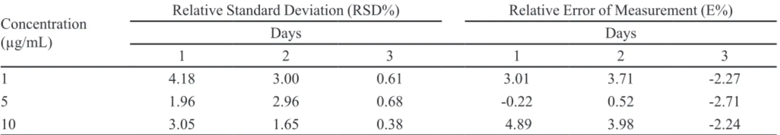
Accuracy and precision are presented in Table I. All values of accuracy and precision were within the accept-able limits, with E% and RSD% values no greater than 5% (FDA, 2000).
NLC characterization
NLCs were prepared with and without CP using the microemulsion technique; the characterization data are shown in Table II.
CP-loaded nanostructured lipid carriers (NLCs) presented low average diameters and low polydispersity index (PdI) values, denoting a uniform size distribution. No signiicant differences in mean diameter, PdI, or zeta potential were observed in NLCs with or without CP, suggesting that the addition of the drug did not alter the original characteristics of the nanoparticle dispersion.
The CP-NLCs presented low drug recovery (Table II) due to formulation adherence to the experimental ap-paratus. Therefore, the use of thermostatic devices is ad
-FIGURE 2 - Chromatograms obtained from the analysis of
TABLE I - Accuracy, repeatability, and intermediate precision of the HPLC method for determining the concentration of CP in organic solutions (n=3)
Concentration (µg/mL)
Relative Standard Deviation (RSD%) Relative Error of Measurement (E%)
Days Days
1 2 3 1 2 3
1 4.18 3.00 0.61 3.01 3.71 -2.27
5 1.96 2.96 0.68 -0.22 0.52 -2.71
10 3.05 1.65 0.38 4.89 3.98 -2.24
TABLE II - Mean diameter, polydispersity index (PdI), zeta potential, entrapment eficiency, drug loading, and drug recovery of
NLCs obtained with and without CP
Mean diameter
(nm) PdI
Zeta potential
(mV) EE% (w/w) DL% (w/w) DR% (w/w)
NLCs 195.5± 17.9 0.22 ±0.05 -49.8 ± 2.5 --- ---
---CP-NLCs 157.9 ± 19.7 0.19 ±0.01 -53.9 ± 1.1 98.6±0.4 2.34±0.1 62.2±3
visable for the transfer procedure to avoid process losses during industrial production of lipid nanoparticles.
Several papers have reported that zeta potential val-ues greater than 30 mV (in modulus) denote good electro-static repulsion between the nanoparticles, improving the physical stability of the system (Freitas, Mueller, 1998). Accordingly, the zeta potential values of the NLCs with and without CP (Table II) suggest that these nanoparticle dispersions have great physical stability. The entrapment eficiency was very high (Table II), which indicates that the lipids and surfactants employed were very suitable for CP nanoencapsulation.
The application of lipid nanoparticles to the skin has been related to several therapeutic beneits, such as the improvement of drug penetration, controlled release, and excellent tolerability (Pardeike, Homoss, Muller, 2009). Small nanoparticles, such as those reported here, ensure close contact of the drug formulation with the stratum corneum and exert occlusive effects, improving skin hydration and the penetration of the drug into the skin (Pardeike, Homoss, Muller, 2009).
In vitro skin penetration of free and CP-loaded lipid nanoparticles
The nanoparticle formulation was added to the donor compartment of a Franz-type cell apparatus (Figure 1), and CP skin penetration was determined.
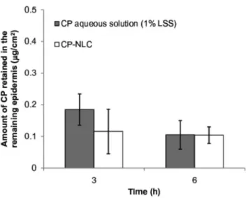
In accordance with theoretical expectations, we ob-served a higher CP accumulation in the stratum corneum
from lipid nanoparticles compared to accumulation from aqueous CP solution (sodium dodecyl sulfate, 1%, w/v) after 6 h of experiment (p<0.01) (Figure 3).In contrast, no signiicant differences were observed in SC samples after 3 hours during the experiment (Figure 3).
Several studies have demonstrated that NLCs can increase drug penetration and retention in the skin, likely due to the occlusive effect caused by the formation of an intact ilm on the skin surface (Marquelle-Oliveira et al., 2010). In addition, NLCs may interact with lipids in the outermost skin layers, resulting in stratum corneum luidi -zation and increased drug penetration. Nanoparticles may
FIGURE 3 - In vitro clobetasol penetration in porcine ear skin
also accumulate in skin appendages, such as hair follicles, which can act as a long-term reservoir for drug release (Knorr et al., 2009). Based on the results presented here, it appears that CP-NLCs accumulate on the skin surface, promoting a controlled release for periods longer than 3 hours. It is important to note that no CP was detected in the receptor media for all formulations tested after 3 or 6 h of the in vitro penetration experiment.
There was no signiicant difference in CP accumula -tion in the remaining skin 3 or 6 hours after the applica-tion of the drug-loaded nanoparticles or aqueous dispersions (Figure 4). Despite this observation, CP nanoencapsulation demonstrated a high potential to improve topical therapy, as the stratum corneum may act as a drug reservoir, and CP higher accumulation in this layer may lead to high CP concentrations in the remaining skin during chronic administration schedules. Indeed, nanoencapsulation may signiicantly increase the concentration of drug available for skin penetration, particularly for drugs with poor aque-ous solubility, which dissolve only a small amount in con-ventional topical vehicles, such as gels and creams. The preparation of higher drug-loading CP lipid nanoparticles is currently under investigation in our laboratory.
CONCLUSIONS
The NLCs obtained in this work by the microemul -sion technique demonstrated high potential for targeting CP to the skin, as they provide drug accumulation in the stratum corneum. We also demonstrated the suitability of the analytical method developed here for determining
CP concentration in two different skin layers, the stratum corneum and remaining skin. The method demonstrated high sensitivity, speciicity, and drug recovery.
ACKNOWLEDGEMENTS
This work was supported by Conselho Nacional de Pesquisa e Desenvolvimento (CNPq- MCTI-Brasil).
REFERENCES
ECHEVARRÍA, L.; BLANCO-PRÍETO, M.J.; CAMPANERO, M.A.; SANTOYO, S.; YGARTUA, P. Development and validation of a liquid chromatographic method for in vitro mupirocin quantiication in both skin layers and
percutaneous penetration studies. J. Chromatogr. B.,v.796,
n.2, p.233-241, 2003.
FOOD AND DRUG ADMINISTRATION. Guidance for Industry. Analytical procedures and methods validation:
chemistry, manufacturing, and controls documentation. Rockville: US Food and Drug Administration, 2000. 33 p.
FONTANA, M.C.; REZER, J.F.P.; CORADINI, K.; LEAL, D.B.R. Improved efficacy in the treatment of contact dermatitis in rats by a dermatologicalnanomedicine
containing clobetasol propionate. Eur. J. Pharm. Biopharm.,
v.79, n.2, p.241-249, 2011.
FREITAS, C.; MULLER, R.H. Effect of light and temperature on zeta potential and physical stability in solid lipid
nanoparticle (SLN™) dispersions. Int. J. Pharm., v.168,
n.2, p.221-229, 1998.
HORN, E.J.; DOMM, S.; KATZ, H.I.; LEBWOHL, M.; MSOWIETZ, V. KRAGBALLE, K. Topical corticosteroids
in psoriasis: strategies for improving safety. J. Eur. Acad.
Dermatol. Venearol., v.24, n.2, p.119-124, 2010.
HU, F.Q.; JIANG, S.P.; DU, Y.Z.; YUAN, H.; YE, Y. Q.; ZENG, S. Preparation and characteristics of monostearin
nanostructured lipid carriers. Int. J. Pharm., v.314, n.1,
p.83-89, 2006.
KALARIYA, M.; PADHI, B.K.; CHOUGULE, M.; MISRA, A. Clobetasol propionate solid lipid nanoparticles cream for effective treatment of eczema: formulation and clinical
implications. Indian J. Exp. Biol., v.43, n.3, p.233-240,
2005.
FIGURE 4 - In vitro clobetasol penetration in the viable
KNORR, F.; LADEMANN, J.; PATZELT, A.; STERRY, W.; BLUME-PEYTAVI, U.; VOGT, A. Follicular transport
route – research progress and future perspectives. Eur. J.
Pharm. Biopharm., v.71, n.2, p.173-180, 2009.
MARQUELE-OLIVEIRA, F.; SANTANA, D.C.A.; TAVEIRA, S.F.; VERMEULEN, D.M.; OLIVEIRA, A.R.M.; SILVA, R.S.; LOPEZ, R.F.V. Development of nitrosyl ruthenium complex-loaded lipid carriers for topical administration: Improvement in skin stability and in nitric oxide release by visible light irradiation. J. Pharm. Biol. Anal., v.53, n.4,
p.843-851, 2010.
MUELLER, B.; ANISSIMOV, Y.G.; ROBERTS, M.S. Unexpected clobetasol propionate proile in human stratum corneum after topical application in vitro. Pharm. Res., v.20, n.11, p.1835-1837, 2003.
PARDEIKE, J.; HOMMOSS, A.; MULLER, R.H. Lipid nanoparticles (SLN, NLC) in cosmetic and pharmaceutical
dermal products. Int. J. Pharm., v.366, n.1-2, p.170-184,
2009.
RAO, G.; MURTHY, R.S.R. Evaluation of liposomal clobetasol propionate topical formulation for intra-dermal delivery.
Indian J. Pharm. Sci., v.62, n.6, p.459-462, 2000.
SENYIGIT, T.; SONVICO, F.; BARBIERI, S.; OZER, O.; SANTI, P.; COLOMBO, P. Lecithin/chitosan nanoparticles of clobetasol-17-propionate capable of accumulation in pig
skin. J. Control. Release, v.142, n.3, p.368-373, 2010.
SOUZA, L.G.; SILVA, E.J.; MARTINS, A.L.L.; MOTA, M.F.; BRAGA, R.C.; LIMA, E.M.; VALADARAES, M.C.; TAVEIRA, S.F.; MARRETO, R.N. Development of topotecan loaded lipid nanoparticles for chemical
stabilization and prolonged release. Eur. J. Pharm.
Biopharm., v.79, n.1, p.189-196, 2011.
WEIGMANN, H.J.; LADEMANN, J.; PELCHRZIM, R.V.; STERRY, W.; HAGEMEISTER, T.; MOLZAHN, R.; SCHAEFER, M.; LINDSCHEID, M.; SCHAEFER, H.; SHAH, V.P. Bioavailability of clobetasol propionate: quantiication of drug concentrationsin the stratum corneum
by dermatopharmacokinetics using tape stripping. Skin
Pharmacol. Appl. Skin Physiol., v.12, n.12, p.46-53, 1999.
ZHANG, J.; SMITH, E. Percutaneous permeation of betamethasone 17-valerate incorporated in lipid
nanoparticles J. Pharm. Sci., v.100, n.3, p.896-903, 2011.
Received for publication on 05th July 2012