Braz. J. Vet. Parasitol., Jaboticabal, v. 27, n. 1, p. 112-117, jan.-mar. 2018 Doi: http://dx.doi.org/10.1590/S1984-296120180012
This is an Open Access article distributed under the terms of the Creative Commons Attribution License, which permits unrestricted use, distribution, and reproduction in any medium, provided the original work is properly cited.
Introduction
Cryptosporidium spp. is a cosmopolitan protozoon described by Tizzer in 1907. However, its pathogenic relevance was only acknowledged during the 1970s, as an important etiology for
diarrhea among cattle (O’DONOGHUE, 1995). Subsequently, this protozoon was reported as a cause of opportunistic infection in immunocompromised HIV-infected humans, and chronic diarrhea caused by Cryptosporidium spp. was noted to be a factor indicative for AIDS (ANGUS, 1990).
Cryptosporidium spp. is considered to be a pathogenic and infectious enteric agent, and it affects various host species, including mammals, reptiles, fish and birds (GUERRANT, 1997; HUNTER
Risk factors and infection due to
Cryptosporidium
spp. in
dogs and cats in southern Rio Grande do Sul
Fatores de risco e infecção por
Cryptosporidium
spp. em cães e gatos no sul do Rio Grande do Sul
Andrios da Silva Moreira1; Cristiane Telles Baptista1; Carolina Litchina Brasil1; Júlia de Souza Silveira Valente1;
Fábio Raphael Pascoti Bruhn2; Daniela Isabel Brayer Pereira1*
1 Laboratório de Micologia, Programa de Pós-graduação em Parasitologia, Departamento de Microbiologia e Parasitologia, Instituto
de Biologia, Universidade Federal de Pelotas – UFPel, Pelotas, RS, Brasil
2 Centro de Controle de Zoonoses, Departamento de Medicina Veterinária Preventiva, Faculdade de Veterinária, Universidade Federal
de Pelotas – UFPel, Pelotas, RS, Brasil
Received September 13, 2017 Accepted February 1, 2018
Abstract
This study investigated the frequency of oocysts of Cryptosporidium spp. in feces from dogs and cats in five municipalities in the southern region of the state of Rio Grande do Sul. The risk factors associated with infection were also investigated. Feces samples from 110 dogs and 18 cats were stained using the auramine method. At the time of feces sampling, a questionnaire with semi-open-ended questions was applied to the animal guardians and all data obtained underwent statistical analysis. The real frequency of oocysts of Cryptosporidium spp. was 24.63% (27 dogs and two cats). Only four samples of dog feces were diarrheic and no presence of oocysts was observed in any of them. Variables that represented risk factors for infection were: homemade food, untreated water, circulation of animals on grassy terrain and living in the same environment as other animals (cattle). The results made it possible to inferring that within the population studied, the frequency of parasitism due to Cryptosporidium spp. in dogs was relevant and emphasize the asymptomatic nature of this infection. The adopting control measures are highlighted, particularly in relation to variables that represent risk factors for this infection.
Keywords: Cryptosporidiosis, diarrhea, auramine, pets.
Resumo
Este estudo verificou a frequência de oocistos de Cryptosporidium spp. em fezes de cães e gatos em cinco municípios da região sul do Rio Grande do Sul e fatores de risco associados à infecção. Amostras de fezes de 110 cães e 18 gatos foram coradas pelo método de auramina. No momento da coleta de fezes aplicou-se um questionário aos tutores dos animais com questões semiabertas e os dados foram submetidos à análise estatística. A frequência verdadeira de oocistos de Cryptosporidium spp. foi de 24,63% (27 cães e 2 gatos). Apenas quatro amostras de fezes caninas eram diarreicas e todas sem oocistos. As variáveis que representaram fatores de risco para a infecção foram: alimentos de preparo caseiro, água não tratada, circulação dos animais em terreno gramíneo e convivência com outros animais, principalmente bovinos. Os resultados sugerem que a frequência de cães parasitados por Cryptosporidium spp. é relevante, reforçando o caráter assintomático da infeção. Destaca-se a importância da adoção de medidas de controle, particularmente das variáveis que representaram fatores de risco à infecção.
Palavras-chave: Criptosporidiose, diarreia, auramina, pets.
*Corresponding author: Daniela Isabel Brayer Pereira. Departamento de Microbiologia e Parasitologia, Instituto de Biologia, Universidade Federal de Pelotas – UFPel, Prédio 18, Sala 14, Campus Universitário Capão do Leão, s/nº, CEP: 96160-000, Pelotas, RS, Brasil.
& THOMPSON, 2005; THOMPSON et al., 2005). Oocysts of Cryptosporidium spp. excreted by infected hosts are sporulated and infectious, and they are resistant to environmental conditions. Infections are acquired through ingestion or inhalation of infectious oocysts (O’DONOGHUE, 1995), and can be transmitted by means of direct host-to-host contact or indirect contact, through food and water.
Studies have shown that cryptosporidiosis in humans can be caused by various genotypes and/or species of Cryptosporidium, including C. parvum, C. canis and C. felis. These last two species alone are responsible for a large proportion of infections in developing countries (XIAO & FENG, 2008). In turn, C. parvum can infect both dogs and humans, and is more recurrent in humans than C. canis (SEVÁ et al., 2010).
Dogs and cats can act as potential transmitters of zoonotic parasites (PEREIRA et al., 2011). In this regard, close contact between dogs and cats and humans increases the risk of transmission of these pathogens. Additionally, feces deposited in public grounds, such as parks and gardens, represent a risk to human health because of the high level of fecal-environmental contamination (ZANZANI et al., 2014). Reports on zoonotic transmission of cryptosporidiosis involving contact with symptomatic or asymptomatic pets or production animals that excreted infecting oocysts have been published (ANGUS, 1983; XIAO & FENG, 2008; BESER et al., 2015).
In order to maintain control over cryptosporidiosis, it is important to establish a specific diagnosis that makes it possible to analyze risk factors, plan interventions and identify outbreaks of the disease (CHALMERS & KATZER, 2013). Among the diagnostic methods available for detecting oocysts in hosts’ feces, the fluorescence staining technique using auramine has shown the highest affinity for oocyst walls, and this method has been considered sensitive and rapid (McPHERSON & McQUEEN, 1993; HANSCHEID et al., 2008). It has thus been used in epidemiological surveys on cryptosporidiosis (QUADROS & ARAÚJO, 2003; QUADROS et al., 2006; ARTIEDA et al., 2012).
The present study investigated the frequency of oocysts of Cryptosporidium spp. in dog and cat feces in municipalities in the southern region of the state of Rio Grande do Sul, and evaluated risk factors associated with the infection.
Material and Methods
From June to September 2016, feces from 110 dogs and 18 cats were sampled in the municipalities of Pelotas (n = 93), Aceguá (n = 3), Hulha Negra (n = 16), Candiota (n = 7) and Piratini (n = 9). Sampling was taken individually for each animal by its guardian, soon after evacuation in properly identified sterile flasks. Then the feces were sent to the laboratory and stored under refrigeration until processing.
At the time of sampling, semi-open-ended questionnaires were applied to the animals’ guardian to obtain the following information: animal’s age, type of feeding, water source, type of housing, type of ground, access to streets, interaction with other animals and presence of diarrhea.
The feces analyzed firstly underwent removal of dirt. Approximately two grams of each sample were diluted in 10 ml of distilled
water and underwent gauze filtration followed by centrifugation for 3 minutes at 3000 rpm. This process was repeated until the supernatant was totally clean. The feces were then transferred into Falcon tubes and were processed using the protocol developed by Ritchie and modified by Young et al. (1979). The sediment was diluted in 2 ml of 10% formalin and 3 ml of ethyl acetate was added. This mixture was centrifuged at 3000 rpm for 10 minutes and the supernatant was discarded. A 10 μl aliquot of sediment was used to prepare smears, which were then stained using the phenolic auramine ‘O’ staining technique (SMITHWICK, 1976). Two slides were made for each sample analyzed.
The SPSS 20.0 software (IBM, 2011) was used to conduct statistical analysis on the data obtained. Presence or absence of oocysts was taken to be the dependent variable and information of epidemiological nature, independent variables. The chi-square test (x2) with a significance level of p < 0.05 and odds ratios (OR) with
a 95% confidence interval, were used to estimate risk. Variables showing associations at the level of p < 0.2 through the x2 test or
Fisher’s exact test were selected in order to construct a multiple logistic regression model, along with calculation of the adjusted OR and its 95% confidence interval (CI95%). A minimum confidence level of 95% was used for all statistical analyses. Estimates of real frequencies were made based on the sensitivity and specificity values of the auramine technique (sensitivity = 92.1%; specificity = 100%), as previously described by Chalmers et al. (2011). The data were calculated based on the method described by Reiczigel et al. (2010), using the EpiTools epidemiological calculators (SERGEANT, 2017).
Results
The real frequency of presentation of oocysts of Cryptosporidium spp. in feces among these animals was 24.63% (CI95% = 17.6;33.3%), including 27 dogs and two cats. The real frequency among dogs was 26.68% ( CI95% = 18.97;36.26%) and among cats, 12.08% (CI95% = 0.33;35.65%). Only four dog feces samples were considered to be diarrheic and none of them presented oocysts of Cryptosporidium spp.
Table 1 shows the results from the analysis on the data extracted from the questionnaires and the univariate analysis on risk factors. It was seen that animals that were exclusively fed with homemade food (p = 0.004; OR = 3.533; CI95% = 1.442;8.652) and those that only circulated on grassy terrain (p = 0.021; OR = 2.778; CI95% = 1.147;6.728) presented higher chances of infection by oocysts of Cryptosporidium spp. Interaction with other animals, notably cattle, was also a risk factor (p = 0.010; OR = 3.333; CI95% = 1.366;8.136). Animals that had access to treated water (p = 0.007; OR = 0.291; CI95% = 0.115;0.738) presented lower chances of infection. Additionally, in the present study, the variable of “rural area” did not act as a risk factor for infection by Cryptosporidium spp. (p = 0.071).
Table 1. Univariate analysis on the variables analyzed and their relationship with infection by Cryptosporidium spp. in dogs (n = 110) and cats (n = 18) in the southern region of the state of Rio Grande do Sul, Brazil.
Variable (+) (-) Total % (+) p OR CI95%
Age*
0-1 year old 4 12 16 14.8 1.0 0.908 [0.267;3.087]
>1 year old 23 76 99 85.2
Feeding: feed
Yes 10 44 54 37.0 0.238 0.588 [0.243;1.426]
No 17 44 61 63.0
Feeding: homemade food
Yes 15 23 38 55.6 0.004 3.533 [1.442;8.652]
No 12 65 77 44.4
Feeding: feed + homemade food*
Yes 2 21 23 7.4 0.096 0.255 [0.056;1.169]
No 25 67 92 92.6
Water
Untreated 19 36 55 70.4 0.007 0.291 [0.115;0.738]
Treated 8 52 60 29.6
Housing: apartment*
Yes 1 9 10 3.7 0.448 0.333 [0.040;2.758]
No 26 78 104 96.3
Housing: house
Yes 5 27 32 18.5 0.232 0.505 [0.173;1.475]
No 22 60 82 81.5
Housing: rural area
Yes 21 51 72 77.8 0.071 2.471 [0.907;6.733]
No 6 36 42 22.2
Type of ground: grass
Yes 15 27 42 55.6 0.021 2.778 [1.147;6.728]
No 12 60 72 44.4
Type of ground: paved
Yes 6 35 41 22.2 0.089 0.424 [0.156;1.158]
No 21 52 73 77.8
Type of ground: grass+ paved
Yes 6 25 31 22.2 0.506 0.709 [0.256;1.963]
No 21 62 83 77.8
Access to streets
Yes 19 53 33 65.5 0.612 1.255 [0.522;3.015]
No 10 35 72 34.5
Interaction with other animals
Yes 19 62 81 70.4 0.993 0.996 [0.387;2.561]
No 8 26 34 29.6
Interaction with other animals: cats*
Yes 3 15 18 15.8 0.535 0.525 [0.134;2.059]
No 16 42 58 84.2
Interaction with other animals: dogs*
Yes 16 42 58 84.2 0.535 1.905 [0.486;7.472]
No 3 15 18 15.8
Interaction with other animals: cattle
Yes 15 24 39 55.6 0.010 3.333 [1.366;8.136]
No 12 64 76 44.4
Presence of diarrhea*
Yes 0 4 4 0.0 0.571 0.757 [0.681;0.841]
No 27 84 111 24.3
Discussion
Infection by Cryptosporidium spp. is an important health problem in developed and developing countries and may represent a risk to life among immunocompromised hosts, children and young animals (ADAMU et al., 2010).
Companion animals are important for maintenance of the disease, given that they can be asymptomatic carriers of the protozoon. Additionally, the increasing sizes of pet populations in urban zones have substantially contributed towards environmental contamination (ZANZANI et al., 2014).
A few studies have been conducted in Brazil with the aim of investigating the prevalence, frequency or occurrence of infection by Cryptosporidium spp. among dogs, cats and livestock (FIGUEIREDO et al., 2004; EDERLI et al., 2005; MOURA et al., 2009; GALVÃO et al., 2012). However, studies involving this infection in companion animals in the southern region of the state of Rio Grande do Sul are scarce.
In the present study, the real frequency of parasitized animals was 24.6%. This result differs from the findings of Quadros et al. (2006), who found that only 10% of the dogs evaluated in the municipality of Lages, state of Santa Catarina, were parasitized with oocysts of Cryptosporidium spp. It can be suggested that this difference is due to the origin of the dogs, since those authors only evaluated samples from dogs living in urban areas. In the present study, 72.5% (21/29) of the animals with presence of oocysts were from “rural areas”. Although “rural area” did not behave as a risk factor in this study (p = 0.071), this factor is closely correlated with consumption of untreated water, living in the same environment as
cattle, and circulation in grassy terrain, which were variables that behaved as risk factors in the present study. On the other hand, similar frequencies were described by Pereira et al. (2011), who reported that oocysts of this protozoon were present in 29.5% of the dogs and 24.7% of the cats that were evaluated in their study. In addition, Balassiano et al. (2009) found oocysts in 26.2% of the dogs that they studied in the municipality of Rio de Janeiro.
No association between host age and infection was observed (p = 0.877), and this finding was similar to what had previously been described by Pivoto et al. (2013). However, the percentage of animals under one year of age that presented oocysts in their feces (25%) was slightly higher than the percentage of animals that were one year old or over (23.2%) (Table 1). Similar results were reported by Olabanji et al. (2016), who found that this variable did not represent a risk factor, although there was higher prevalence of infection among dogs that were between three and six months old. However, Bresciani et al. (2008) described higher occurrence of infection among animals that were between one and four years old. Thompson et al. (2005) stated that, although infections in adult animals may be recurrent, the frequency of infection is higher in young animals.
In the present study, the source of consumed water and the type of feeding behaved as factors associated with infection. These results differ from the ones found by Balassiano et al. (2009), Ederli et al. (2005), Pivoto et al. (2013) and Awadallah & Salem (2015), who did not find any association between infection and water source or type of feeding. However, our findings were in accordance with the results found by Moura et al. (2009). Based on the analysis conducted in the present study, it can be suggested that feeding animals exclusively on homemade food may act as a risk factor for infection by Cryptosporidium spp. It is believed that homemade food may be more susceptible to environmental contamination, thus increasing the chances of infection, as suggested by Moura et al. (2009).
The evaluation on the type of housing showed that this variable did not present any association with infection. However, even though the variable of “rural area” was not statistically associated with infection (p = 0.071), other factors relating to rural areas, including “untreated water sources”, “interaction with cattle” and “circulation in grassy terrain”, were significant and therefore acted as risk factors for infection. Likewise, in a previous study, Pivoto et al. (2013) did not observe any association with the variable of rural area. On the other hand, Awadallah & Salem (2015) reported that 40% of the dogs in rural areas of Egypt were infected with intestinal parasites. They correlated this variable with a high risk of infection. Thompson et al. (2016) stated that the high risk of infection in rural areas was due to low hygiene conditions, living in the same environment as wild species and animals that act as reservoirs, and low levels of veterinary attention to dogs.
The variable of access to streets did not present any significant value in the present study (p = 0.612), and this was concordant with the findings of Balassiano et al. (2009) and Pivoto et al. (2013). However, Olabanji et al. (2016) observed a relationship between this variable and infection, and stated that dogs that were free to roam the streets presented higher exposure to oocysts because they were in contact with contaminated environments and interacted with other infected animals.
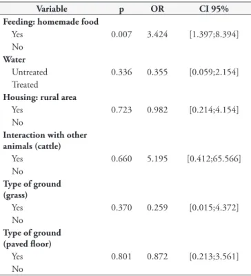
Table 2. Multiple binary logistic regression analysis on the variables analyzed and their relationship with infection by Cryptosporidium spp. in dogs (n = 110) and cats (n = 18) in the southern region of the state of Rio Grande do Sul, Brazil.
Variable p OR CI 95%
Feeding: homemade food
Yes 0.007 3.424 [1.397;8.394]
No Water
Untreated 0.336 0.355 [0.059;2.154] Treated
Housing: rural area
Yes 0.723 0.982 [0.214;4.154]
No
Interaction with other animals (cattle)
Yes 0.660 5.195 [0.412;65.566]
No
Type of ground (grass)
Yes 0.370 0.259 [0.015;4.372]
No
Type of ground (paved floor)
Yes 0.801 0.872 [0.213;3.561]
In comparing interactions with other animals (dogs and cats), the results did not show any significant difference (p = 0.535), thus agreeing with the results from Balassiano et al. (2009) and Pivoto et al. (2013). However, a difference was observed when the animals lived in the same environment as other animal species, especially cattle (p = 0.010). Cattle are the main hosts of C. parvum, although C. canis and C. felis have been sporadically reported in bovine infections (THOMPSON & SMITH, 2011).
Access of animals to environments with the presence of grass behaved as a risk factor and had the capacity to favor infections. On the other hand, “paved floor” behaved as a protection factor. Similar results were described by Silva et al. (2011) in analyzing these variables in relation to calf rearing, and by Kiani et al. (2016). These authors stated that moisture retention by grassy soils increased the duration of oocyst viability.
According to Fayer et al. (1997), diarrhea is the most common symptom of cryptosporidiosis in animals and humans, and a large number of oocysts are excreted in hosts’ feces during acute infection. Pereira et al. (2011) observed high rates of infection in diarrheic feces. However, in the present study, occurrences of diarrhea did not seem to be relevant, since there was no presence of oocysts of Cryptosporidium spp. in the diarrheic feces samples analyzed. Nevertheless, it is important to highlight the fact that the number of diarrheic feces samples analyzed was small (n = 4). Thus, evaluating a larger number of animals with clinical symptoms is necessary in order to obtain a better analysis on the results obtained. Despite this, the results obtained in the present study reinforce the idea that this infection in companion animals is characteristically asymptomatic.
Conclusion
This study is the first to investigate the infection by Cryptosporidium spp. among dogs and cats in the southern region of the state of Rio Grande do Sul. The results made it possible to inferring that within the animal population studied, the frequency of parasitism by Cryptosporidium spp. in dogs was relevant and emphasize the asymptomatic nature of this infection. The adopting control measures are highlighted, particularly in relation to variable homemade food that represented a risk factor for infection by Cryptosporidium spp.
Acknowledgements
The authors thank the “Coordenação de Aperfeiçoamento de Pessoal de Nível Superior (CAPES)” for financial support, and the “Hospital de Clínicas Veterinárias” of the “Universidade Federal de Pelotas” for providing feces samples.
References
Adamu H, Petros B, Hailu A, Petry F. Molecular characterization of
Cryptosporidium isolates from humans in Ethiopia. Acta Trop 2010;
115(1-2): 77-83. http://dx.doi.org/10.1016/j.actatropica.2010.02.003. PMid:20206592.
Angus KW. Cryptosporidiosis in man, domestic animals and birds: a review. J R Soc Med 1983; 76(1): 62-70. PMid:6338228.
Angus KW. Cryptosporidiosis and AIDS. Baillieres Clin Gastroenterol
1990; 4(2): 425-441. http://dx.doi.org/10.1016/0950-3528(90)90010-E. PMid:2282384.
Artieda J, Basterrechea M, Arriola L, Yagüe M, Albisua E, Arostegui N, et al. Outbreak of cryptosporidiosis in a child day-care centre in Gipuzkoa, Spain, October to December 2011. Euro Surveill 2012; 17(5): 1-3. http://dx.doi.org/10.2807/ese.17.05.20070-en. PMid:22321139.
Awadallah MAI, Salem LMA. Zoonotic enteric parasites transmitted from dogs in egypt with special concern to Toxocara canis infection. Vet World
2015; 8(8): 946-957. http://dx.doi.org/10.14202/vetworld.2015.946-957. PMid:27047182.
Balassiano BCC, Campos MR, Menezes RCAA, Pereira MJS. Factors associated with gastrointestinal parasite infection in dogs in Rio de Janeiro, Brazil. Prev Vet Med 2009; 91(2-4): 234-240. http://dx.doi. org/10.1016/j.prevetmed.2009.05.030. PMid:19577316.
Beser J, Toresson L, Eitrem R, Troell K, Winiecka-Krusnell J, Lebbad M. Possible zoonotic transmission of Cryptosporidium felis in a household.
Infect Ecol Epidemiol 2015; 5(1): 28463. http://dx.doi.org/10.3402/iee.
v5.28463. PMid:26446304.
Bresciani KDS, Amarante AFT, Lima VMF, Marcondes M, Feitosa FLF, Táparo CV, et al. Infecções por Cryptosporidium spp. em cães de Araçatuba, SP, Brasil. Vet Zootec 2008; 15(3): 466-468.
Chalmers RM, Campbell BM, Crouch N, Charlett A, Davies A. Comparison of diagnostic sensitivity and specificity of seven Cryptosporidium assays used in the UK. J Med Microbiol 2011; 60(11): 1598-1604. http://dx.doi. org/10.1099/jmm.0.034181-0. PMid:21757501.
Chalmers RM, Katzer F. Looking for Cryptosporidium: the application of advances in detection and diagnosis. Trends Parasitol 2013; 29(5): 237-251. http://dx.doi.org/10.1016/j.pt.2013.03.001. PMid:23566713. Ederli BB, Rodrigues MFG, Carvalho CB. Oocysts of the genus
Cryptosporidium in domiciliated dogs from the city of Campos dos
Goytacazes, the state of Rio de Janeiro. Rev Bras Parasitol Vet 2005; 14(3): 129-131. PMid:16229758.
Fayer R, Speer CA, Dubey JP. The general biology of Cryptosporidium. In: Fayer R. Cryptosporidium and Cryptosporidiosis. Boca Raton: CRC Press; 1997. p. 1-42.
Figueiredo HCP, Pereira DJ Jr, Nogueira RB, Costa PRS. Cryptosporidium
parvum oocyst excretion in healthy dogs from the cities of Lavras and
Viçosa, Minas Gerais State, Brazil. Cienc Rural 2004; 34(5): 1625-1627. http://dx.doi.org/10.1590/S0103-84782004000500049.
Galvão ALB, Ortiz EG, Bresciani KDS, Ferreira GS, Vasconcellos AL, Vieira MC. Importância da Criptosporidiose como Zoonose. Arch Vet Sci 2012; 17(2): 18-28. http://dx.doi.org/10.5380/avs.v17i2.21556. Guerrant RL. Cryptosporidiosis: an emerging, highly infectious threat. Emerg Infect Dis 1997; 3(1): 51-57. http://dx.doi.org/10.3201/ eid0301.970106. PMid:9126444.
Hanscheid T, Cristino JM, Salgado MJ. Screening of auramine-stained smears of all fecal samples is a rapid and inexpensive way to increase the detection of coccidial infections. Int J Infect Dis 2008; 12(1): 47-50. http://dx.doi.org/10.1016/j.ijid.2007.04.008. PMid:17600749.
Hunter PR, Thompson RCA. The zoonotic transmission of Giardia and
Cryptosporidium.Int J Parasitol 2005; 35(11-12): 1181-1190. http://
dx.doi.org/10.1016/j.ijpara.2005.07.009. PMid:16159658.
IBM. Released 2011: IBM SPSS Statistics for Windows. Version 20.0.
Kiani H, Haghighi A, Rostami A, Azargashb E, Tabaei SJS, Solgi A, et al. Prevalence, risk factors and symptoms associated to intestinal parasite infections among patients with gastrointestinal disorders in Nahavand, Western Iran. Rev Inst Med Trop São Paulo 2016; 58(0): 42. http://dx.doi. org/10.1590/S1678-9946201658042. PMid:27253744.
MacPherson DW, McQueen R. Cryptosporidiosis: multiattribute evaluation of six diagnostic methods. J Clin Microbiol 1993; 31(2): 198-202. PMid:8432802.
Moura AB, Teixeira EB, Souza AP, Sartor AA, Bellato V, Stalliviere FM.
Cryptosporidium spp. em cães domiciliados da cidade de Lages, SC. Rev
Ciênc Agrovet 2009; 8(2): 173-178.
O’Donoghue PJ. Cryptosporidium and Cryptosporidiosis in Man and Animals.
Int J Parasitol 1995; 25(2): 139-195.
http://dx.doi.org/10.1016/0020-7519(94)E0059-V. PMid:7622324.
Olabanji GM, Maikai BV, Otolorin GR. Prevalence and risk factors associated with faecal shedding of Cryptosporidium oocysts in dogs in the Federal Capital Territory, Abuja, Nigeria. Vet Med Int 2016; 2016: 4591238. http://dx.doi.org/10.1155/2016/4591238. PMid:26881184. Pereira CRA, Ferreira AP, Koifman RJ, Koifman S. Prevalence of
Cryptosporidium spp. in domestic companion animals of elderly population
in Teresópolis, Rio de Janeiro, Brazil. Rev Bras Geriatr Gerontol 2011; 14(1): 17-25. http://dx.doi.org/10.1590/S1809-98232011000100003.
Pivoto FL, Lopes LFD, Vogel FSF, Botton SA, Sangioni LA. Occurrence of gastrointestinal parasites and parasitism risk factors in domestic cats in Santa Maria, RS, Brazil. Cienc Rural 2013; 43(8): 1453-1458. http:// dx.doi.org/10.1590/S0103-84782013000800018.
Quadros RM, Araújo FAP. Ocorrência de Cryptosporidium sp. Tyzzer, 1907 detectada pelo método de immunofluorescência através da técnica de coloração da auramina em bovinos em propriedades rurais do município de Lages (SC), Brasil. Rev Ciênc Agrovet 2003; 2(1): 68-73.
Quadros RM, Marques SMT, Amendoeira CR, Souza LA, Amendoeira PR, Comparin CC. Detection of Cryptosporidium oocysts by auramine and Ziehl Neelsen staining methods. Parasitol Latinoam 2006; 61(3-4): 117-120. http://dx.doi.org/10.4067/S0717-77122006000200003.
Reiczigel J, Földi J, Òzsvári L. Exact confidence limits for prevalence of a disease with an imperfect diagnostic test. Epidemiol Infect 2010; 138(11): 1674-1678. http://dx.doi.org/10.1017/S0950268810000385. PMid:20196903.
Sergeant ESG. Epitools epidemiological calculators [online]. Australian: Ausvet Pty Ltd.; 2017 [cited 2017 Sep 13]. Available from: http:// epitools.ausvet.com.au
Sevá AP, Funada MR, Souza SO, Nava A, Richtzenhain LJ, Soares RM. Occurrence and molecular characterization of Cryptosporidium spp. isolated from domestic animals in a rural area surrounding Atlantic dry forest fragments in Teodoro Sampaio municipality, State of São Paulo, Brazil.
Rev Bras Parasitol Vet 2010; 19(4): 249-253. http://dx.doi.org/10.1590/
S1984-29612010000400011. PMid:21184703.
Silva FA Jr, Carvalho AHO, Rocha CMBM, Guimarães AM. Risk factors associated with the infection by Cryptosporidium spp. and Giardia
duodenalis in cattle during their growing phase in dairy herds in the
mesoregion of Campo das Vertentes de Minas Gerais, Brazil. Pesq Vet Bras 2011; 31(8): 690-696.
Smithwick RW. Laboratory manual for acid-fast microscopy. 2nd ed. Atlanta: US Department of Health, Education, and Welfare, Center for Disease Control; 1976.
Thompson RCA, Koh WH, Clode PL. Cryptosporidium – What is it?
Food Waterborne Parasitol 2016; 4: 54-61. http://dx.doi.org/10.1016/j.
fawpar.2016.08.004. PMid:26458528.
Thompson RCA, Olson ME, Zhu G, Enomoto S, Abrahamsen MS, Hijjawi NS. Cryptosporidium and cryptosporidiosis. Adv Parasitol 2005; 59: 77-158. http://dx.doi.org/10.1016/S0065-308X(05)59002-X. PMid:16182865.
Thompson RCA, Smith A. Zoonotic Enteric Protozoa. Vet Parasitol
2011; 182(1): 70-78. http://dx.doi.org/10.1016/j.vetpar.2011.07.016. PMid:21798668.
Xiao L, Feng Y. Zoonotic cryptosporidiosis. FEMS Immunol Med
Microbiol 2008; 52(3): 309-323.
http://dx.doi.org/10.1111/j.1574-695X.2008.00377.x. PMid:18205803.
Young KH, Bullock SL, Melvin DM, Spruill CL. Ethyl acetate as a substitute for diethyl ether in the formalin-ether sedimentation technique.
J Clin Microbiol 1979; 10(6): 852-853. PMid:574877.