Original Article
Artigo Original ISSN 2317-1782 (Online version)
This is an Open Access article distributed under the terms of the Creative Commons Attribution License, which permits unrestricted use, distribution, and reproduction in any medium, provided the original work is properly cited.
Characterization of aphasia in aneurysmal
subarachnoid hemorrhage
Caracterização das afasias na hemorragia
subaracnóidea aneurismática
Deborah Karolyne Mendes
Maranhão1
Moysés Loiola Ponte de Souza2
Maria Lúcia Gurgel da Costa1
Ana Cláudia de Carvalho Vieira1
Keywords
Subarachnoid Hemorrhage Aneurysm Middle Cerebral Artery Disorders of Language Aphasia
Descritores
Hemorragia Subaracnoide Aneurisma Artéria Cerebral Média Transtornos da Linguagem Afasia
Correspondence address:
Deborah Karolyne Mendes Maranhão Universidade Federal de Pernambuco – UFPE
Rua Comendador Muniz Machado, Vila da Fábrica 484 A, Camaragibe (PE), Brazil, CEP: 54759-540. E-mail: deborahk_@hotmail.com
Received: March 01, 2017
Accepted: September 02, 2017
Study carried out at Departamento de Fonoaudiologia, Universidade Federal de Pernambuco – UFPE - Recife (PE), Brazil.
1 Universidade Federal de Pernambuco – UFPE - Recife (PE), Brazil. 2 Universidade Federal do Ceará – UFC - Fortaleza (CE), Brazil.
Financial support: nothing to declare. Conflict of interests: nothing to declare.
ABSTRACT
Purpose: Characterize the profile of aphasic syndromes determined by aneurysmal subarachnoid hemorrhage
(aSAH) of the left middle cerebral artery (LMCA). Methods: An analytical, retrospective, cross-sectional study was conducted using a database of cognitive assessments of 193 patients with aSAH admitted to Hospital da Restauração between March 2007 and November 2009. Of these, a total of 26 patients with aSAH in the LMCA
territory confirmed by digital angiography were selected. Aphasia was assessed through the Montreal-Toulouse
Language Assessment Alpha Version Protocol (Alpha Version) and the CERAD Neuropsychological Test Battery
(Verbal fluency). Results: Language and verbal fluency impairments were identified in patients with aSAH in
the LMCA territory when compared with the control population (50 individuals). Of the 26 patients with aSAH, 11 presented aphasic characteristics preoperatively. Conclusion: The results of this research corroborate the literature, showing that the aSAH frame causes cognitive impairments even in the preoperative phase for aneurysm occlusion. Considering the observed aspects, the predominant aphasic syndromes characterize comprehension aphasia due to sequels in the posterior cerebral artery territory.
RESUMO
Objetivo: Caracterizar o perfil dos quadros afásicos determinados pela hemorragia subaracnóidea aneurismática
(HSA) da artéria cerebral média esquerda. Método: Estudo analítico, retrospectivo, transversal, tendo como banco de dados as avaliações cognitivas de 193 pacientes do Hospital da Restauração com HSA aneurismática no
período de março de 2007 a novembro de 2009. Destes, foram selecionados 26 pacientes com HSA em território da ACM-E, confirmada por angiografia digital. Os quadros afásicos foram classificados através do desempenho da linguagem dos pacientes através do Protocolo Montreal-Toulose (Versão Alpha) e a tarefa de fluência verbal
da bateria CERAD. A pesquisa foi realizada no período de junho a agosto de 2015. Resultados: Foi identificado
comprometimento da linguagem e fluência verbal nos pacientes com HSA em território de ACM–E quando
comparados com a população controle (50 indivíduos). Dos 26 pacientes com HSA, 11 apresentaram quadros
afásicos ainda no período pré-operatório. Conclusão: Os resultados da pesquisa corroboram com a literatura,
mostrando que o quadro da HSA promove comprometimentos cognitivos ainda na fase pré-operatória para
INTRODUCTION
Cerebrovascular diseases are the third cause of mortality in Brazil, most of them associated with stroke, which are classified as ischemic or hemorrhagic(1).
Hemorrhagic stroke can occur in several spaces: brain, brain stem (intraparenchymal hemorrhagic stroke), or meninges, and can reach the arachnoid membrane, resulting in a subarachnoid hemorrhage (SAH), which accounts for 5-10% of all cases of stroke(2). For some authors, there is no relation between localization
of brain aneurysm and cognitive impairment; however, others specify and associate cognitive deficits with this localization(3).
The main etiology of SAH consists in the rupture of saccular aneurysms(4-6), which are mostly situated in the arteries of the
polygon of Willis(7), which is defined as an
[...] arterial anastomosis that provides blood flow to the cerebral hemispheres, and is composed of the anterior and posterior cerebral arteries, anterior and posterior communicating arteries, and the internal carotid(8:170).
Aneurysmal SAH (aSAH) can be diagnosed by complementary exams such as computed tomography (CT) without contrast, in which the presence of SAH is detected with the appearance of a hyperdense area, revealing blood leakage in the basal cistern(9). After confirmation of hemorrhage by CT, conventional
angiography should be performed - which is considered the gold standard examination for detecting saccular aneurysms and the most appropriate one to diagnose the causes of non-traumatic SAH(10).
The prognosis of SAH depends on several factors: initial blood volume in the subarachnoid space, presence of new bleeding, and other neurological complications such as delayed cerebral ischemia, hydrocephalus, and cerebral vasospasm(11).
To measure the volume of blood in the subarachnoid space, the Fisher Grading Scale was developed, which correlates the amount of blood identified in the CT with development of vasospasm. This scale comprises four distinct groups (I to IV), with Grade III as the most closely associated with the presence of clinical and angiographic vasospasm(7).
Cognitive changes are one of the main consequences of aSAH. Studies confirm that 30-55% of patients with aSAH present severe or at least marked deficits in one or more functions of the cognitive domain postoperatively(12). Although cognitive
changes represent an important clinical problem, little is still known about the factors that cause these impairments in aSAH.
Due to the need to treat aSAH as early as possible, many authors describe the presence of cognitive impairment postoperatively to aneurysm occlusion(13,14). For this reason, it is not possible
to establish whether the damage was caused by the aSAH or by the surgical or endovascular procedures.
Another unclear point about aSAH is the doubt regarding the presence of cognitive impairment associated with localization of the aneurysm. Some authors claim that there is no correlation between these two aspects(8,15,16), whereas others state the opposite(17).
In the Brazilian health system, the procedure for aneurysm occlusion, avoiding rebleeding, is not always performed in the first days after aSAH. Because of factors such as difficulty in
diagnosing, distance from specialized neurosurgical centers, and overcrowding of hospitals, the procedure for aneurysm occlusion can take several days to be performed(18). In this
context, it is possible to evaluate the patient preoperatively, enabling identification of cognitive damage resulting from aSAH itself. Until the publication by Vieira et al.(19), the literature did
not present studies measuring cognitive skills preoperatively to aneurysm occlusion and, therefore, it was not possible to characterize damages determined by aSAH itself.
Among the cognitive changes, language impairment resulting in aphasia can drastically compromise patients’ quality of life(20).
Aphasia is the loss or impairment of language function caused by brain injury; it involves the ability to interpret and formulate meaningful linguistic elements(21:39).
As aphasia can have its classification determined by the injured area, and because it presents characteristics that can be identified from bedside assessment instruments, this study aimed to characterize the presence of aphasic syndromes in patients with aSAH in the left middle cerebral artery (LMCA) territory preoperatively. In this way, it will be possible to understand the damages in language function caused by aSAH and the influence of aneurysm localization in this function.
METHODS
The present study was conducted at Hospital da Restauração, which is the largest emergency hospital in the state of Pernambuco and has offered a high-complexity care service in neurosurgery since 1976.
Individuals with subarachnoid hemorrhage (SAH) caused by aneurysm in the left middle cerebral artery (LMCA) territory admitted to the Hospital were included in the study. Exclusion criteria were as follows: patients with aneurysmal subarachnoid hemorrhage (aSAH) in other anterior cerebral circulation territories and individuals aged over 75 years.
This is an analytical, retrospective, cross-sectional study conducted using a database of cognitive assessments of 193 patients with aSAH in anterior cerebral circulation territories admitted to the emergency room of Hospital da Restauração between March 2007 and November 2009. Of these, a total of 26 patients with aSAH in the LMCA territory confirmed by digital angiography were selected. The choice of this group is justified by the fact that this artery irrigates the language functional eloquent brain regions(22,23). Data collection was performed through secondary
data obtained from a previous research(18) which enabled the
characterization of aphasia in the assessed individuals in comparison with a control group.
Initially, the individuals underwent computed tomography (CT) scan and/or cerebrospinal fluid (CSF) examination, with confirmation of aneurysm by angiography and diagnosis of aSAH. They were then interviewed and evaluated with respect to language functions in a period of at least eight days after the onset of SAH.
Neurosurgery (NAIN). The evaluation consisted of an interview with the patients and their relatives and application of the Montreal-Toulouse Language Assessment Protocol (Alpha version) and the CERAD Neuropsychological Test Battery (Verbal fluency). The Protocol was first used in Brazil in a multicenter study conducted with individuals from Pernambuco state by Lecours et al.(24). Thus the Protocol presents characteristics
that fit the reality of this population, such as regional matters and schooling.
Data from the language assessment of each patient performed in the initial study were organized, and the characteristics and profiles of aphasia were confirmed according to the Boston classification. Only 26 of the initial 193 patients presented aSAH in the LMCA territory confirmed by digital angiography. The control sample was composed of individuals in the same age range - for convenience, they were relatives and/or companions of the patients, matched for age, schooling, and gender.
Data were analyzed using the SPSS 13.0 software for Windows and Excel 2003. All tests were applied with 95% confidence level. The Kolmogorov-Smirnov test was applied to verify the normality of the quantitative variables. The Student t-test (Normal distribution) and the Mann-Whitney test (Non-normal distribution) were used for the analysis of the two groups. The paired Student t-test was used when the distribution was normal and the Wilcoxon test was applied for non-normal distribution when the same group was analyzed in two moments (pre- and post-operatively).
The present research followed the ethical precepts established in resolution 466/12 of the Conselho Nacional de Saúde, and it was
approved by the Research Ethics Committee of the aforementioned Institution. The realization of this study did not interfere with the treatment offered to the patients, considering that its objective was to analyze previously obtained data. All participants signed an Informed Consent Form (ICF) prior to study commencement.
RESULTS
Aiming to characterize aphasia in aneurysmal subarachnoid hemorrhage (aSAH), 26 patients with ruptured aneurysm in the left middle cerebral artery (LMCA) territory were selected for this study. The performance in the tasks of language and verbal fluency and memory of these patients was compared with that of individuals of a control group. The results were statistically analyzed through the Student t-test (Normal distribution) and the Mann-Whitney test (Non-normal distribution) and are presented in the tables and graph included in this manuscript.
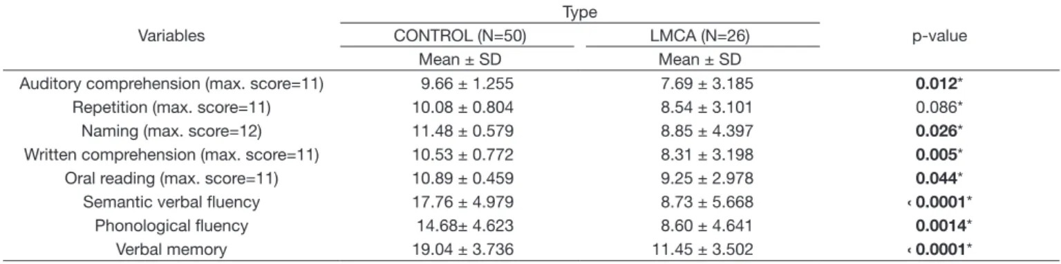
Table 1 shows that the performance in all language functions of the patients with SAH caused by rupture of aneurism in the LMCA was poorer compared with that of individuals in the control group. Only the function of repetition did not present statistically significant difference (p=0.086) between the groups. Evident decrease in the performance of patients with aSAH was observed for the tasks of verbal fluency and memory.
Table 2 presents an analysis of the performance in language and verbal fluency of each patient with aSAH in the LMCA territory. Of the 26 patients investigated, 11 presented profiles characteristic of aphasic syndromes. The individuals were
Table 2. Characterization of the aphasic frames
Participants Gender Age Schooling Dominance Fisher Aphasia
1 M 56 0 D III Transcortical Sensory
2 M 61 8 D III Acquired dyslexia
3 M 73 0 D IV Transcortical Sensory
4 F 55 0 D IV Wernicke’s
5 F 41 5 D III Anomic
6 F 69 0 D - Transcortical Sensory
7 F 55 1 D III Wernicke’s
8 F 38 5 D IV Transcortical Sensory
9 F 43 4 D IV Transcortical Sensory
10 F 51 - D III Global
11 F 63 0 D - Transcortical Sensory
Caption: Fisher: Fisher Grading Scale; D: dextral
Table 1. Performance in the Montreal-Toulouse Language Assessment Protocol of individuals with aSAH in the left middle cerebral artery (LMCA) preoperatively and the control group
Variables
Type
p-value CONTROL (N=50) LMCA (N=26)
Mean ± SD Mean ± SD
Auditory comprehension (max. score=11) 9.66 ± 1.255 7.69 ± 3.185 0.012*
Repetition (max. score=11) 10.08 ± 0.804 8.54 ± 3.101 0.086*
Naming (max. score=12) 11.48 ± 0.579 8.85 ± 4.397 0.026*
Written comprehension (max. score=11) 10.53 ± 0.772 8.31 ± 3.198 0.005* Oral reading (max. score=11) 10.89 ± 0.459 9.25 ± 2.978 0.044*
Semantic verbal fluency 17.76 ± 4.979 8.73 ± 5.668 ‹ 0.0001*
Phonological fluency 14.68± 4.623 8.60 ± 4.641 0.0014*
Verbal memory 19.04 ± 3.736 11.45 ± 3.502 ‹ 0.0001*
predominantly female (72%), aged 30-50 years (27%) and older than 50 years (72%). Seven were illiterate and five had one to eight years of schooling. All participants were dextral. Five patients (45%) presented grade III and four patients (36%) had grade IV on the Fisher scale; the scores of two patients were not found in their medical records. Predominance of aphasic frames was transcortical sensory aphasia with 41.6% followed
by Wernicke’s aphasia with 16.6%. Table 3 shows the semiology of the aphasic frames.
Graph 1 depicts the numerical details of the types of aphasia. Of the 11 patients with aphasic syndrome, six had transcortical sensory aphasia, two presented Wernicke’s aphasia, one had anomic aphasia, one presented global aphasia, and there was one case of acquired dyslexia without major impairment in oral language.
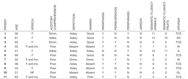
Table 3. Semiology of the aphasic frames
P
A
TIENT
AGE SPEECH AUDITOR
Y
COMPREHENSION REPETITION NAMING PARAPHASIA PERSEVERA
TION
PERIPHRASIS JARGON SEMANTIC FLUENCY ANIMALS SEMANTIC FLUENCY FRUITS APHASIA
1 56 F Dimin. Adeq. Good Y N Y N 11 8 TCS
2 61 F Adeq. Adeq. Good Y N N N 14 10 AD
3 63 F Dimin. Adeq. Good Y Y N N 0 0 TCS
4 55 F and Inc. Poor Absent Absent Y Y N Y 1 2 W
5 41 F Adeq. Adeq. Adeq. N N Y N 10 11 A
6 69 F Poor Adeq. Good N N N N 9 3 TCS
7 55 F and Inc. Poor Dimin. Dimin. Y Y N Y 0 0 W
8 38 F and Inc. Poor Adeq. Absent Y Y N N 0 0 TCS
9 43 F Poor Adeq. Absent Y N N Y 0 0 TCS
10 51 NF Poor Absent Absent Y Y N N 0 0 GL
11 63 F and Inc. Poor Adeq. Poor Y Y N Y 4 0 TCS
Caption: F: Fluent; Inc.: Incoherent; NF: Non-fluent; Y: Yes; N: No; TCS: Transcortical sensory; AD: Acquired Dyslexia; W: Wernicke’s; AN: Anomic; GL: Global; Dimin.: Diminished; Adeq.: Adequate
DISCUSSION
This study aimed to understand the language impairments cause by aneurysmal subarachnoid hemorrhage (aSAH) of the left middle cerebral artery (LMCA) and characterize the possible aphasic syndromes resulting exclusively from SAH, without the interference of procedures conducted to treat the aneurism. This survey intended to provide the scientific community with a better understanding on the possible language impairments that occur as a consequence of aSAH preoperatively and of the influence of aneurysm localization on cognitive functions, such as language performance with characterization of aphasic frames.
Classifying a linguistic-cognitive impairment presents several challenges because the influence of cognitive functions on language is quite evident in certain tests. The rater is responsible for classifying which functions may have influenced the result(20). Thus the choice of material to evaluate an illiterate
population is primordial. In this study, the Montreal-Toulouse Language Assessment Protocol (Alpha version) was in line with our objectives, because it presents characteristics that were in agreement with the reality of our sample, which comprised a significant number of illiterates.
Comparison between the patients with aSAH in the LMCA territory and the individuals in the control group showed reduction in language performance in most tasks, except for repetition, demonstrating that the language function was affected preoperatively in these patients. As the control group was composed of individuals from the same environment as that of the patients, mainly relatives who had similar schooling and sociocultural levels, it was difficult to justify impairments in these functions that were not due to losses resulting from aSAH.
Of the 26 patients with aSAH in the LMCA territory, 14 did not present low performance in the language tasks that characterized aphasic syndromes. The scores of these patients in the Fisher scale were predominantly grade I and II. This finding confirms that not all individuals with SAH present linguistic-cognitive impairments, which may be justified by a smaller volume of blood in the subarachnoid space determined by the Fisher grading scale scores of these patients. As for the 11 patients that presented language impairment, the scores in the Fisher scale were predominantly grade III and IV. These data corroborate the study by Souza et al.(25), who reported
that as the blood volume increases in the subarachnoid cavity, a decline is observed in the cognitive functions of the patient, specifically, the language function is compromised. According to the Fisher scale, grade III is associated with increased risk of vasospasm and ischemia - factors that may lead to greater cognitive impairment(7).
Statistically significant differences in the verbal fluency tasks were observed between the patients with aSAH in the LMCA territory and the individuals in the control group. Although some patients in the SAH group did not present aphasic characteristics, the poor performance of patients with aphasia promoted a decrease in the mean of this group. This poor verbal fluency performance corroborates the findings of a recent study that showed a decrease in the performance of aphasic individuals
compared with that of a control group in the tasks of verbal fluency and memory(26).
The aphasia frames observed in this study show a relevant number of cases of transcortical sensory aphasia. Assessment of these patients showed a poor level of auditory comprehension. In some cases, the speech presented constancy of aphasic characteristics, such as paraphasia, jargon, and perseveration; however, repetition was preserved in all cases. In this sample, when individuals with SAH in the LMCA were compared with those of the control group, repetition was the only task that did not present statistically significant differences. This finding demonstrates that, even though aphasia occurs in the group with aSAH in the LMCA, there is predominance of non-central aphasia, which presents the repetition task preserved(27).
Only three of the 11 aphasic patients presented impairment of the repetition task: two with Wernicke’s aphasia and one with global aphasia. The two patients with Wernicke’s aphasia presented grade IV in the Fisher scale, that is, the maximum blood volume score in the subarachnoid space, which is indicative of greater cognitive impairment(25).
In a study(28) whose findings confirmed a larger number of
cases of aneurysm in the first temporal branches (identified in more than 90% of the cases), the verified results corroborate those demonstrated in the present research, because of the 11 patients with aphasia, six had transcortical sensory aphasia, two presented Wernicke’s aphasia, and one had acquired dyslexia, which characterizes the posterior arm of the middle cerebral artery as a locus of characteristics of aphasic syndromes associated with the temporoparietal region.
CONCLUSION
Subarachnoid hemorrhage in patients with aneurism in the left middle cerebral artery territory causes linguistic-cognitive impairments preoperatively to aneurism occlusion. Reduction in language performance and verbal fluency was observed in the comparison between the groups. Posterior aphasia was predominant among the aphasic frames. The amount of blood in the subarachnoid space may cause greater language impairment. Localization of aneurysm suggests that it occurs in the temporal branch of the M1 segment.
Considering the aspects observed in relation to the Fisher scale, the varying grades present different levels of language impairment. The greater the volume of blood in the subarachnoid space, the greater the predicted language impairment preoperatively to aneurysm occlusion.
Based on the previously mentioned facts, further studies associating aneurysm localization with aphasic syndromes are needed. In addition, a better understanding of health professionals about the cognitive changes caused by aneurysmal subarachnoid hemorrhage is expected.
REFERENCES
2. Bederson JB, Chair MD, Connolly SE, Faha MD, Bajer HH, Dacey GR, et al. Guidelines for the management of aneurysmal subarachnoid hemorrhage: a statement for healthcare professionals from a special writing group of the stroke e council. J Am Heart Assoc. 2009;40(1):994-1025. 3. Ardila A. A proposed reinterpretation and reclassification a of
aphasic syndromes. Aphasiology. 2010;24(3):363-94. http://dx.doi. org/10.1080/02687030802553704.
4. Clinchot DM, Kaplan PE, Murray DM, Pease WS. Cerebral aneurysms and arteriovenous malformations: Implications for rehabilitation. Arch Phys Med Rehabil. 1994;75(12):1342-51. PMid:7993174.
5. Code C. Aphasia and related neurogenic communication disorders. 5. ed. Philadelphia: Burlington; 2012.
6. Weir B, Findlay M. Subarachnoid hemorrhage. In: Carter LP, Spetzler R, Hamilton MG, editors. Neurovascular surgery. New York: McGraw-Hill. 7. Fisher CM, Kistler JP, Davis JM. Relation of cerebral vasospasm to
subarachnoid hemorrhage visualized by computerized tomographic scanning. Neurosurgery. 1980;6(1):1-9. PMid:7354892.
8. Satzger W, Niedermeier N, Schönberger J, Engel RR, Beck OJ. Timing of operation for ruptured cerebral aneurysm and long-term recovery of cognitive functions. Acta Neurochir. 1995;136(3-4):168-74. PMid:8748849. http://dx.doi.org/10.1007/BF01410621.
9. Cancela DMG. O acidente vascular cerebral: classificação, principais
consequências e reabilitação. Psicologia. [Internet]. 2008:1-18 [citado
em 2014 Out 22]. Disponível em: http://www.psicologia.pt/artigos/textos/ TL0095.pdf
10. Sakas DE, Dias LS, Beale D. Subarachnoid haemorrhage presenting as head injury. BMJ. 1995;310(6988):1186-7. PMid:7767157. http://dx.doi. org/10.1136/bmj.310.6988.1186.
11. Flores JCH, Napoles RB. Factores de mal prognóstico en pacientes
com hemorragia subaracnóidea espontânea atendidos em el Hospital
Universitario” Manuel Ascunce Domenéch. Revista Cubana de Neurología y Neurocirurgia. 2016;6(1):1-8.
12. Ogden J, Mee E, Henning M. A prospective study of impairment of cognition and memory and recovery after subarachnoid hemorrhage. Neurosurgery. 1993;33(4):572-87. PMid:8232796. http://dx.doi.org/10.1227/00006123-199310000-00004.
13. Fobe J, Haddad L, Souza A. Desempenho cognitivo em pacientes operados de aneurisma cerebral. Arq Neuropsiquiatr. 1999;57(2A):233-42. PMid:10412523. http://dx.doi.org/10.1590/S0004-282X1999000200011. 14. Hillis A, Anderson N, Sampath P, Rigamonti D. Cognitive impairments after surgical repair of ruptured and unruptured aneurysms. J Neurol Neurosurg Psychiatry. 2000;69(5):608-15. PMid:11032612. http://dx.doi.org/10.1136/ jnnp.69.5.608.
15. Santis A, Laiacona M, Barbarotto R, Divitiis O, Migliore M, Capitani E. Neuropsychological outcome of operated cerebral aneurysms: prognostic factors on 148 patients. Acta Neurol Scand. 1998;97(6):393-7. PMid:9669473. http://dx.doi.org/10.1111/j.1600-0404.1998.tb05972.x.
16. Orbo M, Waterloo K, Egge A, Isaksen J, Ingebrigtsen T, Romner B. Predictors for cognitive impairment one year after surgery for aneurysmal subarachnoid hemorrhage. J Neurol. 2008;255(11):1770-6. PMid:18854912. http://dx.doi.org/10.1007/s00415-008-0047-z.
17. Andersson M, Ystad M, Lundervold A, Lundervold A. Correlations between measures of executive attention and cortical thickness of left posterior middle frontal gyrus: a dichotic listening study. Behav Brain Funct. 2009;5(1):41. PMid:19796388. http://dx.doi.org/10.1186/1744-9081-5-41.
18. Vieira ACC. Estudo comparativo da memória verbal e da linguagem relacionado com o tratamento cirúrgico ou endovascular nos pacientes portadores de aneurismas intracraniano [tese]. Recife: Universidade Federal de Pernambuco; 2009.
19. Vieira ACC, Azevedo-Filho HRC, Andrade G, Costa e Silva IE, Griz MFL, Quinino S, et al. Cognitive changes in patients with aneurysmal subarachnoid hemorrhage before and early posttreatment: differences between surgical and endovascular. World Neurosurg. 2012;78(1-2):95-100. PMid:22120261. http://dx.doi.org/10.1016/j.wneu.2011.09.021.
20. Leśniak M, Bak T, Czepiel W, Seniów J, Członkowska A. Frequency and prognostic value of cognitive disorders in stroke patients. Dement Geriatr Cogn Disord. 2008;26(4):356-63. PMid:18852488. http://dx.doi. org/10.1159/000162262.
21. Murdoch BE. Desenvolvimento da fala e distúrbios da linguagem: uma
abordagem neuroanatômica e neurológica funcional. 2. ed. Rio de Janeiro:
Revinter; 2012.
22. Huberfeld G, Trébuchon A, Capelle L, Badier JM, Chen S, Lefaucheur JP, et al. Preoperative and intraoperative neurophysiological investigations for surgical resections in functional areas. Neurochirurgie. 2017;63(3):142-9. PMid:28506486. http://dx.doi.org/10.1016/j.neuchi.2016.10.008. 23. Jiao Y, Lin F, Wu J, Li H, Chen X, Li Z, et al. Brain arteriovenous
malformations located in premotor cortex: surgical outcomes and risk factors for postoperative neurological deficits. World Neurosurg. 2017;105:432-40. PMid:28583455. http://dx.doi.org/10.1016/j.wneu.2017.05.146. 24. Lecours AR, Mehler J, Parente MA, Caldeira A, Cary L, Castro MJ, et al.
Illiteracy and brain damage - 1. Aphasia testing in culturally contrasted populations (control subjects). Neuropsychologia. 1987;25(1):231-45. PMid:2437493. http://dx.doi.org/10.1016/0028-3932(87)90134-5. 25. Souza MLP, Vieira ACC, Quinino GAS, Griz MFL, Azevedo-Filho
HRC. Fisher grading scale associated with language disorders in patients with anterior circulation aneurysmal subarachnoid hemorrhage. World Neurosurg. 2015;84(2):308-13. PMid:25797076. http://dx.doi.org/10.1016/j. wneu.2015.03.017.
26. Bonini MV, Radanovic M. Cognitive deficits in post-stroke aphasia. Arq Neuropsiquiatr. 2015;73(10):840-7. PMid:26465401. http://dx.doi. org/10.1590/0004-282X20150133.
27. Ulm AJ, Fautheree GL, Tanriover N, Russo A, Albanese E, Rhoton AL Jr, et al. Microsurgical and angiographic anatomy of middle cerebral artery aneurysms: prevalence and significance of early branch aneurysms. Neurosurgery. 2008;62(5, Supl 2):344-53. PMid:18596514.
28. Tanriover N, Kawashima M, Rhoton AL Jr, Ulm AJ, Mericle RA. Microsurgical anatomy of the early branches of the middle cerebral artery: morphometric analysis and classification with angiographic correlation. Neurosurg. 2003;98(6):1277-90. PMid:12816276.
Author contributions