LETTERS TO THE EDITOR
Injection of crack cocaine: a
case report
Rev Bras Psiquiatr. 2015;37:80 doi:10.1590/1516-4446-2014-1480
A 26-year-old American man was referred to our service for methadone replacement therapy and treatment of crack/cocaine addiction. His addiction history started with use of opioids in early adolescence. As his tolerance escalated, he began injecting cocaine with morphine, also known as ‘‘speedball.’’ Since he started methadone replacement therapy and discontinued morphine use, he reported a substantial increase in cocaine injection, as well as crack injection, the latter motivated by lower prices and wider availability. He fulfills DSM-5 diagnosis criteria for severe dependence on both opioids and cocaine. A multidisciplinary team managed his case.
In 2013, the United Nations Office on Drugs and Crime estimated the number of injecting drug users (IDU) worldwide at 14 million.1The prevalence of crack cocaine injection (CCI) is unknown; however, some local studies have assessed it and observed variable frequencies. A PubMed search using the terms ‘‘injection crack AND intravenous crack’’ yielded 44 papers, eight of which reported actual CCI. None concerned the Brazilian context. This is, to our knowledge, the first Brazilian case report of CCI.
One paper reports an increase of CCI between 1990 and 1993 in London, from 1 to 27%.2 Another study presented the results of a 1997-1999 cohort of 2,198 homeless IDUs recruited from six U.S. cities and estimated the frequency of CCI as 15% among partici-pants.3A later U.S. study with 989 participants reported a 9% lifetime prevalence of CCI among IDUs,4 and a Canadian study with 4,088 IDUs reported a 15.2% rate of CCI.5
CCI is accomplished by dissolving crack in vinegar or lemon juice and does not require the application of heat. The shift from cocaine use to CCI can be explained by several reasons, including changes in illicit drug markets and a desire for greater psychoactive effects. This is particularly important as some anecdotal unpublished reports state that cocaine may be present in greater amounts in crack than in its powder form.
CCI may be a marker of high-risk behaviors, and correlates with use of shooting galleries, initiation of others into drug injection, and serologic evidence of hepatitis B virus and hepatitis C virus infection.3Furthermore, crack injectors reported higher rates of abscesses and mental illness.4HIV infection is also a real risk for all IDUs. All of these factors can have an important impact on public health issues related to drug use, and must be considered in policy development.
Furthermore, this population may require broader treatment strategies. For instance, since severe mental
illness may be more prevalent in crack cocaine injectors, hospitalization can be necessary for acute management of high-risk symptomatology (suicidality, aggressiveness, agitation). Moreover, health maintenance programs (i.e., management of infectious diseases), first-line social assistance (shelter, food, hygiene), and access to sustainable livelihood programs (housing, vocational training) can be vital for comprehensive treatment.
CCI deserves a more comprehensive investigation among the Brazilian drug user population, as it may be a marker of high-risk behaviors. Public health surveillance of this practice is of great importance to provide a better understanding of the behavior of users who self-administer crack cocaine via this route and to enable implementation of prevention and harm reduction policies.
Melina Mendonc¸a, Dartiu X. Silveira, Thiago M. Fidalgo Addiction Unit (PROAD), Department of Psychiatry, Universidade Federal de Sa˜o Paulo (UNIFESP), Sa˜o Paulo, SP, Brazil
Submitted May 28 2014, accepted Jul 24 2014.
Disclosure
The authors report no conflicts of interest.
References
1 United Nations Office on Drugs and Crime (UNODC). World Drug Report 2013. United Nations publication, Sales N6. E.13.XI.6 [Internet]. 2013 [cited 2014 Ago 14]. http://www.unodc.org/unodc/ secured/wdr/wdr2013/World_Drug_Report_2013.pdf
2 Hunter GM, Donoghoe MC, Stimson GV. Crack use and injection on the increase among injecting drug users in London. Addiction. 1995;90:1397-400.
3 Santibanez SS, Garfein RS, Swartzendruber A, Kerndt PR, Morse E, Ompad D, et al. Prevalence and correlates of crack-cocaine injection among young injection drug users in the United States, 1997-1999. Drug Alcohol Depend. 20057;77:227-33.
4 Buchanan D, Tooze JA, Shaw S, Kinzly M, Heimer R, Singer M. Demographic, HIV risk behavior, and health status characteristics of ‘‘crack’’ cocaine injectors compared to other injection drug users in three New England cities. Drug Alcohol Depend. 2006;81:221-9. 5 Roy E, Leclerc P, Morissette C, Arruda N, Blanchette C, Blouin K,
et al. Prevalence and temporal trends of crack injection among injection drug users in eastern central Canada. Drug Alcohol Depend. 2013;133:275-8.
Subarachnoid hemorrhage
misdiagnosed as
adjustment disorder
Rev Bras Psiquiatr. 2015;37:80––82doi:10.1590/1516-4446-2014-1502
A 38-year-old woman with a history of recent grief and anxiety disorder, treated with alprazolam, was brought directly to the psychiatric emergency room after experi-encing a sudden change in behavior. The patient was Revista Brasileira de Psiquiatria. 2015;37
agitated and dysphoric, with no other changes at physical examination, except for nausea and vomiting. No other relevant personal, family (including intracranial aneur-ysms), or social history was known.
Adjustment disorder with anxiety symptoms, secondary to a recent diagnosis of cancer in a close family member, seemed the most plausible diagnosis to prehospital emergency providers. However, as the psychiatric team felt that an organic cause had not been clearly excluded, routine blood work and a computed tomography (CT) scan of the head were obtained.
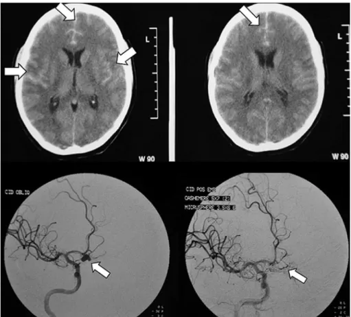
Head CT revealed a frontal subarachnoid hemorrhage (SAH). Posterior cerebral angiography found two bilobar saccular aneurysms, one measuring 4 mm at the left middle cerebral artery bifurcation and a second one measuring 6 mm at the anterior communicating artery (Figure 1).
After imaging, the patient developed headache, menin-gism, and upper limb paresis. She was rapidly transferred to the Neurosurgery Department and underwent endo-vascular coiling of the larger aneurysm. Following treat-ment in a neurosurgery ward, the patient was discharged with no neurological deficit (National Institute of Health Stroke and Modified Rankin Scales). There were no psychological sequelae that justified psychiatric treatment. Bizarre presentations of SAH have been recorded in the literature, including kleptomania, akinetic mutism, amnesic-confabulatory syndrome, Capgras misidentifica-tion syndrome, auditory hallucinamisidentifica-tions, and persecutory
delusion.1 However, there are very few reports of SAH
presenting with psychiatric manifestations in the absence of classic symptoms such as acute headache or menin-gism.
SAH occurs in 9 per 100,000 persons/year, mainly in women.2Up to 85% of cases are aneurysmatic in origin, and the mortality rate is 50%, with 15% dying before any hospital treatment.3 Severe headache, vomiting, and neck rigidity are common symptoms.4Surgical or endovas-cular ablation are the best therapeutic available options.5
We believe the frontal localization of the SAH in this patient led to sudden behavioral changes that were mis-understood by the ambulance staff. This case reminds us that, in the emergency setting, careful triage is necessary to avoid fatal outcomes, especially in hurried or high-risk referrals to psychiatry.
Joa˜o Gama-Marques, Filipa Palhava˜, Sofia Brissos Schizophrenia Department, Centro Hospitalar Psiquia´trico de Lisboa, Lisbon, Portugal
Submitted Jun 25 2014, accepted Sep 18 2014.
Disclosure
The authors report no conflicts of interest.
Figure 1 Head computed tomography scan revealing frontal subarachnoid hemorrhage and cerebral angiography showing two aneurysms, before and after endovascular coiling.
Letters to the Editor 81
References
1 Mobbs RJ, Chandran KN, Newcombe RL. Psychiatric presentation of aneurysmal subarachnoid haemorrhage. ANZ J Surg. 2001;71: 69-70.
2 de Rooij NK, Linn FH, van der Plas JA, Algra A, Rinkel GJ. Incidence of subarachnoid haemorrhage: a systematic review with emphasis on region, age, gender and time trends. J Neurol Neurosurg Psychiatry. 2007;78:1365-72.
3 Van Gijn J, Kerr RS, Rinkel GJ. Subarachnoid haemorrhage. Lancet. 2007;369:306-18.
4 Cohen-Gadol AA, Bohnstedt BN. Recognition and evaluation of nontraumatic subarachnoid hemorrhage and ruptured cerebral aneurysm. Am Fam Physician. 2013;88:451-6.
5 Connolly ES Jr, Rabinstein AA, Carhuapoma JR, Derdeyn CP, Dion J, Higashida RT, et al. Guidelines for the management of aneurysmal subarachnoid hemorrhage: a guideline for healthcare professionals from the American Heart Association/American Stroke Association. Stroke. 2012;43:1711-37.
Marchiafava-Bignami
disease as a cause of visual
hallucinations
Rev Bras Psiquiatr. 2015;37:82––83 doi:10.1590/1516-4446-2014-1529
Alcohol is one of the most used addictive substances worldwide and its dependence constitutes one of the most important causes of morbidity and mortality, accounting for 5.9% of all deaths.1Alcohol has two types of effects on the brain: direct, by acting on neurotransmitters and electro-lytes; and indirect, such as through encephalopathy or coagulopathies. Some conditions, such as Marchiafava-Bignami disease (MBD), are associated with chronic alcoholism, but it is still not clear whether directly or indirectly.2
A 52-year-old woman was admitted to our institution with a 10-day history of visual hallucinations –– complain-ing of dead people and cameras inside her house ––and cognitive impairment. There were no focal neurological signs nor associated delirium. An extensive clinical interview revealed a sustained pattern of excessive daily alcohol consumption ––predominantly red wine ––for more than 5 years, arising after her daughter’s marriage. Computed tomography (CT) of the head showed a hypoattenuating lesion affecting the genu and splenium of the corpus callosum. Further investigation by magnetic resonance imaging revealed a T2-hyperintense lesion with restricted diffusion involving the entire corpus callosum, without swelling or enhancement, suggestive of MBD (Figure 1). The patient was treated with B-complex vitamins (thiamine, 300 mg three times a day for 14 days, and folic acid, 5 mg/day). Low-dose quetiapine was also administered, later replaced by olanzapine 5 mg/day, and contributed to slight improvement. The patient continued to experience episodes of visual hallucinations after
discharge, although less frequently. Oral thiamine therapy (300 mg/day) was maintained after discharge.
MBD is a rare condition associated with chronic alcoho-lism, with only a few reports in non-alcoholic individuals. It most commonly affects middle-aged men with a history of chronic alcohol abuse or malnourishment. MBD is charac-terized by progressive demyelination and thinning of the corpus callosum, affecting mainly the genu and the splenium, that can even progress to focal necrosis.3
In acute MBD, patients may present with seizures or coma, whereas patients with chronic MBD usually exhibit cognitive deficits, hallucinations, or depression lasting for several months. MBD can also coexist with Wernicke’s encephalopathy, Korsakoff’s syndrome, osmotic demye-lination syndrome, and Morel’s laminar necrosis, which are also associated with chronic alcoholism.2
Imaging is crucial to the diagnosis. CT may reveal focal or diffuse hypoattenuating lesions involving the genu and the splenium as well as in the periventricular area. Mag-netic resonance studies usually depict non-edematous T2-hyperintense lesions, sometimes with focal areas of necrosis. In the acute phase, there can be peripheral enhancement on postcontrast studies, and lesions exhibit restricted diffusion.3
Heinrich et al.4proposed an imaging-based classifica-tion in which the type A corresponds to diffuse callosal involvement, whereas type B, which carries a better prognosis, includes only partial lesions.
Acute stroke, extrapontine myelinolysis, lymphoma, and psychiatric disorders should be considered in the differ-ential diagnosis.
No specific, proven therapy is available for MBD. Treatment is mainly symptomatic, with the administration of B-complex vitamins and folate. The role of antipsycho-tic drugs is not established. In the patient reported herein, their use contributed to slight symptomatic improvement. Alcohol avoidance is mandatory.5
Prognosis is variable, ranging from a very unusual complete recovery to death.5Despite meeting criteria for type-A disease, with a worse prognosis, our patient experienced a slight recovery, with some episodes of visual hallucinations persisting.
This rare case, made even more unusual by its occurrence in a female patient, highlights the importance of a thorough clinical evaluation and imaging studies in the detection of such an infrequent cause of psychiatric symptoms.
Luı´s Augusto,1Rita Figueiredo,1Henrique Costa,2,3 Carina Reis,1Maria Luı´s Silva1 1Neuroradiology Department, Centro Hospitalar de S. Joa˜o, Porto,
Portugal.2Neurology Department, Centro Hospitalar de S. Joa˜o, Porto, Portugal.3Porto Medical School, Porto, Portugal
Submitted Jul 31 2014, accepted Oct 09 2014.
Disclosure
The authors report no conflicts of interest. Letters to the Editor
82