ISSN 1980-5098 DOI: http://dx.doi.org/10.5902/1980509832058
MICROPROPAGATION OF Pinus tecunumanii MICROPROPAGAÇÃO DE Pinus tecunumanii
Laudiane Bruna Zanella1 Luziane Franciscon2 Renata Lúcia Grunennvaldt3 Jéssica de Cássia Tomasi3 Juliana Degenhardt-Goldbach4
ABSTRACT
Pinus tecunumanii is one of the most important tropical species of Pinus in Brazil. This work aimed to develop a micropropagation protocol for Pinus tecunumanii from the apical shoots of young plants grown in a greenhouse. To optimise the aseptic treatment protocol, the effect of 0.05 or 0.1% mercuric chloride in combination with 0 or 1 g L-1 fungicide and NaOCl was evaluated. Next, the effect of 2 or 4 µM BA (6-benzyl adenine) on in vitro multiplication was evaluated over three subcultures. Supplementation with 1.5 g L-1 activated charcoal was tested for elongation. For rooting, the effects of WV5 salts (at half strength) and 20 g L-1 sucrose were evaluated on media supplemented with 2.68 µM naphthalene-acetic acid and 0.44 µM BA. Aseptic treatment of explants with 0.05% mercuric chloride and sodium hypochlorite (with or without fungicide) was effective to establish 83.3% of explants in vitro. On media supplemented with 2 or 4 µM BA, shoot multiplication was promoted. After the third subculture on medium supplemented with 2 µM BA, 70.83% of the explants exhibited new shoots with an average of 3.7 new shoots per explant. On medium containing activated charcoal, elongation was observed in 59.76% of the explants, while on medium without activated charcoal only 33.33% elongated. Rooting was observed in 14.8% of explants on medium supplemented with WV5 salts.
Keywords: shoot multiplication; elongation; rooting.
RESUMO
Pinus tecunumanii é uma das espécies tropicais mais importantes do gênero Pinus no Brasil. Este trabalho teve por objetivo desenvolver um protocolo de micropropagação para Pinus tecunumanii a partir de gemas apicais de plantas jovens mantidas em casa de vegetação. Para a assepsia foi avaliado o efeito de 0,05 ou 0,1% de cloreto de mercúrio em combinação com 0 ou 1 g L-1 de fungicida e NaOCl. O efeito de 2 ou 4 µM BA (6-benzil adenina) foi avaliado na multiplicação in vitro ao longo de três subcultivos. O efeito da adição de 1,5 g L-1 de carvão ativado foi avaliado no meio de alongamento. Para o enraizamento, o efeito dos sais WV5 meia força e de 20 g L-1 de sacarose foi avaliado em meio suplementado com 2.68 µM ácido naftaleno acético (ANA) e 0.44 BA. A assepsia dos explantes com 0,05% de cloreto de mercúrio, hipoclorito de
sódio, com ou sem fungicida foi eficiente no estabelecimento in vitro de até 83,3% dos explantes. Em
meio suplementado com 2 ou 4 µM BA ocorreu multiplicação de brotações adventícias e após o terceiro subcultivo, 70,83% dos explantes formaram novos brotos em meio suplementado com 2 µM BA, com uma média de 3,7 novos brotos por explante. Em meio suplementado com carvão ativado foi observado alongamento em 59,76% dos explantes, enquanto no meio sem carvão 33,33% dos explantes alongaram.
1 Biotecnóloga, MSc., Doutoranda em Agronomia-Produção Vegetal, Universidade Federal do Paraná, Rua dos Funcionários, 1540, CEP 80035-050, Curitiba (PR), Brasil. [email protected]
2 Estatística, MSc., Analista EMBRAPA Florestas, Estrada da Ribeira, Km 111, CEP 83411-000, Colômbo (PR), Brasil. [email protected]
3 Engenheira Florestal, MSc., Doutoranda em Agronomia-Produção Vegetal, Universidade Federal do Paraná, Rua dos Funcionários, 1540, CEP 80035-050, Curitiba (PR), Brasil. [email protected] / jehtomasi@hotmail. com
4 Engenheira Agrônoma, Dra., Pesquisadora EMBRAPA Florestas, Estrada da Ribeira, Km 111, CEP 83411-000, Colombo (PR), Brasil. [email protected]
Foi observado enraizamento em 14,8% dos explantes em meio suplementado com sais WV5. Palavras-chave: multiplicação de brotações; alongamento; enraizamento.
INTRODUCTION
Reforestation with exotic species not only produces biomass, but also preserves native species by
reducing indiscriminate exploitation of the natural flora. However, in order to be economically viable, the
use of exotic species such as Eucalyptus and Pinus require focused breeding programs.
In the last three decades, the use and application of Pinus lumber has increased substantially, and these species have become an essential raw material that drives a productive sector of the Brazilian economy (VASQUES et al., 2007). Pinus tecunumanii is the most valued tropical species of Pinus in Brazil. It can be found naturally from southern Mexico to central Nicaragua and can reach up to 50 m in height with a straight trunk that is usually free of branches until a height of 20–30 m. The wood of excellent quality, is produced in high yields and has characteristics that facilitate mechanical processing. Moreover, Pinus tecunumanii grows rapidly in the tropical region of Brazil. Embrapa Forestry has a Pinus breeding program that focuses on Pinus taeda, Pinus maximinoii, Pinus caribeaa var. hondurensis, Pinus elliottii and Pinus tecunumanii. This program aims to improve each species as well as produce hybrids. The main selection criteria for the Pinus tecunumanii breeding program are vigour, stem form, wood quality and adaptation to planting areas. Furthermore, the timber has to have a suitable density for the sawmill (Ananda Virginia de Aguiar, breeder at Embrapa Forestry, personal communication).
Major limitations to improvement are the long intervals between flowerings and selection, and the
poor rooting of cuttings from older trees. Furthermore, seeds of high genetic quality are still not available
in sufficient quantities. Therefore, alternative techniques for vegetative propagation of these species are
required (STOJICIC et al., 1999).
Micropropagation is a useful tool in breeding programs, not only for cloning superior genotypes, but also to clone genotypes originating from controlled crosses. Tissue culture studies of Pinus tecunumanii are scarce, and micropropagation protocols are genotype dependent, requiring protocols to be developed for
each specific species or clone (CUESTA et al., 2008).
Micropropagation by apical shoots is the most usual method for mass production of superior clones by tissue culture, and it is advantageous because clones with known characteristics can be produced on a
large scale (HARTMANN et al., 2011). Plants from regenerated shoot tips or axillary buds usually show
higher genetic stability and have a lower risk of somaclonal variation when compared to plants regenerated
by organogenesis (ABDULLAH et al., 1986).
In the last two decades, several protocols have been developed for in vitro culture of Pinus species
(DUMAS; ONTEUUIS, 1995; EWALD, 1998; CHANG et al., 2001; PARASHARAMI et al., 2003; PREHN et al., 2003; RENAU-MORATA et al., 2005). However, in most cases, regeneration from adult
material has limited practical applications, and most Pinus micropropagation protocols reported in the literature are based on organogenesis from the cotyledons of seeds germinated in vitro (ALONSO et al.,
2006; ÁLVAREZ; MAJADA; ORDÁS, 2009; HUMÁNEZ et al., 2011; STOJICIC et al., 2012). Reports of
protocols based on introduction of apical and axillary buds isolated from seedlings grown in greenhouses or adult plants are rare (CORTIZO et al., 2009; OLIVEIRA et al., 2012).
The success of micropropagation depends on the sequence of several steps, where each step depends on the success of the previous one. Initial establishment of an in vitro culture requires efficient, aseptic collection of explants (GEORGE et al., 2008).
The addition of an exogenous cytokinin to the culture media is favourable for in vitro multiplication of pine. 6- Benzyl adenine (BA) is a plant growth regulator that is widely used to induce formation of axillary shoots and its concentration is often variable among species (ALONSO et al., 2006; ÁLVAREZ;
MAJADA; ORDÁS, 2009; ZHU et al., 2010; OLIVEIRA et al., 2012). An additional step in the in vitro
culture may be necessary to elongate explants prior to rooting. Activated charcoal has been successfully used for elongation in protocols for micropropagation of Pinus species. The required concentrations range from 1.0–3.0 g L-1 (ÁLVAREZ; MAJADA; ORDÁS, 2009; CORTIZO et al., 2009; ZHU et al., 2010;
Rooting is the most critical step in micropropagation of the Pinus species and losses during acclimatisation often limit the use of micropropagation for cloning on a commercial scale (OLIVEIRA et al., 2003). The maturation of conifers decreases their ability to form adventitious roots and is responsible for the low rates of rooting observed in the micropropagation of conifers from adult plants (DUMAS; ONTEUUIS, 1995; RENAU-MORATA et al., 2005). In the micropropagation of 25-year-old Pinus pinea plants, Cortizo et al. (2009) observed that only 8.4% of explants formed roots.
This work aimed to establish a micropropagation protocol for Pinus tecunumanii from apical shoots of 1.5-year-old greenhouse-grown plants.
MATERIALS AND METHODS
Plant material
Apical shoots from young Pinus tecunumanii plants kept in a greenhouse at Embrapa Forestry were used as explants. The plants belonged to a seed orchard and were 1.5 years old when the explants were collected. The seed orchard belongs to the genetic breeding program of Embrapa Forestry carried out in partnership with the Forestry Company Vale do Corisco, and is located in Ventania, PR state, Brazil. The experiments were carried out in the Laboratory of Tissue Culture and Transformation at Embrapa Forestry.
Aseptic treatment of explants
Prior to explants harvest, 1g L-1 of the fungicide Cercobin® was applied once a week for three weeks and daily for three days in the greenhouse. Apical shoots were collected (approximately 3.5 cm
long), and approximately 90% of their needles were removed. The explants were then placed in flasks containing distilled water. In the laboratory, the apical shoots were manipulated in a laminar flow hood and
divided into four treatments: T1 - 0.05% mercuric chloride for 5 min + 0.5% NaOCl for 5 min; T2 - 0.1% mercuric chloride for 5 min + 0.5% NaOCl for 5 min; T3 - 1 g L-1 fungicide for 10 min + 0.05% mercuric chloride for 5 min + 0.5% NaOCl for 5 min; or T4 - 1 g L-1 fungicide for 10 min + 0.1% mercuric chloride for 5 min + 0.5% NaOCl for 5 min.
The shoots were then washed three times in sterile distilled water and cut into approximately 1.5 cm long explants. The explants were inoculated vertically in test tubes containing 13 mL of WV5 culture medium (COKE, 1996) supplemented with 20 g L-1 sucrose and 7 g L-1 Difco Bacto-Agar. Each treatment group consisted of 30 explants. Explants were evaluated after four weeks to determine the extent of bacterial and fungal contamination, oxidation and survival rate.
Effect of BA on shoot proliferation
The aseptic treatment procedure for explants was carried out as described above for treatment T3. Afterwards, the explants (approximately 1.5 cm long) were inoculated on WV5 culture medium containing 20 g L-1 sucrose, 7 g L-1 Difco Bacto-Agar and 0 (control), 2 or 4 µM of BA. The explants were transferred every month to fresh medium of the same composition. The percentage of oxidation, axillary
shoot formation and elongation was assessed after three, four and five months in culture (the second, third
and fourth subcultures, respectively). Each treatment group consisted of 10 vessels with four explants each for a total of 40 explants per treatment.
Elongation
After five months in culture, explants in the multiplication stage were cut into 1.5 cm segments and
for both treatment groups for a total of 92 explants per treatment.
Rooting and acclimatisation
After seven months on media with 0, 2 or 4 µM of BA, explants were transferred to WV5 medium supplemented with 20 g L-1 sucrose, 7 g L-1 agar, and without plant growth regulators and grown for 8 weeks. After this period, the explants were divided into three treatment groups according to Oliveira et al. (2012): T1- ½ WV5 + 20 g L-1 sucrose + 2.68 µM naphthalene-acetic acid (NAA) + 0.44 µM BA; T2 - water/agar + 2.68 µM NAA + 0.44 µM BA; or T3 - water/agar + 20 g L-1 sucrose + 2.68 µM NAA + 0.44 µM BA.
After nine days, the explants were transferred to the same media without growth regulators. One month later, the rooting rate and the number of roots were evaluated. The rooted explants were transferred
to a substrate (Plantmax:vermiculite, 1:1, v/v) in plastic cups covered with PVC film and kept in the growth room. Each day the PVC film cover was opened for increasingly longer periods (from 1 to 5 hours), and after one week the film was completely removed. A month later, the survival rate was assessed. We used six
vessels with four explants each for a total of 24 explants per treatment.
Culture conditions
The pH of the culture media was adjusted to 5.8 prior to autoclaving. At all stages, the cultures were
maintained in a growth chamber at 23 ± 2°C with a 16 h photoperiod and a light intensity of 40 µmol m-2.s-1.
Statistical analysis
The experimental design was completely randomised for all experiments. The experiments were repeated once and all data were used in the analysis. Data analysis was carried out using deviance analysis. Binomial distributions were used for binary variables and Poisson distributions for quantitative variables. Means were compared using Tukey’s test (p < 0.05) and a comparison among subcultures was carried out using a t-test (p < 0.05).
RESULTS AND DISCUSSION
Aseptic treatment
The first step in the establishment of an in vitro culture is to aseptically treat enough material to
carry out all treatment variations with sufficient replicates. For this reason, the aseptic procedure needs to
be able to control contamination without oxidising the explants.
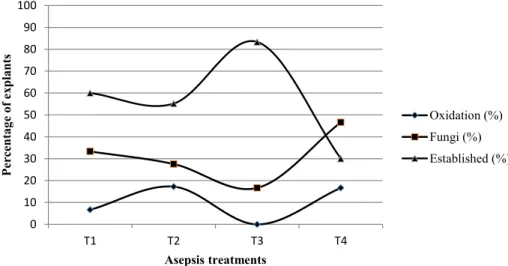
One month after introduction, establishment of up to 83.33% of explants was observed for the treatment that combined fungicide with the lower concentration of mercuric chloride (T3) (Figure 1). In this treatment, no oxidation was observed and fungal contamination was lower than in the other treatments,
although the differences were not statistically significant.
The higher concentration of mercuric chloride was detrimental to establishment and increased oxidation relative to T3. Along with 0.1% mercuric chloride, the explants also exhibited higher contamination rates, probably due to excessive injury of the tissues. Additionally, we found that immersion in fungicide was even more damaging. Using the same concentrations of mercuric chloride and sodium hypochlorite, Oliveira et al. (2012) were able to establish up to 86% of Pinus taeda explants. The concentration of mercuric chloride used to establish adult explants of Pinus pinea was 0.2% with a contamination rate of 44% and 65% of explants established (CORTIZO et al., 2009). Up to 0.3% mercuric chloride was used for introduction of Pinus brutia, although sodium hypochlorite was not used (ABDULLAH; YEOMAN; GRACE, 1987).
The majority of the contamination was caused by fungi, which is similar to the findings in a report
bacterial contamination, which was not observed in three of the treatment groups and only in 6.6% of explants in T4 (data not shown).
YEOMAN; GRACE, 1987).
However, lower concentrations of mercuric chloride were able to control contamination, and
However, this technique is still 0
10 20 30 40 50 60 70 80 90 100
T1 T2 T3 T4
P
er
ce
nta
g
e
o
f ex
pla
nts
Asepsis treatments
Oxidation (%) Fungi (%) Established (%)
FIGURE 1: Effect of aseptic treatments on in vitro establishment, contamination and oxidation of apical shoots of
Pinus tecunumanii after 30 days in culture. Means and confidence intervals (labelled with letters) do not
differ significantly according to Tukey’s test (p < 0.05). T1 - 0.05% mercuric chloride for 5 min + 0.5% NaOCl for 5 min; T2 - 0.1% mercuric chloride for 5 min + 0.5% NaOCl for 5 min; T3 - 1 g L-1 fungicide for 10 min + 0.05% mercuric chloride for 5 min + 0.5% NaOCl for 5 min; or T4 - 1 g L-1 fungicide for 10 min + 0.1% mercuric chloride for 5 min + 0.5% NaOCl for 5 min.
FIGURA 1: Efeitos da assepsia no estabelecimento in vitro, contaminação e oxidação de ápices caulinares de Pinus tecunumanii após 30 dias no cultivo in vitro. Médias e intervalos de confiança seguidos da mesma letra
não apresentam diferença estatística significativa de acordo com o teste de Tukey (p < 0,05). T1 – 0,05% de cloreto de mercúrio por 5 min + 0,5% de NaOCl por 5 min; T2 – 0,1% de cloreto de mercúrio por 5 min + 0,5% de NaOCl por 5 min; T3 – 1 g L-1 de fungicida por 10 min + 0,05% de cloreto de mercúrio por 5 min + 0,5% de NaOCl por 5 min ou T4 – 1 g L-1 de fungicida por 10 min + 0,1% de cloreto de mercúrio por 5 min + 0,5% de NaOCl por 5 min.
Although asepsis protocols are frequently overlooked during the optimisation of micropropagation
protocols, Cortizo et al. (2009) demonstrated that this step influenced not only survival but also propagation
rates in Pinus pinea. In our work, contamination was the main limitation on establishment, and it affected
almost 50% of explants in T4 (Figure 1). However, lower concentrations of mercuric chloride were able to
control contamination, and fungi were observed in less than 20% of explants.
Effect of BA on multiplication
Micropropagation by establishment of apical shoots is a successful technique for cloning plants of superior genotypes for several forest species. However, this technique is still limited in conifers (ORGANIZAÇÃO DAS NAÇÕES UNIDAS PARA ALIMENTAÇÃO E AGRICULTURA, 2004). In the present work, we observed shoot development of Pinus tecunumanii in the presence of BA.
After the second subculture, 56.81% axillary shoot formation was observed in explants grown with 4 µM BA, which was statistically higher than control explants or those grown with 2 µM BA (Table 1).
Oxidation was not influenced by the BA concentration and ranged from 17.14% (CI 12.86) in the treatment
TABLE 1: Percentage of Pinus tecunumanii explants with axillary buds after growth on WV5 medium supplemented with BA after three, four and five months of micropropagation.
TABELA 1: Porcentagem de explantes de Pinus tecunumanii com gemas axilaresem meio WV5 suplementado com BA após três, quatro e cinco meses de micropropagação.
Treatment 2nd subculture 3rd subculture 4th subculture Control 10.52 ± 10.05 aA 15.7 ± 9.39 aA 21.43 ± 23.52 aA 2 µM BA 28.57 ± 15.42 aA 41.20 ± 14.18 bAB 70.83 ± 19.14 bB 4 µM BA 56.81 ± 15.08 bA 31.40 ± 13.1 abA 33.33 ± 18.61 aA
Means ± Confidence interval (CI). Values within a column followed by the same lower case letter and means within a row followed by the same upper case letter do not differ significantly according to Tukey’s test and a t-test (p < 0.05), respectively. BA - 6-benzyl adenine.
Elongation was not influenced by the BA concentration and occurred in less than 45% of the
explants. We observed that oxidation was more intense in the third subculture and ranged from 37.30% (± 13.4) in 4 µM BA to 60.80% (± 13.5) in the control samples. The average number of shoots formed after the fourth subculture was 1.57 for the control explants and 3.7 or 2.2 for explants on media supplemented with 2 or 4 µM BA, respectively (Figure 2).
After five months inculture, axillary shoot multiplication was highest in explants grown with 2 µM BA (Table 1). Oxidation was reduced when compared to the third subculture and ranged from 8.33% in the treatment with 2 µM BA and 35.71% in control.
FIGURE 2: Left – Multiplication of Pinus tecunumanii on WV5 medium supplemented with 2 µM BA; Centre – Explant of Pinus tecunumanii elongated on WV5 medium supplemented with 1.5 g L-1 activated charcoal. Right – Explant of Pinus tecunumanii rooted on medium containing ½ WV5 + 20 g L-1 sucrose + 2.68 µM NAA + 0.44 µM BA.
FIGURA 2: Esquerda – Multiplicação de Pinus tecunumanii em meio WV5 suplementado com 2 µM BA; Centro – Explantes de Pinus tecunumanii alongados em meio WV5 suplementado com 1,5 g L-1 de carvão ativado; Direita – Explante de Pinus tecunumannii enraizado em meio contendo ½ WV5 + 20 g L-1 sacarose + 2.68 µM NAA + 0.44 µM BA.
Most micropropagation protocols for Pinus species induce shoot growth from cotyledon explants of in vitro germinated seeds (ALONSO et al., 2006; ÁLVAREZ; MAJADA; ORDÁS, 2009; ZHU et al., 2010;
SOJICIC et al., 2012). In most of these reports, BA was the most efficient cytokinin at promoting axillary
shoot formation. BA was effective at inducing shoot multiplication in the three subcultures evaluated in
this work, and the response from the two doses were statistically different (Table 1). However, even in
the absence of cytokinin we observed axillary shoot formation in the explants suggesting that endogenous cytokinin also contributes to this response, as observed for Pinus taeda (OLIVEIRA et al., 2012).
Comparison among subcultures showed that axillary shoot multiplication increased significantly on
the medium supplemented with 2 µM BA, while for the control and 4 µM BA subcultures, the multiplication
of axillary shoots was not statistically different (Table 1). However, we did not evaluate whether this increase
Ci. Fl., v. 28, n. 2, abr .- jun., 2018
Oliveira et al. (2012) evaluated the impact of BA on the in vitro multiplication of Pinus taeda over three subcultures. These authors observed that 1 and 2 µM BA were more effective at inducing
shoot multiplication in the first subculture. The rates ranged from 30.3% to 72.7%, with shoots in the first subculture and from 58.8% to 77.5% in the two other subcultures. However, in the last two subcultures the concentrations of BA did not influence the multiplication rates. Similar responses were observed in other
studies of Pinus using different concentrations of BA (GUPTA; DURZAN 1985; ZEL et al., 1988; LAPP et al., 1996; NANDWANI; KUMARIA; TANDON, 2001; WATT et al., 2010).
In Pinus peuce, 2 µM BA was also more efficient at shoot induction than higher concentrations. The authors observed up to 15.12 shoots per cotyledon explant (STOJICIC et al., 2012). Also in cotyledon explants, Álvarez, Majada and Ordás (2009) tested 1, 10 or 100 µM BA and observed that the highest percentage of cotyledons formed buds in the medium containing 10 µM BA (81.3%).
After the fourth subculture, the average number of shoots varied between 1.57 and 3.7. These values are similar to those observed for Pinus taeda (OLIVEIRA et al., 2012). In their work the number
of shoots was not influenced by the BA concentration but was higher in the second and third subcultures
(2.4–3.2 shoots). After four subcultures, Alonso et al. (2006) observed a maximum of 4.64 shoots per Pinus pinea explant.
Elongation
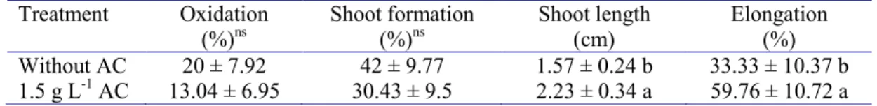
Activated charcoal in the culture medium induced elongation of the explants (Table 2). In the presence of activated charcoal nearly twice as many explants elongated. While explants on medium without activated charcoal were on average 1.57 cm in height, in the presence of activated charcoal they reached 2.23 cm. Axillary shoot multiplication and oxidation did not differ between the two treatments.
TABLE 2: Effect of activated charcoal on shoot elongation, oxidation and axillary shoot multiplication in Pinus tecunumanii.
TABELA 2: Efeito do carvão ativado no alongamento de brotações, oxidação e multiplicação de brotações axilares de Pinus tecunumanii.
Treatment Oxidation (%)ns
Shoot formation (%)ns
Shoot length (cm)
Elongation (%) Without AC 20 ± 7.92 42 ± 9.77 1.57 ± 0.24 b 33.33 ± 10.37 b 1.5 g L-1 AC 13.04 ± 6.95 30.43 ± 9.5 2.23 ± 0.34 a 59.76 ± 10.72 a
al., 2006; KALIA et al., 2007; ZHU et al., 2010; OLIVEIRA et al., 2012). Elongation was also
the development and survival of shoots (YASSEEN, 2001; VAN WINKLE et al.
However, in our study, the addition of activated charcoal was not detrimental to axillary shoot
CHRISTIE et al., 1985), and normal roots were Means ± Confidence interval. Values within a column followed by the same letter do not differ significantly according to Tukey’s test (p < 0.05). AC – activated charcoal.
The average number of shoots in the responsive explants was 2.14 for treatment with activated charcoal and 2.05 in the control. The addition of activated charcoal was also effective for other Pinus
species (STOJICIC et al., 1999; SUL; KORBAN, 2004; ALONSO et al., 2006; KALIA et al., 2007; ZHU
et al., 2010; OLIVEIRA et al., 2012). Elongation was also observed in shoots induced from cotyledons in the presence of 2 g L-1 activated charcoal in Pinus pinaster (ÁLVAREZ; MAJADA; ORDÁS, 2009), Pinus peuce (STOJICIC et al., 2012) and Pinus heldreichii (STOJICIC et al., 1999; STOJICIC; BUDIMIR, 2004).
Although the mechanism of action remains unknown, activated charcoal is capable of adsorbing cytokinins and other compounds such as polyphenols, inorganic cations and 5-hydroxymethyl furfural (an inhibitory compound released by autoclaving sucrose) that inhibit the development and survival of
shoots (YASSEEN, 2001; VAN WINKLE et al., 2003). However, in our study, the addition of activated
Rooting and acclimatisation
One month after transferring to rooting medium without plant growth regulators, root formation was only observed in the media in which salt concentration was half that of WV5 medium, which occurred in 14.28% of the explants with an average of the roots of all explants necrosd after 45 days on the substrate. The explants remained alive for 45 days; after this period the roots became necrotic, although the shoots remained green (Figure 2).
One of the major bottlenecks in conifer micropropagation is in vitro adventitious root formation. In stone pine, although micropropagation by organogenesis from cotyledons already has an advanced protocol (NANDWANI; KUMARIA; TANDON, 2001; ALONSO et al., 2006), the rooting of explants still needs to be improved (SUL; KORBAN, 2004).
Low concentrations of salts in the medium, especially nitrates, are beneficial for root formation in
several plant species (ORDÁS et al., 1985). In our experiments, macro and microelements were essential for in vitro root formation in Pinus tecunumanii, although the observed rate was low compared to other species such as Pinus taeda (OLIVEIRA et al., 2012). The rooting of Pinus pinea was also dependent on
the concentration of macroelements and on modifications to the medium (½ LP) (AITKEN-CHRISTIE et
al., 1985), and normal roots were observed in up to 68% of the explants (ALONSO et al., 2006). Media containing only water/agar failed to induce root formation, although success with this medium has been reported for Pinus taeda (OLIVEIRA et al., 2012).
For Pinus taeda, a nine-day induction period in medium with regulators (2.68 uM NAA and 0.44 uM BA), and subsequent subculture onto medium containing only water/agar without sucrose, favoured rooting in 35% of explants after six weeks of cultivation with an average of 2.3 roots per explant (CÉZAR et al., 2015). Montalbán, De Diego and Moncaleán (2011) observed that the in vitro use of IBA for elongated shoots of Pinus radiata was more efficient for plant production because explants rooted in this auxin had better survival rates in the greenhouse when compared to using NAA.
Protocols that use cotyledon explants have higher rooting rates compared to micropropagation based on nodal segments of adult plants. Rooting of Pinus pinea shoots induced from cotyledons ranged between 19.62% and 39.40% for six families and acclimatisation varied from 55 to 97% depending on the
genotype (CUESTA et al., 2008). However, when Pinus pinea was introduced from adult plants, only 8.4%
of the explants rooted on medium supplemented with 20 µM NAA (CORTIZO et al., 2009).
Although the rates were low, our results showed that it was possible to obtain in vitro rooted shoots from 1–1.5-year old Pinus tecunumanii plants, encouraging further investigation into the rooting and acclimatisation steps of the protocol.
Acclimatisation of rooted plants was not observed. The roots of all explants developed necrosis after 45 days on the substrate. Acclimatisation is often not as critical as rooting for other pine species (ALONSO et al., 2006; OLIVEIRA et al., 2012; STOJICIC et al., 2012), although it is considered a major
bottleneck in the successful micropropagation of conifers (MOHAMMED; VIDAVER, 1988).
To our knowledge, this is the first report of successful micropropagation of Pinus tecunumanii. The results presented here will provide a foundation for future protocol improvements aimed at maximising output and increasing rooting rates.
CONCLUSIONS
Our micropropagation protocol for Pinus tecunumanii commenced with aseptic treatment of explants, using fungicide, 0.05% mercuric chloride and sodium hypochlorite, which resulted on establishment rates
higher than 80%. Higher multiplication rates were induced on WV5 medium supplemented with 2 µM BA,
and elongation was enhanced on medium containing activated charcoal. Although the rooting protocol still requires optimisation, a rate of 14.28% was observed in WV5 salts supplemented with NAA and BA.
By using this protocol, we were able to clone and maintain more than 20 genotypes selected for
their micropropagation potential. These clones will be planted in the field to evaluate their commercial
REFERENCES
ABDULLAH, A. A. et al. Rapid micropropagation of Calabrian pine from primary and secondary buds on
shoot explants. Canadian Journal of Forest Research, Ottawa, v. 16, n. 3, p. 637-641, jan. 1986.
ABDULLAH, A. A.; YEOMAN, M. M.; GRACE, J. Micropropagation of mature Calabrian pine
(Pinus brutia Ten.) from fascicular buds. Tree Physiology, Victoria, v. 3, n. 2, p. 123-136, jun. 1987.
AITKEN-CHRISTIE, J. et al. Explant developmental state and shoot formation in Pinus radiate cotyledons. Botanical Gazette, Chicago, v. 146, n. 2, p. 190-203, jun. 1985.
ALONSO, P. et al. An improved micropropagation protocol for stone pine (Pinus pinea L.). Annals of Forest Science, Paris, v. 63, n. 8, p. 879-885, may 2006.
ÁLVAREZ, J. M.; MAJADA, J.; ORDÁS, R. J. An improved micropropagation protocol for maritime pine (Pinus pinaster Ait.) isolated cotyledons. Forestry, Oxford, v. 82, n. 2, p. 175-184, jan. 2009.
CÉZAR, M. T. et al. Influence of culture medium, explant length and genotype on micropropagation of
Pinus taeda L. Ciência Florestal, Santa Maria, v. 25, n. 1, p. 13-22, jan./mar. 2015.
CHANG, S. H. et al. Micropropagation of Taxus mairei from mature trees. Plant Cell Reports, Berlin,
v. 20, n. 1, p. 496-502, jul. 2001.
COKE, J. E. Basal nutrient medium for in vitro cultures of loblolly pines. USA Patent 5.534.433. 1996. Disponível em: <http://www.freepatentsonline.com/5534433.pdf>. Acesso em: 1996.
CORTIZO, M. et al. Micropropagation of adult Stone Pine (Pinus pinea L.). Trees, Santa Monica, v. 23, n. 1, p. 835-842, apr. 2009.
CUESTA, C. et al. Clonal micropropagation of six selected half-sibling families of Pinus picea and somaclonal variation analysis. Plant Cell, Tissue and Organ Culture, Dordrecht, v. 95, n. 1, p. 125-130, jul. 2008.
DUMAS, E.; ONTEUUIS, O. In vitro rooting of micropropagated shoots from juvenile and mature Pinus pinaster explants: influence of activated charcoal. Plant Cell, Tissue and Organ Culture, Dordrecht, v. 40, n. 3, p. 231-235, mar. 1995.
EWALD, D. Advances in tissue culture of adult larch. In Vitro Cellular & Developmental Biology-Plant, Columbia, v. 34, n. 4, p. 325-330, oct. 1998.
ORGANIZAÇÃO DAS NAÇÕES UNIDAS PARA ALIMENTAÇÃO E AGRICULTURA. Preliminary review of biotechnology in forestry, including genetic modification. Forest genetic resources working paper FGR/59E. Rome: Forest Resources Development Service, Forest Resources, 2004.
GEORGE, E. F. et al. Plant Propagation by Tissue Culture. Netherlands: Springer, 2008. 504 p.
GUPTA, K.; DURZAN, D. J. Shoot multiplication from mature trees of Douglas-fir (Pseudotsuga menziesii)
and sugar pine (Pinus lambertiana). Plant Cell Reports, Berlin, v. 4, n. 4, p. 177-179, aug.1985.
HARTMANN, H. T. et al. Hartmann and Kester’splant propagation: principles and practices. 8th ed.
New Jersey: Prentice Hall, 2011. 912 p.
HUMÁNEZ, A. et al. Thidiazuron enhances axillary shoot proliferation in juvenile explants of Mediterranean
provenances of maritime pine Pinus pinaster. In Vitro Cellular & Developmental Biology-Plant, Columbia, v. 47, n. 5, p. 569-577, oct. 2011.
KALIA, R. K. et al. Plantlet regeneration from fascicular buds of seedlings shoot apices of Pinus roxburghii Sarg. Biologia Plantarum, Praha, v. 51, n. 4, p. 653-659, dec. 2007.
LAPP, M. S. et al. Microculture of western white pine (Pinus monticola) by induction of shoots on bud explants from 1-to-7-year-old-trees. Tree Physiology, Victoria, Canada, v. 16, n. 4, p. 447-451, apr. 1996.
MOHAMMED, G. H.; VIDAVER, W. E. Root production and plantlet development in tissue-cultured
conifer. Plant Cell, Tissue and Organ Culture, Dordrecht, v. 14, n. 3, p. 137-160, jan. 1988.
MONTALBÁN, I. A.; DE DIEGO, N.; MONCALEÁN, P. Testing novel cytokinins for improved in vitro adventitious shoots formation and subsequent ex vitro performance in Pinus radiata. Forestry, Oxford, v. 84, n. 4, p. 363-373, oct. 2011.
NANDWANI, D.; KUMARIA, S.; TANDON, P. Micropropagation of Pinus kesiya Royle ex Gord (Khasi pine). Gartenbauwissenschaft, [s. l.], v. 66, n. 2, p. 68-71, 2001.
OLIVEIRA, P. et al. Sustained in vitro root development obtained in Pinus pinea L. inoculated with ectomycorrhizal fungi. Forestry, Oxford, v. 76, n. 5, p. 579-587, 2003.
ORDÁS, R. J. et al. Desarrollo de técnicas de cultivo “in vitro” para la micropropagación de variedades de manzana sidrera. Edafol Agrobiotecnology, [s. l.], v. 43, p. 905-917, 1985.
PARASHARAMI, V. A. et al. Bud break and plantlet regeneration in vitro from mature trees os
Pinus roxburghii Sarg. Current Science, Bangalore, v. 84, n. 2, p. 203-207, jan. 2003.
PREHN, D. et al. Regeneration of whole plants from apical meristems of Pinus radiate. Plant Cell, Tissue and Organ Culture, Dordrecht, v. 73, n. 1, p. 91-94, apr. 2003.
RENAU-MORATA, B. et al. Factors influencing axillary shoot proliferation and adventitious budding in cedar. Tree Physiology, Victoria, v. 25, n. 4, p. 477-486, feb. 2005.
STOJICIC, D.; BUDIMIR, S. Cytokinin-mediated axillary shoot formation in Pinus heldreichii. Biologia Plantarum, Praha, v. 48, n. 3, p. 477-479, sep. 2004.
STOJICIC, D. et al. Micropropagation of Pinus heldreichii. Plant Cell, Tissue and Organ Culture, Dordrecht, v. 59, n. 2, p. 147-150, nov. 1999.
STOJICIC, D. et al. Micropropagation of Pinus peuce. Biologia plantarum, Praha, v. 56, n. 2, p. 362-364, jun. 2012.
SUL, I. W.; KORBAN, S. S. Effects of salt formulations, carbon sources, cytokinins, and auxins on shoot organogenesis from cotyledons of Pinus pinea L. Plant Growth Regulation, Dordrecht, v. 43, n. 3, p. 197-205, jul. 2004.
VASQUES, A. G. et al. Uma síntese da contribuição do gênero Pinus para o desenvolvimento sustentável no sul do Brasil. Floresta, Curitiba, v. 37, n. 3, p. 445-450, sep. 2007.
VAN WINKLE, S. C. et al. The impact of Gelrite and activated carbon on the elemental and the organogenic response in Pinus pinea cotyledons. Plant Cell Reports, Berlin, v. 21, n. 12, p. 1175-1182, aug. 2003. WATT, M. et al. Micropropagation via axillary bud proliferation from seedlings and juvenile shoots of Pinus patula Schiede et Deppe. Southern African Forestry Journal, Pretoria, v. 181, n. 1, p. 1-6, sep. 2010.
YASSEEN, M. Y. Influence of agar and activated charcoal on uptake of gibberellin and plant morphogenesis
in vitro. In Vitro Cellular & Developmental Biology-Plant, Columbia, v. 37, n. 2, p. 204-205, mar. 2001. ZEL, J. et al. Micropropagation of Pinus sylvestris. Plant Cell, Tissue and Organ Culture, Dordrecht, v. 14, p. 169-175, 1988.
ZHU, L.-H. et al. Micropropagation of Pinus massoniana and mycorrhiza formation in vitro. Plant Cell,