BIOMECHANICAL COMPARISON OF MEDIAL VERSUS
LATERAL SIDED PLATING IN FEMORAL FRACTURES
COMPARAÇÃO BIOMECÂNICA DE PLACA ÓSSEA MEDIAL
VERSUS LATERAL EM FRATURAS DO FÊMUR
F
irata
l1, B
ilgehant
osun2, t
amers
inmazcelik3, m
ustaFao
zmen3 1. Golcuk Necati Celik State Hospital, Kocaeli, Turkey.2. School of Medicine, Kocaeli University, Kocaeli, Turkey. 3. Mechanical Engineering, Kocaeli University, Kocaeli, Turkey.
Citation: Al F, Tosun B, Sinmazcelik T, Ozmen M. Biomechanical comparison of medial versus lateral sided plating in femoral fractures. Acta Ortop Bras. [online]. 2018;26(4):265-70. Available from URL: http://www.scielo.br/aob.
Work conducted at the Kocaeli University, Kocaeli, Turkey.
Correspondence: Bilgehan Tosun, M.D. Kocaeli University School of Medicine, Department of Orthopedics, Uctepeler Mvkii, Umuttepe Kampusu, 41100, İzmit, Kocaeli, Turkey.
bilgehantosun@yahoo.com
O
riginala
rticleDOI: http://dx.doi.org/10.1590/1413-785220182604191645
All authors declare no potential conflict of interest related to this article.
Article received in 02/16/2018, approved in 05/29/2018. ABSTRACT
Objective: The aim of the present study was to determine whether the side of application of the plate itself affects the mechanical stability of the fixation. The specific question addressed is whether or not a lateral or medial plate application is biomechanically better, for the treatment of distal diaphysis fractures of the femur. Methods: Stability and stiffness of medial sided plating relative to the conventional lateral sided plating in distal diaphysis of the femur were measured by analyzing axial loading forces leading to implant failure. Sixty synthetic femurs were tested in physiological bending, to calculate the yield and ultimate load to displacement following fixation of distal diaphysis fractures of the femur by either medial or lateral sided plating. Axial loading was applied to samples using a uniaxial testing machine. Results: There was more implant deformation in the lateral sided plating group – a difference with statistical significance. Conclusion: Medial sided plating was found to be as stiff as lateral plating. Medial plating may be a reasonable treatment option that can be used safely in selected cases. Level of Evidence I, Therapeutic Studies Investigating the Results of Treatment
Keywords: Femoral fractures. Bone plates. Stress, mechanical.
RESUMO
Objetivo: O objetivo deste estudo foi determinar se o lado de aplicação da placa em si afeta a estabilidade mecânica da fixação. A questão específica abordada é se a aplicação da placa lateral ou medial é melhor ou não em termos biomecânicos para o tratamento das fraturas da diáfise distal do fêmur. Métodos: A estabilidade e a rigidez da placa medial com relação à lateral, convencional na diáfise distal do fêmur, foram medidas pela análise das forças de carga axial que levam à falha do implante. Sessenta fêmures sintéticos foram testados em flexão fisiológica, para calcular a tolerância e a carga final para o deslocamento após a fixação das fraturas diafisárias distais do fêmur com placa medial ou lateral. A carga axial foi aplicada às amostras usando máquina de teste uniaxial. Resultados: Verificou-se maior deformação do implante no grupo de placa lateral – diferença com significância estatística. Conclusão: Constatou-se que a placa medial era tão rígida quanto a lateral. A placa medial pode ser uma opção de tratamento razoável e segura em casos selecionados.Nível de evidência I, Estudos terapêuticos - Investigação dos resultados do tratamento.
Descritores: Fraturas do fêmur. Placas ósseas. Estresse mecânico.
INTRODUCTION
The standard treatment for femoral shaft fractures is intramedullary nailing.1 However, a plate osteosynthesis is particularly advantageous
in certain situations. Patients with fractures of the proximal or distal shaft,2,3 an excessively narrow intramedullary canal,4,5 polytrauma
pa-tients,6 and those with vascular injury associated with femoral fractures7
constitute the spectrum of the indications for plate osteosynthesis. From previous investigations, it is known that the tension side of the femur, which is the lateral side, transfers to the anterior side at the distal part.8,9 Also, the compression side of the femur is the
medial aspect proximally, whereas it is the dorsal aspect distally. Based on this information we hypothesized that a plate applied to the lateral or medial side of the femur in distal diaphyseal femoral fractures acts as a compression device. Therefore, we expected
similar biomechanical results when the plate was applied either medially or laterally in distal femoral diaphyseal fractures. To our knowledge, there is no previous study that compares the stability of medial versus lateral femoral plating.
The purpose of this study was to compare the biomechanical behaviors of medial and lateral sided plating in synthetic femurs with fractured distal diaphysis, by analyzing axial loading forces leading to implant failure.
MATERIALS AND METHODS
condyle, and 30 mm in diameter at the mid-diaphysis. The femur was divided into three parts, the proximal femur, diaphysis and distal femur. These parts were delineated as the proximal femur, ending at the distal part of the lesser trochanter, the middle – from this point to the distal metaphysis - and the distal femur, including the distal metaphysis and epiphysis. The diaphysis was further divided into proximal, mid-diaphysis and distal diaphysis. Fracture was created in 30 samples, at the junctional area between the diaphysis and distal metaphysis, 150 mm (36%) proximal from the distal joint surface. Fractures were also created in a further 30 samples, just inside the distal 1/3 portion of the diaphysis, 180 mm (43%) proximal from the distal joint surface. Thus, the models were categorized into four groups according to their osteotomy levels and fixation methods. Group 1 had an osteotomy 150 mm from the distal joint surface that was stabilized by medial sided plating (M-15, n=15); Group 2, 180 mm osteotomy level - medial sided plating (M-18, n=15); Group 3, 150 mm osteotomy level - lateral sided plating (L-15, n=15) and Group 4, had 180 mm osteotomy level - lateral side plating (L-18, n=15).
4.5 mm low-contoured broad locked dynamic compression plates with 8 holes were bent so that they fit the femoral curve during plate application. The plates were first provisionally applied to the lateral or medial surface of the femur, to dictate the screw position. Screw holes were prepared by drilling centrally through the oval holes of the plate. The plate was then placed either on the medial or lateral side, in neutral mode. After drilling the screw holes, 4.5 mm tapping was done to facilitate insertion of the screws. Eight cortices were screwed by 4.5 mm non-locking cortical screws on each side of the simulated fracture. Each model was then anatomically positioned on the uniaxial testing machine (Shimadzu Autograph AG-X, 2007, Kyoto, Japan) to accept load along their mechanical axes (Figure 1). To secure the distal femur, 8 pointed screws were inserted with a jig, into the femoral condyles. The femoral head was also stabilized by a bolt pin to the spherical connection adaptor (Figure 2). After calibrating the testing machine, which was repeated before each test, static tests were performed in compression mode of 10 kN load capacity (Shimadzu AG-X tensile test machine) according to the ASTM D69510 testing standard, with a crosshead velocity
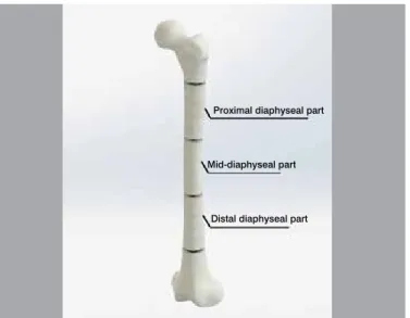
of 5 mm/min. Fifteen synthetic femur specimens for each case were tested. The femurs were loaded progressively at a speed of 5 mm/min until the occurrence of a subsequent fracture. During the tests, load-displacement (deformation) curves (Figure 3) were recorded online using the Trapexium X software, and analyzed for the following structural bone properties: yield load (the force causing the first bone damage visible in the load displacement curve, the force at which the load displacement curve broke from linearity), ultimate load (the force causing bone fracture), displacement at yield (defined as the amount of bone deformation at the yield point), and displacement at fracture (deformation at the fracture point). According to the producer, the measurement error of the method was ±1% of the recorded value. Testing after each experimental period was performed on the same day, by the same operator. The level of the subsequent fracture was determined by the distance from the upper end of the greater trochanter to the fracture line on the lateral cortex. The first 60 mm from the upper end of the greater trochanter was defined as the proximal part of the femur. Thus, the following 258 mm femoral diaphysis was divided equally into proximal, mid and distal diaphyseal parts (Figure 4). Some models showed two fracture lines. According to the level and number of subsequent fracture, six groups of subsequent fractures were observed. Implant deformation was also evaluated for plastic deformation of the plate, by inspecting each model.
The configuration of subsequent fracture was separated into four groups, based on the fracture line. A transverse fracture line originating from the
Figure 1. The experimental setup with one specimen is shown. The spec-imen is positioned anatomically on the uniaxial testing machine.
Figure 3. Symbolic load-displacement curve of a synthetic femur specimen. Figure 2. Stabilization of the models by securing the distal femur to the spherical connection adaptor with 8 pointed screws and the femoral head by a bolt pin.
Ultimate load-the force causing bone fracture
Yield load-the force causing the first damage visible
Displacement (mm)
lateral cortex that had a reverse oblique pattern on the medial cortex was placed in Group A. Group B consisted of a transverse fractures originating from the lateral cortex that had an oblique pattern on the medial cortex; Group C consisted of transverse fractures originating from the medial cortex that had a reverse oblique pattern on the lateral cortex. Group D had a fracture line related to a screw entry point (Figure 5).
Statistical Methods
The normality of distribution of the parametric data was checked using the Kolmogorov-Smirnov test. The distribution was accepted as parametric if the results of the Kolmogorov-Smirnov test were not significant. The results for yield and ultimate load to displacement, and comparisons of the subsequent fracture levels between 15
and 18 cm osteotomy groups, were subjected to the t test. Multiple pairwise comparisons were performed by the Mann- Whitney U test. The number of subsequent fracture and implant deformations was analyzed by the Chi-Square test. Kruskal-Wallis one-way analysis of variance was used to measure statistical differences between modalities, and P values < 0.05 were considered significant.
RESULTS
Fifty-three synthetic composite femurs were available for the current study. Of the sixty models, two models with inexact osteotomy levels and five models with improper data acquisition during testing were excluded from the study.
As the experiment progressed and stiffness increased, the synthetic femurs began to break at different sides in different models.
Stability and Stiffness
In all the specimens, it was noted that there was a significant differ-ence in yield displacement values between medial sided plating and the conventional lateral sided plating in the distal diaphysis of the femur specimens (Figure 6). The yield displacement values of M15, M18, L15 and 18 were 13.79 mm, 14.92 mm, 10.75 mm and 10.18 mm, respectively. The ultimate load values of M15, M18, L15 and 18 were 2826.59 N, 2556.61 N, 2456.41 N and 2326.1 N, respectively. The ultimate load values of the medial sided plating specimens at osteotomy levels 15 and 18 were higher than those of the lateral sided plating specimens (Table 1). Yield (P15=0.409, P18=0.427)
and ultimate loads to displacement (P15=0.357, P18=0.701) values
were not statistically significantly different between groups.
Figure 4. Level of the subsequent fracture. Femoral diaphysis was divided equally into proximal, mid and distal diaphyseal parts.
Figure 6. Load-displacement curves according to four different types of fixation and osteotomy levels.
Figure 5. Types of subsequent fractures. A. Transverse fracture line origi-nating from the lateral cortex had a reverse oblique pattern on the medial cortex. B. Transverse fracture originating from the lateral cortex had an oblique pattern on the medial cortex. C. Transverse fracture originating from the medial cortex had a reverse oblique pattern on the lateral cortex. D. Fracture line related to a screw entry point.
Table 1. Values for mean yield load, ultimate load and displacement at fracture according to the type of fixation and osteotomy levels.
Group Number of specimen
Mean values
Standard deviation (+)
Standard deviation (+)
Yield load (N) M15
15
2333.58 65 60
L15 1627.32 80 70
M18 1978.45 45 45
L18 1778.31 55 70
Ultimate load (N)
M15
15
2826.59 85 90
L15 2456.41 70 75
M18 2556.61 80 65
L18 2326.1 60 55
Displacement at fracture
M15
15
21.40 0.25 0.5
L15 24.80 0.3 0.6
M18 24.45 0.4 0.3
L18 60.2 0.9 0.85
M15 L15 M18 L18
Displacement (mm) Proximal diaphyseal part
Mid-diaphyseal part
Distal diaphyseal part
Load (N)
A
B
B
C
C
D
Features of Subsequent Fracture
The samples demonstrated at least one subsequent fracture. Sub-sequent fractures mostly occurred at the proximal femoral region. A single subsequent fracture was seen in 41 samples (77.3%), while 12 samples (22.7%) showed two subsequent fractures. The second frac-ture was always at the first or eighth screw hole (91.7%). In the models with two subsequent fractures, the mean distance from the greater trochanter to the first fracture line was 164.6 mm, while the distance to the second fracture line was 289.5. Subsequent fractures seen in lateral sided plating (L15 and L-18) group demonstrated a predilection for the proximal femur. In the group of medial sided plating (M-15 and M-18), subsequent fractures mostly occurred in the mid-diaphyseal region (Table 2). There were no statistically significant differences in terms of the number and location of subsequent fractures.
For the subsequent fractures, a transverse fracture line originating from the lateral cortex that had a reverse oblique pattern on the medial cortex was the most frequent fracture configuration (58.5%). This was followed by a transverse fracture originating from lateral cortex with an oblique pattern on the medial cortex (13.2%); a transverse fracture originating from the medial cortex with a reverse oblique pattern on the lateral cortex (1.9%), and a fracture line at a screw hole (26.4%), respectively.
Implant Deformation
Of the 53 synthetic femurs, plastic deformation of the plate was seen in 21 samples (39.6%) after accomplishing axial loading. Surprisingly, these deformed implants, with the exception of one at the 18 cm osteotomy, all belonged to lateral sided plating group (Figure 7). The implant deformation rate was 71.4% in the L15 group, and 76.9% in the L18 group (Table 3). There was more implant deformation in lateral sided plating group, with statistical significance (P<0.0001).
DISCUSSION
For fractures of the femoral diaphysis, from the lesser trochanter to 10 cm proximal to the knee joint, locked intramedullary nailing is the standard treatment modality. However, a plate osteosynthesis is particularly advantageous in certain situations. Patients with an excessively narrow intramedullary canal,4,5 polytrauma patients,6
and those with vascular injury associated with the femoral fractures7
constitute good indications for plate osteosynthesis.
Orthopaedic surgeons tend to attach a lateral plate in almost all cases, due to the simplicity of the procedure, and better preser-vation of the muscles, nerves and vascularization. Medial sided femoral plating has been reported less frequently for the clinical management of lower limb deformities after correction osteoto-mies.11-15 Despite its occasional use, no clinical and mechanical
information on this technique in the trauma setting has been reported. From a mechanical standpoint, our results showed no significant difference between lateral and medial sided plating by means of axial loading.
Superficial femoral vessels are injured with greater frequency than common femoral vessels in femoral fractures.16 In the setting
of femoral fractures associated with vessel injury, the vessels should be explored via a posteromedial approach to the femur. A posteromedial approach allows medial plating to the distal femur diaphyseal fracture. Therefore, fractures located in the distal diaphyseal region were used for the study.
The loading model described by Koch is used to investigate the biomechanical behavior of the femur models.17 Koch carried out
a detailed analysis of a femur without muscles and soft tissues to show that the lateral side of the femur was in tension and the medial side was in compression. An analysis of Koch’s model by Fetto et al.18 revealed that the tensile load on the lateral side
changed to a compressive load when the actions of the iliotibial band and vastus lateralis- gluteus complex were included. It was the contribution of Pauwels19 that introduced the tension band
principle, which states that tensile forces on the convex side of a curved tube can be converted to compressive forces by applying an implant to the convex side of the tube. The tension band prin-ciple works when there is anatomic apposition of cortices on the opposite side of application of plate. It does not apply when there is comminution on the opposite cortex or in comminuted fractures. Ascenzi et al.20 had demonstrated the different collagen orientation
within the femoral diaphysis as to the aspect of compression and tension. This principle was strongly propagated by AO. They offered the typical application of this principle by fixing a plate to the femur on the lateral side of the diaphysis.21 However, Cordey
et al. speculated that the tension side of the femur, which is at the lateral aspect, particularly at the proximal part, turns around the anterior aspect distally. The compression side of the femur also turns from the medial to the posterior aspect distally.8,9 This
means that when a plate is fixed to the lateral aspect of the distal femur, this plate may not be applied according to the tension
Table 2. Subsequent fracture location. Lateral sided plating group has a predilection for the proximal femur. In the group of medial sided plating, subsequent fractures mostly occurred in the mid-diaphyseal region.
Group Sample size Mean (mm) Standard deviation
Fracture location M15 13 211.69 111.70 Mid-diaphyseal
L15 14 142.00 70.31 Proximal
diaphyseal M18 13 169.69 70.56 Mid-diaphyseal
L18 13 126.23 26.24 Proximal
diaphyseal
Table 3. Implant deformation according to the groups. All deformed implants, except for one belong to lateral sided plating group.
Group Sample size Implant deformation Implant deformation
- +
M15 13 13 0 0%
L15 14 4 10 71.4%
M18 13 12 1 7.7%
L18 13 3 10 76.9%
Figure 7. Implant deformation in the lateral sided plating group. B. Excellent plate contour in the medial sided plating group.
Figure 8. Diagrammatic representation of load sparing in femoral diaphy-seal fractures. A. Medial femoral plating. By medializing the plate fixation, some of the load supported by the plate is shared by bone fragments. B. Intramedullary nailing. C. Traditional lateral femoral plating.
band principle. A plate that is fixed to the lateral or medial side of the distal femur may act as a compression device, due to the rotation of the axis, in order to produce tension at the anterior aspect and compression at the posterior aspect within the distal diaphysis of the femur.
Cordey et al.9 evaluated the strain within the diaphysis during axial
loading. They also measured high bending forces and stresses in the diaphysis, when the tension band effect of the iliotibial tract has been neglected. High bending forces were explained by the higher bending moment in the frontal plane. Because effect of the soft tissues on stability and stiffness was not taken into account, subsequent fractures mostly occurred at the proximal femoral region in the lateral sided plating group. The lack of muscle attachments will probably have a dramatic effect on the secondary fracture pattern.
The plates applied to the lateral side of the femur have more stress, as the bending moment experienced by the plate is directly related to the force of application and the distance of the implant from the force of application. The line of application of the weight-bearing force is approximately 1 to 2 cm distant from the force of appli-cation with plate fixation of the femur. Stress applied to the femur passes directly up the femoral shaft and bypasses the femur by means of absorption of stress through the distal screws into the plate, and back into the femur through the proximal screws.22
The placement of the plate relative to the loading direction will determine the proportion of the load supported by the plate. By medializing the plate fixation, some of the load supported by the plate will be shared by bone fragments (Figure 8). Thus, lateral sided plates were the only ones that experienced plastic deformation. This is because the loading path was more medial, meaning that the lateral plates were subjected to bending that the medial plates were spared.
The variability of cadaveric specimens has always been a problem, requiring enormous sample sizes to obtain satisfactorily significant results. For this reason, synthetic femurs were chosen for this study. Synthetic femurs have a standardized geometry, very small speci-men to specispeci-men variability, and material behavior approximating that of bone.23 Schoenfeld et al. looked at the pullout strength and
load to failure properties of self-tapping cortical screws in synthetic and cadaveric environments representing healthy and osteoporotic bone, and found that although the trends may be similar, screw performance in the synthetic models was markedly different from that in cadavers.24 As distal diaphyseal femoral fractures are mostly
seen in young adults, due to high energy related traumas, part of the experiment was to represent bone quality by the synthetic composite femurs. Therefore, we used a thoroughly validated model of human femur to remove these undesirable characteristics seen in cadaveric specimens.
In a situation of a vascular injury, the first step is to shunt the limb, followed by rapid external fixation, then vascular repair, immedi-ately followed by the definitive fixation, provided the patient is well enough. A midlateral approach is traditionally used for femoral fractures in which plates and screws are used for the fixation. In such cases, especially with a vascular injury, this procedure requires two separate incisions; a medial approach for the vascular repair, and a lateral approach for the fracture treatment. The soft tissue disruption associated either with open reduction and internal fixation, or with external fixation, may be reduced by using a single medial approach that allows bone stabilization under direct visualization of the repaired vessels.
In the current study, medial sided plating constructs were compara-ble in stiffness to the conventional lateral sided plating constructs. In selective cases, medial plating may be a reasonable treatment option that can be used safely.
In this study, only the primary fixation strength in a composite femur model was tested by axial loading. Torsional testing could also have been tested. It is also unknown how the results of this study translate to actual bone healing rates, loss of correction, and clinical outcome. As a limitation of our study, the failure of fixation rarely occurs with one-time high level loading. It would have been more appropriate to perform the testing in cyclic loading.
REFERENCES
1. Winquist RA, Hansen ST Jr, Clawson DK. Closed intramedullary nailing of femoral fractures. A report of five hundred and twenty cases. J Bone Joint Surg Am. 1984;66(4):529-39.
2. Krettek C, Schandelmaier P, Tscherne H. [Distal femoral fractures. Transarticular reconstruction, percutaneous plate osteosynthesis and retrograde nailing]. Unfallchirurg. 1996;99(1):2-10.
3. Kinast C, Bolhofner BR, Mast JW, Ganz R. Subtrochanteric fractures of the femur. Results of treatment with the 95 degrees condylar blade-plate. Clin Orthop Relat Res. 1989;(238): 122-30.
4. Sink EL, Hedequist D, Morgan SJ, Hresko T. Results and technique of unstable pediatric femoral fractures treated with submuscular bridge plating. J Pediatr Orthop. 2006;26(2):177-81.
AUTHORS’ CONTRIBUTIONS: Each author made significant individual contributions to this manuscript. FA (0000-0003-4208-8162)*and BT (0000-0002-0184-8850)* were the main contributors in the drafting of the manuscript. FA and Mustafa Ozmen (0000-0001-6795-2856)* evaluated the data from the statistical analysis. FA, BT and TS (0000-0002-3276-5820)* performed the literature search and review of the manuscript, and contributed to the intellectual concept of the study.. *ORCID (Open Researcher and Contributor ID).
5. Kanlic EM, Anglen JO, Smith DG, Morgan SJ, Pesántez RF. Advantages of Submuscular Bridge Plating for Complex Pediatric Femur Fractures. Clin Orthop Relat Res. 2004;426:44-51.
6. Apivatthakakul T, Chiewcharntanakit S. Minimally invasive plate osteosynthesis (MIPO) in the treatment of the femoral shaft fracture where intramedullary nailing is not indicated. Int Orthop. 2009;33(4):1119-26.
7. Sher MH. Principles in the management of arterial injuries associated with fracture/dislocations. Ann Surg. 1975;182(5):630-4.
8. Hommel GJ, Lobrano C, Ogden AL, Mukherjee DP, Anissian L, Marymont JV. A quantitative analysis of tension band plating of the femur diaphysis. Arch Orthop Trauma Surg. 2011;131(10):1325-30.
9. Cordey J, Borgeaud M, Frankle M, Harder Y, Martinet O. Loading model for the human femur taking the tension band effect of the ilio-tibial tract into account. Injury. 1999;30 Suppl 1:26-30.
10. ASTM D695-10. Standard Test Method for Compressive Properties of Rigid Plastics. ASTM. 2008;8:1-8
11. Backstein D, Morag G, Hanna S, Safir O, Gross A. Long-term follow- up of distal femoral varus osteotomy of the knee. J Arthroplasty. 2007;22(4 Suppl 1):2-6. 12. Preston CF, Fulkerson EW, Meislin R, Di Cesare PE. Osteotomy about the knee:
applications, techniques, and results. J Knee Surg. 2005;18(4):258-72. 13. Wang JW, Hsu CC. Distal femoral varus osteotomy for osteoarthritis of the knee.
J Bone Joint Surg Am. 2005;87(1):127-33.
14. Aglietti P, Menchetti PP. Distal femoral varus osteotomy in the valgus osteoarthritic knee. Am J Knee Surg. 2000;13(2):89-95.
15. McDermott AG, Finkle JA, Farine I, Boynton EL, MacIntosh DL, Gross A. Distal
femoral varus osteotomy for valgus deformity of the knee. J Bone Joint Surg Am. 1988;70(1):110-6.
16. Asensio JA, Kuncir EJ, García-Núñez LM, Petrone P. Femoral vessel injuries: Analysis of factors predictive of outcomes. J Am Coll Surg. 2006;203(4):512-20. 17. Koch JC. The laws of bone architecture. Am J Anat. 1917;21(2):177. 18. Fetto J, Leali A, Moroz A. Evolution of the Koch model of the biomechanics of
the hip: clinical perspective. J Orthop Sci. 2002;7(6):724-30.
19. Pauwels F. Die Bedeutung der Bauprinzipien des Stütz und Bewegungsap-parates für die Beanspruchung der Röhrenknocken. Z Anat Entw Gesch. 1948;114:129-66.
20. Ascenzi A, Improta S, Portigliatti Barbos M, Carando S, Boyde A. Distribution of lamellae in human femoral shafts deformed by bending with inferences on mechanical properties. Bone. 1987;8(5):319-25.
21. Müller ME, Allgöwer M, Schneider R, Willenegger H. Manual of internal fixation. 2nd ed. Berlin: Springer, 1979.
22. Mooney V, Claudi B. Fractures of the shaft of the femur. In: Fractures. Rockwood, CA. Jr.; Green, DP., eds. Philadelphia, JB Lippincott; 1975.p.1093. 23. Papini M, Zdero R, Schemitsch EH, Zalzal P. The biomechanics of
hu-man femurs in axial and torsional loading: comparison of finite element analysis, human cadaveric femurs, and synthetic femurs. J Biomech Eng. 2007;129(1):12-9.