Application of synthetic peptides to the diagnosis
of neurocysticercosis
Agne`s Fleury1,2, Constantino Beltran3, Elizabeth Ferrer4, Teresa Garate4, Leslie J. S. Harrison5, R. Michael E. Parkhouse6, Esperanza Garcia1, Gladis Fragoso3, Julia Costa-Cruz7, Germano Biondi8, Svetlana Agapejev9 and Edda Sciutto3
1Instituto Nacional de Neurologı´a y Neurocirugı´a, Mexico, D.F. Mexico
2Institut d’Epidemiologie Neurologique et de Neurologie Tropicale, Limoges, France
3Instituto de Investigaciones Biomedicas, Universidad Nacional Autonoma de M exico, Mexico, D.F. Mexico 4Instituto de Salud Carlos III, Majadahonda, Madrid, Spain
5Department of Tropical Animal Health, University of Edinburgh, Roslin, UK 6Instituto Gulbenkian de Ciencia, Oeiras, Portugal
7Laboratorio de Parasitolog ıa, Universidad Federal de Uberlandia, Uberlandia, Brazil 1
8Faculdade de Medicina Veterinaria e Zootecnia, Universidad Estadual Paulista, Botucatu, Brazil 9Faculdade de Medicina, Universidad Estadual Paulista, Botucatu, Brazil
Summary We tested the possible diagnostic utility of fiveTaenia saginataoncosphere-derived synthetic peptides in T. soliumneurocysticercosis (NC). The five peptides correspond to protein sequences with high antigenic indexes that were cloned from aT. saginataoncosphere cDNA library. The test samples consisted of cerebrospinal fluid (CSF) samples randomly collected from patients referred from Mexican and Brazilian neurological institutes. Indirect enzyme-linked immunosorbent assays (ELISA) were carried out with the peptides either unconjugated or coupled to carrier proteins, and were compared with results obtained usingT. soliumcyst fluid as a positive control. For active inflammatory NC, the higher sensibility (93%) and specificity (85%) was obtained with peptides HP6-2 and Ts45W-1, respectively, coupled to ovalbumin, in both Mexican and Brazilian patients. Examining the results of the individual peptide assays in combination, in some instances, improved the sensitivity to 100%.
keywords peptides,Taenia solium, neurocysticercosis, diagnosis
Introduction
Neurocysticercosis (NC) is the most common worldwide parasitic disease of the central nervous system and its prevalence is highest in developing countries (Del Brutto et al.1998). In recent years, human cases of NC have emerged in developed countries, such as the USA and Europe, mostly because of migrant workers (Shandera et al.1994; Rousseauet al.1999
2,3 ; Carangeloet al.2001
2,3 ;
Terrazaet al.2001). Accurate diagnosis of NC still requires cranial imaging technology such as computerized tomography (CT) and/or magnetic resonance imaging (MRI), techniques that are expensive and frequently inaccessible to people of endemic areas (Sciuttoet al. 2000
4 ). The diagnosis of NC without imaging studies is difficult because of the highly variable clinical symptoms, ranging from asymptomatic to a very severe neurological
syndrome with intracranial hypertension or dementia, with a variety of non-specific mild clinical symptoms in between.
For immunological diagnosis, the enzyme-linked immunosorbent assay (ELISA) and the enzyme-linked immunotransfer blot (EITB) are the most widely used techniques (Rosas et al. 1986; Garcı´aet al. 1995
5 ).
employed. There is therefore an inherent advantage of reproducibility in diagnostic tests based on recombinant proteins or synthetic peptides (Gevorkianet al. 1996; Herna´ndez et al. 2000; Ferreret al. 2003). Such a permanent source of diagnostic reagents would also be useful in traditionally non-endemic countries at risk of sporadic incursions of the parasites, or where the collection ofT. solium cysticerci is difficult.
Recently, synthetic peptides based on antigenic sequences identified in immunogentic oncosphere proteins of Taenia saginatahave been shown to function well in the diagnosis of bovine cysticercosis (Ferrer et al. 2003), and hence we evaluated them for the diagnosis of human NC using samples from Mexico and Brazil. The five peptides consist of: one peptide (HP6-3) derived from the sequence of the 18 kDaT. saginata surface/ secreted oncospheral adhesion antigen identified by McAb-HP6, two peptides (Ts45W-1 and Ts45W-5) derived from the sequence of the T. saginatahomologue of theT. ovis 45W protective gene family, one peptide (TS45S-10) derived from a T. saginata sequence with significant similarity to theT. ovis 45S protective antigen, and one peptide (TEG-1) derived from the sequence of theT. saginata homologue of Echinococcus spp. main surface protein. The corresponding proteins, which may also be potential protective antigens, consist of: the 18 kDa T. saginata surface/secreted oncospheral adhesion antigen identified by McAb-HP6 (Benı´tez et al. 1996; Bonay et al. 2002; Ferrer et al. 2003), the T. saginatahomologue of the T. ovis 45W protective gene family (Johnsonet al. 1989; Waterkeyn et al. 1995; Ferreret al. 2003), the T. saginata
homologue ofT. ovis 45S protective antigen (Waterkeyn et al. 1995; Ferreret al. 2003) and the T. saginata homologue ofEchinococcus spp. surface protein (Benı´tezet al. 1998).
Material and methods Cerebrospinal fluid
Cerebrospinal fluid (CSF) samples were collected in Mexico and Brazil. The Mexican samples formed three clinically distinct groups: 27 CSF from patients (12 men and 15 women, mean age 40 ± 13 years) with radiologi-cally diagnosed NC taken before any treatment, 16 CSF from patients with clinically diagnosed NC taken 1–12 months after specific drug treatment (albendaz-ole +prednisona) and 31 CSF from non-NC control patients. We also collected and tested 11 CSF of the 27 NC patients 1–8 months after cysticidal treatment (albendaz-ole + prednisona).
The CSF samples from Brazil were obtained by sub-occipital puncture at the Neurological Department of the Medicine, University of Botucatu and consisted of 24 CSF samples from control, non-NC neurological patients and 32 from diagnosed NC patients (24 men and eight women, mean age 38.6 ± 9.4 years), 1–360 days after drug treat-ment (albendazole).
Clinical details and characteristics of each CSF sample, including the number of cells, were obtained from the medical records. The diagnosis of NC, as determined by neuroimaging, was classified as ‘active’ when at least one viable cysticercus or arachnoiditis was present, and cases were classified as either single- or multiple-viable cysts. Alternatively, when neuroimaging studies only detected calcifications or meningeal fibrosis, which are the per-manent sequelae of previous infections (Sotelo et al. 1985), the disease was classified as ‘inactive’. Active cysticercosis patients were further classified as ‘inflam-matory’ or ‘non-inflam‘inflam-matory’ by counting mononuclear cells in their CSF; inflammatory being defined as having more than six mononuclear cells per cubic millimetre. Controls were patients for whom the diagnosis of NC had been excluded by neuroimaging, and who were seen in neurological institutes for inflammatory, vascular, infectious and degenerative disease.
Peptides
The peptides (Ferreret al.2003) examined in this study and described in the introduction are summarized in Table 1. Peptides were synthesized by stepwise solid-phase synthesis with Na
-tert-butyloxycarbonyl (BOC) derivatives ofl-amino acids on phenyl-acetamidomethyl (PAM) resin (Sigma Chemical Co, St Louis, MO). The peptides were 95% pure as judged by high-performance liquid chroma-tography (HPLC) on analytical C18reversed-phase column (3.9·150 mm; Delta Pak, Waters). The correct amino acid sequence of each peptide was confirmed by protein sequencing on a pulsed liquid-phase protein sequencer (Applied Biosystems) at the National Institute of Cardiol-ogy, Mexico City. Peptides were coupled to maleimide activated ovalbumin (OVA) and keyhole limpet haemo-cyanin (KLH) (Pierce Warriner Ltd.) following the manufacturer’s instructions.
Antibody detection by ELISA
check for reproducibility. A sample was considered positive if the specific optical density (OD) values were greater than the mean of the values obtained with negative CSF samples plus two standard deviations (SD). Five to 10 negative CSF from confirmed non-NC neuro-logical patients different from those included in the study to determine the specificity were used in each plate to determine the cut-off value.
Statistical analysis
Sensitivity was calculated as the number of proven NC cases that were ELISA positive, and specificity as the number of negative control samples that were ELISA negative. Dichotomous data were compared using the chi-square Pearson test or the Yates corrected chi-square test. Differences in mean values between groups were Table 1 The amino acid sequences and Genebank numbers of the peptides selected for study
12
Protein description and Genebank numbers
Sequence designation
Peptide designation
Number of
amino acids Amino acid sequence
Major secreted ‘functional’ antigen
identified by McAb HP6 (Genebank No. X95983)
HP6 HP6-3 15 CRFVKTPSKKKTKSR
Taenia ovis45W gene family homologue (Genebank no. AJ430567)
Ts45W Ts45W-1 24 AYEQPIERTVVGHQTLRDIFVWGC
Ts45W-5 10 CGGTEESVVTASRS
Taenia ovis45S homologue (Genebank no. AJ430566)
Ts45S Ts45S 10 14 CGGTEESVVTASRS
Echinococcussurface protein homologue (Genebank no. X97000)
TEG TEG-1 23 CRDPSKMRDIDRHHEYNVREGND
Table 3 Sensitivity of ELISA in CSF using VFA or different free synthetic peptides as a source of antigen (Mexican and Brazilian drug-treated patients)
13
Mexico Brazil
Active non-inflammatory (n¼8)
Active inflammatory (n¼8)
Active non-inflammatory (n¼3)
Active inflammatory
(n¼9) Inactive (n¼7)
Vesicular fluid 87.5 (0.88 ± 0.8) 100 (1.3 ± 0.58) 100 (0.68 ± 0.24) 100 (1.28 ± 0.34) 57 (0.42 ± 0.5) Ts45W-1 12.5 (0.13 ± 0.08) 62.5 (0.34 ± 0.55) 0 (0.09 ± 0.007) 77.8 (0.71 ± 0.45) 0 (0.086 ± 0.013) Ts45W-5 12.5 (0.12 ± 0.08) 50 (0.34 ± 0.5) 0 (0.12 ± 0.02) 77.8 (0.72 ± 0.55) 0 (0.094 ± 0.02) HP6-3 12.5 (0.19 ± 0.08) 50 (0.38 ± 0.43) 0 (0.18 ± 0.02) 77.8 (0.84±0.58) 0 (0.14 ± 0.03) TEG-1 12.5 (0.11 ± 0.05) 50 (0.29 ± 0.45) 0 (0.1 ± 0.04) 77.8 (0.5 ± 0.45) 0 (0.09 ± 0.02) Ts45S 10 12.5 (0.11 ± 0.09) 62.5 (0.26 ± 0.27) 0 (0.13 ± 0.01) 77.8 (0.63 ± 0.49) 0 (0.11 ± 0.02)
The values are sensitivity values and those in parentheses are OD values (mean ± SD). VFA, vesicular fluid antigen.
Table 2 Range of age of Mexican and Brazilian drug-treated patients with different clinical NC
Range of age (years)
Mexico Brazil
Active inflammatory
Active
non-inflammatory Active inflammatory
Active
non-inflammatory Inactive
0–10 0 0 0 0 0
11–20 1 0 0 0 2
21–30 1 4 1 0 3
31–40 6 2 2 3 2
41–50 6 1 17 0 1
51–60 3 1 0 0 0
analysed by Student’st-test or by a non-parametric test (Mann–Whitney).
Results
The five peptides were first tested in a free unconjugated form and the most obvious conclusion is that the peptides function best for antibody detection in cases of active inflammatory NC (Table 3), especially in patients aged 30–50 years (Table 2).
As different peptides do not necessarily bind with equal efficiencies to ELISA plates, as an attempt to standardize conditions, the five peptides were coupled to protein carriers, OVA and KLH. These preliminary experiments indicated that there was no significant difference in the results obtained using free simple peptides or peptides coupled to the carrier proteins (Table 4).
These peptide carrier protein conjugates were used as probes for the Mexican and Brazilian CSF samples. Employing Mexican active inflammatory NC, the sensi-tivity using Ts45W-5 and HP6-3 coupled to KLH or Ts45W-1, HP6-3, TEG-1 and Ts45S coupled to OVA was as good as that obtained using cyst fluid antigen. Similarly, with the Mexican active non-inflammatory NC, peptide HP6-3 OVA gave the same sensitivity as cyst fluid antigen (Table 5). Finally, using Ts45W-1, Ts45W-5 and HP6-3 coupled to OVA with the Brazilian active inflammatory NC, the same sensitivity was obtained as with cyst fluid antigen (Table 5).
As immune responses to parasites are characteristically highly heterogeneous, the effect of combining the results from certain individual peptide assays was examined. With the Mexican samples, a combination of HP6-3 OVA and Ts45W-1 OVA gave a sensitivity and specificity quite similar to those obtained with cyst fluid (Tables 5 and 6). Similarly, with the Brazilian samples, combinations of Ts45W-5 OVA and Ts45S OVA or TEG-1 OVA also indicate an improved sensitivity (Table 5) with a good specificity (Table 6), which were between 93% and 100%. Table 4 Sensitivity (%) of ELISA using free unconjugated peptides or coupled with carrier proteins (Brazilian CSF samples from eight treated patients)
Free unconjugated
peptide KLH OVA
Ts45W-1 75 75 87.5
Ts45W-5 75 75 87.5
HP6-3 87.5 75 87.5
TEG-1 75 75 87.5
Ts45S 10 75 75 87.5
Sensitivity¼number of positives/number of sample tested.
Interestingly, cysticidal and corticosteroid treatment did not significantly modify CSF levels of antibodies or sensitivity of the assay employing the different peptides as a source of antigen (Table 7).
Discussion
Diagnosis of human, bovine and porcine cysticercosis still relies on the detection of antibodies using parasite extracts
or multi-component glycoprotein fractions. The use of sufficiently sensitive and specific recombinant proteins or synthetic peptides as antigens in such assays would provide the much needed reproducibility and circumvent reliance on the less reproducible parasite extracts. Five peptides of high antigenic indices from the sequences of four poten-tially protectiveT. saginataproteins already shown to have utility in the diagnosis ofT. saginatacysticercosis (Ferrer et al.2003) were tested as possible reagents for the diagnosis of human neurocysticercosis. It is worth mentioning that the HP6-3 peptides were taken from the sequence of a oncospheralT. saginataantigen with proven protective capacity against bovineT. saginatacysticercosis (LJS Harrison and RME Parkhouse, unpublished data
6 ),
and will be of interest to determine the relevance of homologous anti-HP6-3 antibodies in the human disease.
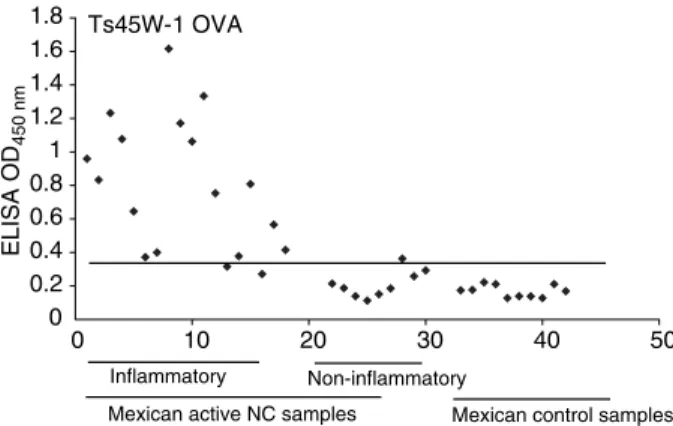
Most of the data presented was obtained with peptide– protein conjugates. As seen in Table 4 these peptides gave comparable results in soluble form or coupled to proteins (OVA or KLH). Peptides HP6-3 and Ts45W-1 coupled to OVA detected the majority of the active inflammatory NC patients [83.3% (Figure 1) and 88.9% (Figure 2) of the Mexican, respectively, and 85% of the Brazilian]. Notably, as active NC was mainly found in patients between 30 and 50 years of age, the test could be more effective in this age group. The fact that the results obtained with the Mexican samples mirror those of the Brazilian samples suggests that similar antigenic epitopes are shared by T. soliumfrom these two countries. More than 45% of these patients also recognized the other four peptides that were tested, a fact that points to a low variability of cysticercal antigens and an important cross-immunity Table 6 Specificity (%) of ELISA in CSF using VFA or different
synthetic peptides coupled with carrier protein as a source of antigen (Mexican and Brazilian subjects)
Neurological patients (No NC)
Mexican (n¼31) Brazilian (n¼24)
Vesicular fluid 96.8 (0.11 ± 0.05) 95.8 (0.08 ± 0.03) Ts45W-1 KLH 96.8 (0.16 ± 0.04) 100 (0.25 ± 0.04) Ts45W-5 KLH 93.5 (0.15 ± 0.04) 94.4 (0.1 ± 0.06) HP6-3 KLH 93.5 (0.13 ± 0.03) 95.8 (0.25 ± 0.1) TEG-1 KLH 100 (0.14 ± 0.04) 95.8 (0.18 ± 0.13) Ts45S 10 KLH 96.8 (0.14 ± 0.04) 95.8 (0.13 ± 0.07) Ts45W-1 OVA 93.5 (0.18 ± 0.06) 91.7 (0.16 ± 0.1) Ts45W-5 OVA 96.8 (0.18 ± 0.05) 91.7 (0.16 ± 0.1) HP6-3 OVA 93.5 (0.17 ± 0.05) 100 (0.38 ± 0.16) TEG-1 OVA 96.8 (0.22 ± 0.1) 95.8 (0.08 ± 0.04) Ts45S 10 OVA 93.5 (0.2 ± 0.07) 95.8 (0.1 ± 0.08) HP6-3 OVA +
Ts45W-1 OVA
90.3
Ts45w-5 OVA + Ts45S 10 OVA or TEG-1 OVA
87.5
Values in parentheses are OD values (mean ± SD).
Table 7 Sensitivity of the antibody detection assay before and after specific treatment in 11 Mexican NC patients
Sensitivity (%)
Before treatment After treatment*
Vesicular fluid 100 (1.74 ± 0.72) 100 (1.45 ± 0.75) Ts45W-1 KLH 45.4 (0.55 ± 0.26) 72.7 (0.58 ± 0.20) Ts45W-5 KLH 63.6 (0.56 ± 0.31) 72.7 (0.57 ± 0.20) HP6-3 KLH 81.8 (1.21 ± 0.58) 72.7 (1.01 ± 0.28) TEG-1 KLH 63.6 (0.56 ± 0.26) 54.5 (0.51 ± 0.18) Ts45S 10 KLH 63.6 (0.37 ± 0.13) 81.8 (0.38 ± 0.13) Ts45W-1 OVA 72.7 (0.64 ± 0.43) 72.7 (0.55 ± 0.27) Ts45W-5 OVA 45.4 (0.56 ± 0.38) 54.5 (0.53 ± 0.26) HP6-3 OVA 81.8 (1.30 ± 0.52) 81.8 (1.05 ± 0.36) TEG-1 OVA 81.8 (0.36 ± 0.16) 54.5 (0.33 ± 0.11) Ts45S 10 OVA 72.7 (0.65 ± 0.41) 63.6 (0.57 ± 0.24)
* These CSF were collected 1–8 months after treatment. Values in parentheses are OD values (mean ± SD).
HP6-3 OVA
0 0.5 1 1.5 2 2.5 3
0 10 20 30 40 50 60
ELISA OD
450 nm
Mexican active NC samples Inflammatory Non-inflammatory
Mexican control samples
Figure 1
between cestodes. A combination of two peptides [HP6-3 (OVA) and Ts45W-1 (OVA)] reached a sensitivity of 100% and a specificity as good as that obtained using cyst fluid and thus merits serious consideration for the design of a diagnostic kit.
In summary, this study has identified peptides of potential use in NC diagnosis. Simple and reproducible antibody detection assays for NC would find utility in hospitals and in epidemiological studies in endemic areas and would also be a useful resource in non-endemic countries to determine possible exposure to the parasite.
Acknowledgements
This investigation was partially supported by Consejo Nacional de Ciencia y Tecnologı´a, G25955m, British Council, Direccion General de Personal Academico 212401, Universidad Nacional Autonoma de Mexico, the European Union INCO-DC STD4 Programme (Project CT95-0002), Programme ECOS/ANUIES M99SO3, The Howard Hughes Medical Institute and the French Embassy in Mexico.
References
Benı´tez L, Garate T, Harrison LJS, Kirkham P, Brookes SM & Parkhouse RME (1996) Cloning and sequencing of the gene encoding the principal 18-kDa secreted antigen of activated oncospheres ofTaenia saginata.Molecular Biochemistry and Parasitology78, 265–268.
Benı´tez L, Harrison LJS, Parkhouse RME, Gonzalez LM, Gottstein B & Garate T (1998) Sequence and immunogenicity of the
Taenia saginatahomologue of the major surface antigen of Echinococcusspp.Parasitology Research84, 426–431. Bonay P, Gonzalez LM, Benı´tez Let al.(2002) Genomic and
functional characterisation of a secreted antigen ofTaenia saginataoncospheres.Molecular Biochemistry and Parasitology 121, 269–273.
Carangelo B, Erra S, Del Basso De Caro MLet al.(2001) Neurocysticercosis. Case report.Journal of Neurosurgery Science1, 43–46.
Del Brutto OH, Sotelo J & Roman GC (1998)Neurocysticercosis. A Clinical Handbook. Swets & Zeitlinger, Netherlands
7 .
Ferrer E, Benı´tez L, Foster Met al.(2003)Taenia saginata derived synthetic peptides with potential for the diagnosis and immunoprophylaxis of bovine cysticercosis.Veterinary Parasitology111, 83–94
8 .
Garcı´a E, Ordon˜ez G & Sotelo J
9 (1995) Antigens fromTaenia
crassicepscysticerci used in complement fixation, enzyme-linked immunosorbent assay, and Western blot (immunoblot) for diagnosis of neurocysticercosis.Journal of Clinical Microbiology33, 3324–3325.
Gevorkian G, Manoutcharian K, Larralde Cet al.(1996) Immuno-dominant synthetic peptides ofTaenia crassicepsin murine and human cysticercosis.Immunology Letters49, 185–189. Herna´ndez M, Beltra´n C, Garcı´a Eet al.(2000) Cysticercosis:
towards the design of a diagnostic kit based on synthetic peptides.Immunology Letters71, 13–17.
Johnson KS, Harrison GB, Lightowlers MWet al.(1989) Vac-cination against ovine cysticercosis using a defined recombinant antigen.Nature338, 585–587.
Ordon˜ez G, Medina MT & Sotelo J (1996) Immunoblot analysis of sera and CSF from patients with various forms of neurocys-ticercosis.Neurology and Infectious Epidemiology1, 57–61. Ramos-Kuri M, Montoya RM, Padilla Aet al.(1992)
Immuno-diagnosis of neurocysticercosis. Disappointing performance of serology (enzyme-linked immunosorbent assay) in an unbiased sample of neurological patients.Archives of Neurology49, 633–636.
Rosas N, Sotelo J & Nieto D (1986) ELISA in the diagnosis of neurocysticercosis.Archives in Neurology43, 353–356. Rousseau MC, Guillotel B & Delmont J (1999) Neurocysticercosis
in the South-East of France 1988–1998.Presse Medicale 39, 2141–2144.
Sciutto E, Fragoso G, Fleury Aet al.(2000).Taenia solium disease in humans and pigs: an ancient parasitosis disease rooted in developing countries and emerging as a major health problem of global dimensions.Microbes Infection2, 1875–1890.
Shandera WX, White AC Jr, Chen JC, Diaz P & Armstrong R (1994) Neurocysticercosis in Houston, Texas. A report of 112 cases.Medicine73, 37–52.
Sotelo J, Guerrero V & Rubio F (1985) Neurocysticercosis: a new classification based on active and inactive forms. A study of 753 cases.Archives of Internal Medicine145, 442–445.
Terraza S, Pujol T, Gascon J & Coracha´n M (2001)
Neurocysticercosis: an imported disease?Medicina Clinica116, 261–263.
Ts45W-1 OVA
0 0.2 0.4 0.6 0.8 1 1.2 1.4 1.6 1.8
0 10 20 30 40 50
ELISA OD
450 nm
Mexican active NC samples
Inflammatory Non-inflammatory
Mexican control samples
Tsang VC, Brand JA & Boyer AE (1989) An enzyme-linked immunoelectrotransfer blot assay and glycoprotein antigens for diagnosing human cysticercosis (Taenia solium).Journal of Infectious Diseases159, 50–59.
Waterkeyn JG, Lightowlers MW, Coppel R & Cowman AF (1995) Characterization of the gene family encoding a host-protective antigen of the tapewormTaenia ovis.Molecular Biochemistry and Parasitology73, 123–131.
Authors 10
Agne`s FleuryandEsperanza Garcia, Instituto Nacional de Neurologı´a y Neurocirugı´a, Insurgentes Sur 3877, 14269 Mexico, D. F. Mexico. Tel./Fax: (52-55) 5606-4040; E-mail: afleury@biomedicas.unam.mx
Constantino Beltran,Gladis FragosoandEdda Sciutto(corresponding author), Instituto de Investigaciones Biomedicas, UNAM, A.P. 70228, 04510 Mexico, Mexico. Tel.: +52-55-5622-3818; Fax: +52-55-5622-3369; E-mail: edda@servidor.unam.mx, gladis@servidor.unam.mx
Elizabeth FerrerandTeresa Garate, Instituto de Salud Carlos III, Majadahonda, Crta. Majadahonda Pozuelo Km2, 28220 Madrid, Espan˜a. Tel.: +34-91-509 7901; Fax: +34-91-509 7966; E-mail: tgarate@isciii.es
Leslie J. S. Harrison, University of Edinburgh, Department of Tropical Animal Health, Sir Alexander Robertson Centre for Tropical Veterinary Medicine, Easter Bush Veterinary Centre, Easter Bush, Roslin EH25 9RG, UK. Tel./Fax: +44-131-650 6246; E-mail: leslie.harrison@ed.ac.uk
R. Michael E. Parkhouse, Instituto Gulbenkian de Ciencia, Rua Quinta Grande 6, PO Box 2781, Oeiras, Portugal. Tel.: +35-121-440 7900; Fax: +35-121-440 7970; E-mail: parkhouse@igc.gulbenkian.pr
Julia Costa-Cruz, Laboratorio de Parasitologı´a, Universidad Federal de Uberlandia, Av. Para´ 1720, 38400-90Z, Uberlandia MG, Brazil. Tel./Fax: +55-34-3218 2333; E-mail: costacruz@ufu.br