Metabolites of interest for food technology produced by microalgae
from the Northeast Brazil
1Metabólitos de interesse à tecnologia de alimentos produzidos por microalgas do
Nordeste do Brasil
Katharina Kardinele Barros Sassi2*, João Andrade da Silva2, Clediana Dantas Calixto3, Roberto Sassi3 and Cristiane Francisca da Costa Sassi3
ABSTRACT - There is an increasing demand for bioprospection focusing on microalgae isolated from the northeastern region of Brazil with potential importance for food industries. To attend that need, we evaluated the characteristics of 12 regional species of microalgae grown under controlled cultivation conditions (temperature = 24 ± 1 ºC, illumination 150 µmol photons m-2 s-1, photoperiod of 12 h) in terms of their nutritional quality and lipid profiles. Significant differences in growth
characteristics and chemical compositions were observed among the species investigated. High carbohydrate contents (> 25 g 100 g-1) were recorded in various strains of Chlorococcum and the marine microalgaAmphidinium carterae;
high protein contents (> 35 g 100 g-1) were observed inScenedesmus acuminatusandPediastrum tetras; and high lipid
contents (> 25 g 100 g-1) inA. carterae and some strains ofChlorococcum sp. (cf.hypnosporum).Chlamydomonas sp.
demonstrated the greatest production of carotenoids (64.92 mg g-1), chlorophyll-a (234.74 mg g-1), and chlorophyll-b
(59.34 mg g-1). The lipid profiles of Chlorella cf.minutissima, four strains ofChlorococcum sp. (cf. hypnosporum),
P. tetras, Planktothrix isothrix, and S. acuminatus indicated the presence of palmitic, oleic (ω-9), linoleic (ω-6) and
α-linolenic (ω-3) acids, with more than 50% omegas in the total composition of their fatty acids. In terms of chemical nutrients, the microalgae cited were found to be potential sources of omegas, carotenoids, and chlorophylls that could be used in food industries.
Key words: Polyunsaturated fatty acids. Carotenoids. Omegas. Microalgae cultivation.
RESUMO - Existe uma demanda por pesquisas prospectivas de microalgas isoladas da região Nordeste do Brasil que possam ser potencialmente importantes à indústria de alimentos. Nesta pesquisa foram avaliadas as características da cinética de crescimento sob condições controladas de cultivo (temperatura = 24 ± 1 ºC, iluminação 150 µmol fótons m-2 s-1, fotoperíodo de
12 h), a qualidade nutricional e o perfil lipídico de doze espécies regionais de microalgas visando suprir esta lacuna. Diferenças nas características de crescimento e na composição química foram observadas nas espécies pesquisadas, com maiores teores de carboidratos (> 25 g 100 g-1) registrados em várias cepas deChlorococcume na microalga marinhaAmphidinium carterae,
de proteína (> 35 g 100 g-1) emScenedesmus acuminatusePediastrum tetras e de lipídios (> 25 g 100 g-1) emA. carteraee em
algumas cepas deChlorococcumsp. (cf. hypnosporum). AChlamydomonassp. apresentou os maiores teores de carotenoides
(64,92 mg g-1), clorofila-a (234,74 mg g-1) e clorofila-b (59,34 mg g-1). O perfil lipídico evidenciou a presença dos ácidos
palmítico, oleico (ω-9), linoleico (ω-6) e α-linolênico (ω-3), com mais de 50% de ômegas em sua composição total de ácidos graxos, emChlorellacf. minutissima, em quatro cepas deChlorococcumsp. (cf. hypnosporum),P. tetras,Planktothrix isothrix
eS. acuminatus. No que concerne aos nutrientes químicos foi observado que as microalgas citadas são fontes potenciais de
produção de ômegas, carotenoides e clorofilas para serem utilizados na indústria de alimentos. Palavras-chave: Ácidos graxos poli-insaturados. Carotenoides. Ômegas. Cultivo de microalga. DOI: 10.5935/1806-6690.20190007
*Author for correspondence
Received for publication in 22/02/2016; approved in 02/04/2018
1Parte da Tese de Doutorado da primeira autora apresentada ao Programa de Pós-Graduação em Ciência e Tecnologia de Alimentos - Universidade
Federal da Paraíba/UFPB
2Programa de Pós-Graduação em Ciência e Tecnologia de Alimentos, Centro de Tecnologia, Universidade Federal da Paraíba,Campus I, João
Pessoa-PB, Brasil, 58.051-900, kardinele@yahoo.com.br, joaoctdr@gmail.com
3Laboratório de Ambientes Recifais e Biotecnologia com Microalgas/LARBIM, Centro de Ciências Exatas e da Natureza, Universidade Federal da
INTRODUCTION
Recent research has analyzed the chemical compositions of microalgae and certified their positive contributions to human health and possible uses as food resources (ANDRADEet al., 2018; HAYESet al., 2018; SATHASIVAM; KI, 2018). Theoretically, microalgae are capable of producing more lipids than any other conventional crop, and numerous species can synthesize considerable quantities of essential fatty acids (EFA), especially omega-3 (ω-3) and 6 (ω-6) (the two most abundant), as well as α-linolenic acid (ALA, C18:3 ω -3) and linoleic acid (AL, C18:2 ω-6) (BELLOU et al., 2016; HO et al., 2014) – both precursors in the human body to long chain (≥ C20) polyunsaturated fatty acids (PUFA) (RINCÓN-CERVERA et al., 2016). Those microorganisms could be used as sustainable sources of EFA for human consumption or for use in animal rations as replacements for fish oil, which is now their principal source (RYCKEBOSCHet al., 2014).
Western diets are currently excessive in terms of ω-6, but deficient in ω-3, resulting in a imbalance in the ω-6:ω-3 ratio that can interfere in the conversion of ω-3 ALA into eicosapentaenoic acid (EPA, C20:5) and docosahexaenoic acid (DHA, C22:6). That lack of conversion can result in increased levels of arachidonic acid (AA, C20:4 ω-6) in the phospholipid membranes that, over time, result in the excessive production of pro-inflammatory eicosanoids and the consequent hardening and contraction of blood vessels, increasing pain transmission, immunosuppression and, the pathogenesis of cardiovascular, inflammatory, and autoimmune diseases, as well as cancer – as increasing levels of ω-3 exert a suppressor effect (RUBIO-RODRÍGUEZ et al., 2010; SUBASH-BABU; ALSHATWI, 2018). There is not yet a consensus, however, in respect to optimal ω-6:
ω-3 ratios, nor sufficient evidence to define the maximum tolerable dose, although some researchers suggest that the ratios between those acids should lie between 4-5:1, but never above 10:1 (CANDELA; LÓPEZ; KOHEN, 2011; WARNERet al., 2017).
The present work sought to characterize and compare the lipid profiles of 12 regional strains of microalgae isolated from marine and Different freshwater environments in northeastern Brazil to determine if they produce significant levels of omegas ω-3, ω-6, and ω-9 and could serve as alternative sources of compounds of interest to the food industry. The nutritional qualities of those algal strains were also investigated in terms of their percentage contents of carbohydrates, proteins, lipids, carotenoid pigments and chlorophyll-a and b, as well as their growth characteristics in mono-specific cultures under controlled conditions.
MATERIALS AND METHODS
Biomass production and growth characteristics
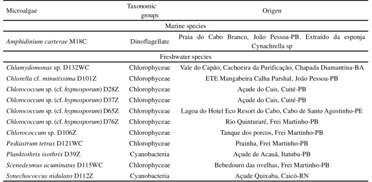
Twelve regional strains of microalgae maintained that the Microalgae Collection of the Laboratory of Reef Environments and Biotechnology with Microalgae (LARBIM/UFPB) were investigated, including 11 freshwaters and one marine species. All of the strains were isolated from distinct aquatic environments in northeastern Brazil: nine from Paraíba State (PB), one from Bahia State (BA), one from Pernambuco State (PE), and one from Rio Grande do Norte State (RN) (Table 1).
The species were cultivated in triplicate in flat bottomed 6 L flasks containing 5 L of Conway media (WALNE, 1966) for the cultivation of the marine microalga, or 5 L of Zarrouk medium (ZARROUK, 1966) for cultivating Chamydomonas, Pediastrum, and Scenedesmus, or WC medium (GUILLARD; LORENZEN, 1972) for cultivating the other freshwater strains. The microalgae were grown in culture chambers (24 ± 1 ºC) under a light intensity of approximately 150 µmol photons m-2 s-1, furnished by 40 W fluorescent lamps, under a 12 h photoperiod, with continuous forced air (0.1 L min-1) injection.
Culture growth was accompanied by measuring “in vivo” fluorescence (Turner Design Fluorometer) and by cell counts using Fuchs–Rosenthal or Sedgewick-Rafter chambers (for filamentous forms) as viewed under a Leica binocular microscope. Growth curves were prepared for each species, allowing calculations of their growth velocity (k), expressed as the number of cell divisions per day (STEIN 1973), the duration (in days) of their log phase, maximum cell density (MCD), culture time, final biomass yield, and biomass productivity per day. Upon reaching their respective stationary phases, the cultures were interrupted and the biomasses produced were concentrated by centrifuging (at 18oC), frozen at -30 °C, and subsequently lyophilized. The dry biomasses were weighed using an analytical balance and maintained under refrigeration (9 ºC) until analyzed.
Chemical analyses
Table 1 -List of the microalgae investigated in the present project, citing their respective taxonomic groups and origins
Statistical analyses
All of the data obtained from the analyses were submitted to statistical treatments usingStatistica 7.0 software, at a 5% level of significance. The homoscedasticity of the variances of all of the variables analyzed were confirmed using the Levene test. The differences among the variables analyzed among the species were compared using one-way ANOVA and the Tukey HSD a posteriori test.
RESULTS AND DISCUSSION
Kinetic growth characteristics
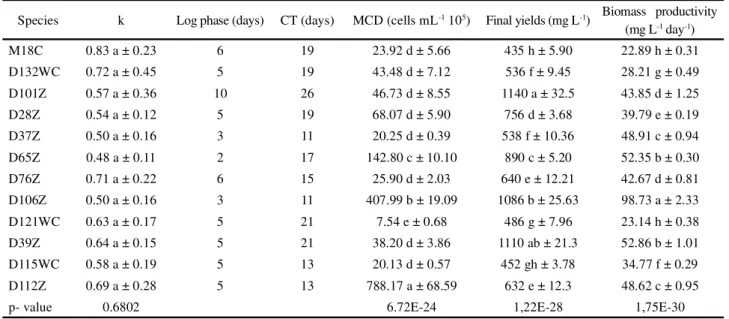
The kinetic growth characteristics of the microalgae species investigated here are listed in Table 2. The statistical analyses verified significant differences in terms of their maximum cell density values (MCD) (F = 363,7311; gl = 11; p<0.01; Table 2), final yields (F = 907,6998; gl = 11; p<0.01; Table 2), and biomass productivity (F = 1294,2997; gl = 11; p<0.01; Table 2). The growth velocities (k) did not significantly differ between the different species analyzed (F = 0.7561; gl = 11; p = 0.6802; Table 2).
Not all of the species that demonstrated highk values produced large quantities of biomass or rapidly reached their log phase. Similarly, not all of the species with large k values produced the highest cell concentrations (Table 2). Those data indicated that each species responded differently to the culture conditions, and reinforced the importance of
determining the unique behaviors of the different strains potentially useful for biotechnological applications under controlled conditions. Significant differences were observed between clones of the same species, such as clones D28Z, D37Z, D65Z and D76Z of Chlorococcum sp. (cf. hypnosporum) (Table 2), which demonstrated wide variations in their kinetic growth characteristics -reinforcing the idea that various factors (possibly genetic) can affect microalgae growth in addition to the physical and/or chemical conditions in the environment.
The values encountered here corroborated data published by other authors in terms of the biomass productivities of various microalgae. The maximum biomass production of 98.73 mg L-1d-1 for the chlorophyte Chlorococcum sp. D106Z seen here was greater than that reported by Rodolfi et al. (2009) (21.8 mg L-1d-1). Those authors also evaluatedS. acuminatus, and encountered values greater than those reported here (35.1-53.9 mg L-1 d-1). Chiu et al. (2008) cultivated C. minutissima and obtained a biomass productivity of 143 mg L-1d-1, while Nakanishi et al. (2014) reported the productivity ofChlamydomonas sp. as 169.1 mg L-1 d-1, both values greater than those reported here. The data encountered in the literature for different microalgae confirm the concept that different intrinsic and extrinsic factors can alter the kinetics of microalgae growth.
Chemical analyses
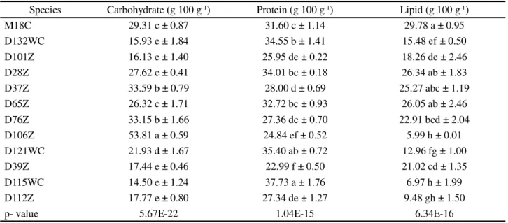
Carbohydrate analyses revealed values varying between 14.50 g 100 g-1 of biomass (S. acuminatus
Microalgae Taxonomic
groups Origen
Marine species
Amphidinium carterae M18C Dinoflagellate Praia do Cabo Branco, João Pessoa-PB. Extraído da esponja
Cynachrella sp Freshwater species
Chlamydomonas sp. D132WC Chlorophyceae Vale do Capão, Cachoeira da Purificação, Chapada Diamantina-BA Chlorella cf.minutissima D101Z Chlorophyceae ETE Mangabeira Calha Parshal, João Pessoa-PB Chlorococcum sp. (cf.hypnosporum) D28Z Chlorophyceae Açude do Cais, Cuité-PB
Chlorococcum sp. (cf.hypnosporum) D37Z Chlorophyceae Açude do Cais, Cuité-PB
Chlorococcum sp. (cf.hypnosporum) D65Z Chlorophyceae Lagoa do Hotel Eco Resort do Cabo, Cabo de Santo Agostinho-PE Chlorococcum sp. (cf.hypnosporum) D76Z Chlorophyceae Rio Quinturaré, Frei Martinho-PB
D115WC) to 53.81 g 100 g-1 (Chlorococcumsp.D106Z), with expressive values (greater than 25 g 100 g-1) noted for Chlorococcum sp. (cf. hypnosporum) strains D28Z, D37Z, D65Z and D76Z and for the microalgaA. carterae M18C (F = 250.4351; gl = 11; p<0.01; Table 3).
In terms of carbohydrates, Kiran et al. (2015) reported values inferior to those encountered here with Chlorococcum sp. when exposed to different culture media nitrogen concentrations, and they obtained yields of 18 g 100 g-1 in culture medium containing 100 mg of sodium nitrate per liter (equivalent to the nitrogen concentration in the Zarrouk medium used here). The yields obtained in the present work for that microalga were greater, varying from 26.32 to 53.81 g 100 g-1. The carbohydrate contents of microalgae are important not only in terms of producing supplements and rations for animals, but also for the production of biofuels through fermentation (CHEWet al., 2017).
The species studied here showed statistically different protein concentrations (F=73,1832; gl=11; p<0.01; Table 3). The species S. acuminatus D115WC (37.73 g 100 g-1) andP. tetrasD121WC (35.40 g 100 g-1) had the highest protein concentrations, whileP. isothrixD39Z (22.99 g 100 g-1) andChlorococcumsp.D106Z (24.84 g 100 g-1) had the lowest. Expressive values (greater than 30 g 100 g-1) were encountered in the marine microalga
A. carterae M18C and in the freshwater chlorophytes Chlamydomonassp. D132WC andChlorococcumsp. (cf. hypnosporum) strains D28Z and D65Z. It is important to note that carbohydrate and protein contents were significantly different between the D28Z and D37Z clones of Chlorococcum sp. (cf. hypnosporum) that had been isolated from the same locality, showing that different lineages of the same species can demonstrate significant differences in their metabolisms.
Lipid analyses demonstrated differences between the species investigated (F = 76,4348; gl = 11; p<0.01, Table 3), with the highest lipid percentages being found in A. carteraeM18C andChlorococcumsp. (cf. hypnosporum) strains D28Z, D37Z, and D65Z. Those results were similar to those described by Mahapatra and Ramachandra (2013) forChlorococcumsp. (30.55 g 100 g-1), by Hoet al. (2014) forChlamydomonassp. (15.3 g 100 g-1), and by Lemahieu et al. (2013) forChlorella (14.7 g 100 g-1). It is important to note, however, that culture conditions and the phase of development at harvesting can be manipulated to direct microalgae metabolism to produce certain desired metabolites. Nitrogen depletion, for example, will force microalgae metabolism to diminish protein or peptide concentrations but increase the percentages of energy-rich compounds such as carbohydrates and lipids, or polysaccharides and fatty acids (HO et al., 2014).
Table 2 - Kinetic characteristics of the growth of the 12 microalgae species investigated
Species k Log phase (days) CT (days) MCD (cells mL-1 105) Final yields (mg L-1) Biomass productivity
(mg L-1 day-1)
M18C 0.83 a ± 0.23 6 19 23.92 d ± 5.66 435 h ± 5.90 22.89 h ± 0.31
D132WC 0.72 a ± 0.45 5 19 43.48 d ± 7.12 536 f ± 9.45 28.21 g ± 0.49
D101Z 0.57 a ± 0.36 10 26 46.73 d ± 8.55 1140 a ± 32.5 43.85 d ± 1.25
D28Z 0.54 a ± 0.12 5 19 68.07 d ± 5.90 756 d ± 3.68 39.79 e ± 0.19
D37Z 0.50 a ± 0.16 3 11 20.25 d ± 0.39 538 f ± 10.36 48.91 c ± 0.94
D65Z 0.48 a ± 0.11 2 17 142.80 c ± 10.10 890 c ± 5.20 52.35 b ± 0.30
D76Z 0.71 a ± 0.22 6 15 25.90 d ± 2.03 640 e ± 12.21 42.67 d ± 0.81
D106Z 0.50 a ± 0.16 3 11 407.99 b ± 19.09 1086 b ± 25.63 98.73 a ± 2.33
D121WC 0.63 a ± 0.17 5 21 7.54 e ± 0.68 486 g ± 7.96 23.14 h ± 0.38
D39Z 0.64 a ± 0.15 5 21 38.20 d ± 3.86 1110 ab ± 21.3 52.86 b ± 1.01
D115WC 0.58 a ± 0.19 5 13 20.13 d ± 0.57 452 gh ± 3.78 34.77 f ± 0.29
D112Z 0.69 a ± 0.28 5 13 788.17 a ± 68.59 632 e ± 12.3 48.62 c ± 0.95
p- value 0.6802 6.72E-24 1,22E-28 1,75E-30
Table 3 - Carbohydrate, protein, and lipid contents in the biomasses of the 12 species of microalgae investigated
Significant differences (F=146,0379; gl=11; p<0.01; Table 4) were also observed between the 12 species in terms of their productions of carotenoids, with minimum and maximum amounts of those compounds being produced byChlorococcumsp.D106Z (1.57 mg g-1) and Chlamydomonassp. D132WC (64.92 mg g-1) respectively.
In terms of chlorophyll-a and b production, Chlorococcum sp D106Z demonstrated significantly lower concentrations (3.61 mg g-1 of chlorophyll-a and 1.18 mg g-1 of chlorophyll-b) than Chlamydomonas sp. D132WC (234.74 and 59.34 mg g-1 respectively) (chlorophyll-a: F = 191,8190; gl = 11; p<0.01 and chlorophyll-b: F = 77,0232; gl = 8; p<0.01; Table 4).
According to Bouman et al. (2018), the photosynthetic pigments present in microalgae reflect chromatic adjustments needed to maximize energy capture under distinct solar irradiation conditions. From a human health point of view, carotenoids (including both carotenes and xanthophylls) act as antioxidants, providing protection against oxidative stress (SATHASIVAM; KI, 2018). Some xanthophylls, such as violaxanthin, antheraxanthin, zeaxanthin, neoxanthin, and lutein occur both in microalgae and higher plants, although microalgae also produce different types of xanthophylls, such as
loroxanthin, astaxanthin, and canthaxanthin (synthesized by green algae), and diatoxanthin, diadinoxanthin, and fucoxanthin (synthesized by brown algae or diatoms) (BARREDO, 2012). Chlorophylls likewise have beneficial
effects for human health due to their anticancer properties and anti-inflammatory and anti-oxidant activities, and can help prevent arteriosclerosis as well as atherothrombotic cardiovascular diseases (PEMMARAJUet al., 2018).
Fatty acid compositions
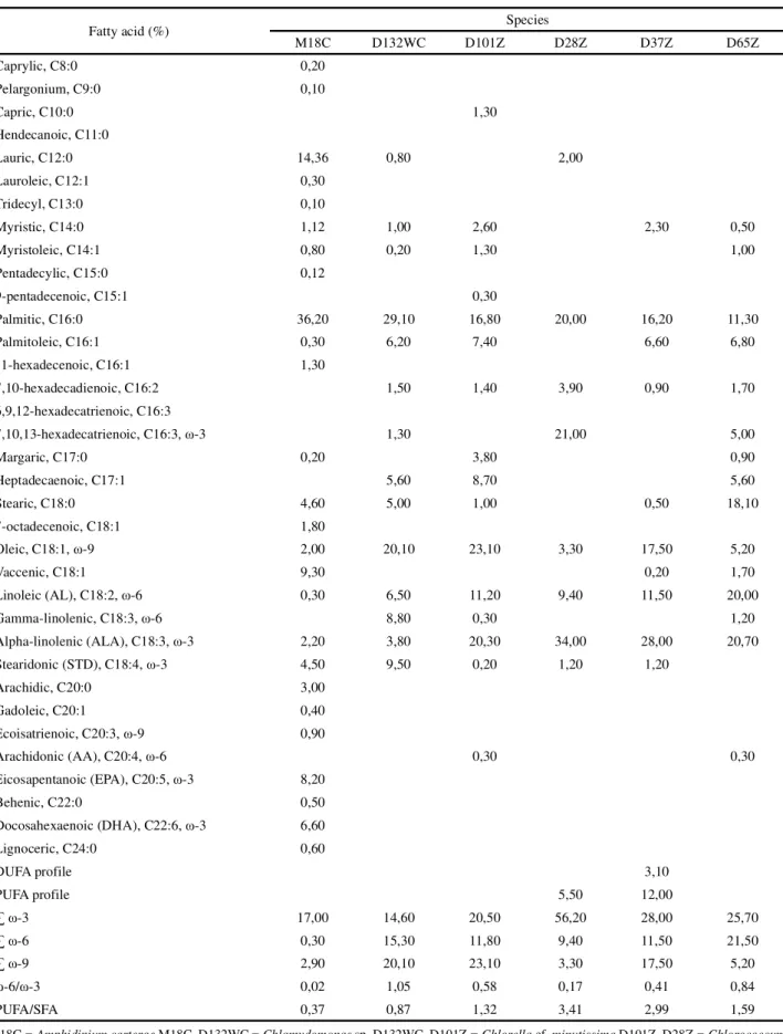
The fatty acid profiles of the microalgae examined here are presented in Tables 5 and 6. Fatty acids with carbon chains varying from C8 to C24, both saturated and unsaturated (with 1 to 6 unsaturated bonds), were identified. The most frequent fatty acids were: myristic (C14:0), palmitic (C16:0), palmitoleic (C16:1), stearic (C18:0), oleic (C18:1, ω-9), linoleic (AL, C18:2 ω-6), α-linolenic (ALA, C18:3 ω-3), and stearidonic (STD, C18:4 ω-3). Among those, palmitic acid stood out as the principal saturated fatty acid (SFA), with the microalgae examined producing that fatty acid at concentrations varying from 11.3% (Chlorococcumsp. (cf. hypnosporum) D65Z) to 38.0% (S. nidulans D112Z) of all fatty acid methyl esters (FAME) produced. The same pattern of palmitic acid predominance was reported by Nakanishiet al. (2014) for Chlamydomonas sp., by Ambrozova et al. (2014) for eight microalgae, and by Campos et al. (2010) for 10 species of marine microalgae.
The unsaturated fatty acids (USFA) present in all of the microalgae studied were oleic (ω-9), AL (ω-6), and ALA (ω-3), with percentages varying from 2% (A. carterae Species Carbohydrate (g 100 g-1) Protein (g 100 g-1) Lipid (g 100 g-1)
M18C 29.31 c ± 0.87 31.60 c ± 1.14 29.78 a ± 0.95
D132WC 15.93 e ± 1.84 34.55 b ± 1.41 15.48 ef ± 0.50
D101Z 16.13 e ± 1.40 25.95 de ± 0.22 18.26 de ± 2.46
D28Z 27.62 c ± 0.41 34.01 bc ± 0.18 26.34 ab ± 1.83
D37Z 33.59 b ± 0.79 28.00 d ± 0.69 25.27 abc ± 1.19
D65Z 26.32 c ± 1.71 32.72 bc ± 0.93 26.05 ab ± 2.46
D76Z 33.15 b ± 1.66 27.36 de ± 0.70 22.91 bcd ± 2.04
D106Z 53.81 a ± 0.59 24.84 ef ± 0.52 5.99 h ± 0.01
D121WC 21.93 d ± 1.67 35.40 ab ± 0.72 12.96 fg ± 1.00
D39Z 17.44 e ± 0.46 22.99 f ± 0.50 21.02 cd ± 1.35
D115WC 14.50 e ± 1.24 37.73 a ± 1.76 6.97 h ± 1.99
D112Z 17.77 e ± 0.80 27.34 de ± 1.27 9.48 gh ± 1.50
p- value 5.67E-22 1.04E-15 6.34E-16
M18C) to 32.5% (P. tetrasD121WC), 0.3% (A. carterae M18C) to 30% (P. isothrixD39Z), and 1.7% (P. isothrix D39Z) to 34% (Chlorococcum sp. (cf. hypnosporum) D28Z) of the total FAME produced respectively. The species that produced the highest levels of oleic acid were Chlorococcum sp. (cf. hypnosporum) strains D37Z and D76Z, C. minutissimaD101Z, S. acuminatus D115WC, P. tetras D121WC, and Chlamydomonas sp. D132WC. In terms of AL, the highest levels were observed in P. isothrix D39Z, Chlorococcum sp. (cf. hypnosporum) D65Z,Chlorococcumsp.D106Z, andS. nidulansD112Z. In terms of ALA, the species that demonstrated the highest concentrations wereChlorococcumsp. (cf. hypnosporum) strains D28Z, D37Z, D65Z, and D76Z, C. minutissima D101Z, andChlorococcumsp.D106Z (Tables 5 and 6). Humans do not synthesize ALA or AL as they do not produce the enzymes Δ-12 and Δ-15 desaturase, and those acids (precursors of ω-3 and ω-6 respectively) must be obtained in our diet (LEEet al., 2016).
Radmann and Costa (2008) analyzed S. nidulans and reported low percentages of AL (2.71%) and AA (0.49%), and slightly higher percentages of ALA (7.61%). Evaluations ofChlamydomonassp. cultivated at different nitrate concentrations by Ho et al. (2014) showed percentages of oleic acid varying from 9.1-26.6%, of AL varying from 21.7-25.3%, and of ALA varying from 14.4-5.4%. Campos, Barbarino and Lourenço (2010) reported 4.7% AL, 17.5% ALA, and 0.8% EPA
in C. minutissima. Mahapatra and Ramachandra (2013) reported 1.59%, 14.3% and 7.07% of oleic acid, AL, and ALA inChlorococcumsp respectively; in that same report, those same fatty acids varied between 5.2-22.5%, 8.6-30%, and 1.7-28.3%, respectively, in all of the strains ofChlorococcumsp.
The microalgae examined in the present work did not demonstrate good ω-6/ω-3 ratios (Table 5 and 6) when compared to the suggested healthy ratio of 4-5:1 (CANDELA; LÓPEZ; KOHEN, 2011; WARNER et al., 2017); even microalgae that demonstrated high concentrations of ω-6 had inadequate ratios, as observed inChlamydomonassp. D132WC (1.05), P. isothrixD39Z (28.24), andS. nidulansD112Z (2.44). The same low ω -6/ω-3 ratios were observed by Ryckeboschet al. (2014), varying from 0.053 to 2.0 among nine different species examined, although the ratio seen in fish oil is 0.071, indicating that even the current principal source of omegas has a low ratio of those compounds.
PUFA/SFA ratios can be used to rapidly evaluate the fatty acid profiles of the microalgae analyzed. According to Ambrozova et al. (2014), the larger that value, the greater will be the potential benefits to human health. The highest ratios were observed here in the species C. minutissima D101Z; Chlorococcum sp. (cf. hypnosporum) strains D28Z, D37Z, D65Z and D76Z, Chlorococcum sp. D106Z, and P. isothrix D39Z (Table 5). Ambrozova et al. (2014) analyzed the PUFA/SFA
Table 4 - Carotenoid and chlorophyll-a and b contents in the biomasses of the 12 microalgae species investigated
ND = not determined, M18C = Amphidinium carteraeM18C, D132WC =Chlamydomonas sp. D132WC, D101Z = Chlorellacf. minutissimaD101Z, D28Z = Chlorococcumsp. (cf. hypnosporum) D28Z, D37Z = Chlorococcum sp. (cf. hypnosporum) D37Z, D65Z = Chlorococcumsp. (cf. hypnosporum) D65Z, D76Z = Chlorococcumsp. (cf. hypnosporum) D76Z, D106Z = Chlorococcumsp.D106Z, D121WC = Pediastrum tetrasD121WC, D39Z = Planktothrix isothrixD39Z, D115WC = Scenedesmus acuminatusD115WC, D112Z = Synechococcus nidulansD112Z; Values expressed as means with standard errors. Means followed by the same letters in the same column do not statistically differ (ANOVA and Tukey Test, p ≥ 0.05)
Species Carotenoid (mg g-1) Chlorophyll-a (mg g-1) Chlorophyll-b (mg g-1)
M18C 17.64 cd ± 4.40 102.57 b ± 6.52 ND
D132WC 64.92 a ± 3.94 234.74 a ± 23.22 59.34 a ± 7.76
D101Z 13.47 de ± 1.88 42.53 cd ± 5.14 15.89 c ± 2.13
D28Z 21.02 c ± 1.57 56.84 c ± 3.17 22.18 c ± 4.93
D37Z 12.26 de ± 1.88 37.99 cd ± 5.07 14.92 c ± 2.22
D65Z 13.30 de ± 0.89 33.51 d ± 2.57 15.53 c ± 1.07
D76Z 10.40 e ± 2.13 30.31 d ± 5.36 13.03 cd ± 2.56
D106Z 1.57 f ± 0.19 3.61 f ± 0.27 1.18 e ± 0.37
D121WC 10.48 e ± 2.91 23.96 de ± 1.84 4.65 de ± 0.64
D39Z 8.68 e ± 1.39 29.89 d ± 1.51 ND
D115WC 34.79 b ± 2.64 97.71 b ± 5.17 34.61 b ± 2.42
D112Z 13.99 de ± 1.11 25.79 de ± 4.11 ND
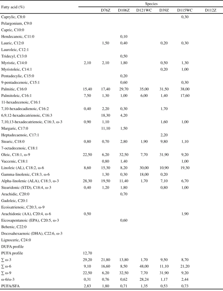
Table 5 - Fatty acid profiles of the microalgae species cultivated (percentage values in relation to the total fatty acid methyl esters)
M18C = Amphidinium carteraeM18C, D132WC =Chlamydomonas sp. D132WC, D101Z = Chlorellacf. minutissimaD101Z, D28Z = Chlorococcum sp. (cf. hypnosporum) D28Z, D37Z = Chlorococcum sp. (cf. hypnosporum) D37Z, D65Z = Chlorococcumsp. (cf. hypnosporum) D65Z
Fatty acid (%) Species
M18C D132WC D101Z D28Z D37Z D65Z
Caprylic, C8:0 0,20
Pelargonium, C9:0 0,10
Capric, C10:0 1,30
Hendecanoic, C11:0
Lauric, C12:0 14,36 0,80 2,00
Lauroleic, C12:1 0,30
Tridecyl, C13:0 0,10
Myristic, C14:0 1,12 1,00 2,60 2,30 0,50
Myristoleic, C14:1 0,80 0,20 1,30 1,00
Pentadecylic, C15:0 0,12
9-pentadecenoic, C15:1 0,30
Palmitic, C16:0 36,20 29,10 16,80 20,00 16,20 11,30
Palmitoleic, C16:1 0,30 6,20 7,40 6,60 6,80
11-hexadecenoic, C16:1 1,30
7,10-hexadecadienoic, C16:2 1,50 1,40 3,90 0,90 1,70
6,9,12-hexadecatrienoic, C16:3
7,10,13-hexadecatrienoic, C16:3, ω-3 1,30 21,00 5,00
Margaric, C17:0 0,20 3,80 0,90
Heptadecaenoic, C17:1 5,60 8,70 5,60
Stearic, C18:0 4,60 5,00 1,00 0,50 18,10
7-octadecenoic, C18:1 1,80
Oleic, C18:1, ω-9 2,00 20,10 23,10 3,30 17,50 5,20
Vaccenic, C18:1 9,30 0,20 1,70
Linoleic (AL), C18:2, ω-6 0,30 6,50 11,20 9,40 11,50 20,00
Gamma-linolenic, C18:3, ω-6 8,80 0,30 1,20
Alpha-linolenic (ALA), C18:3, ω-3 2,20 3,80 20,30 34,00 28,00 20,70
Stearidonic (STD), C18:4, ω-3 4,50 9,50 0,20 1,20 1,20
Arachidic, C20:0 3,00
Gadoleic, C20:1 0,40
Ecoisatrienoic, C20:3, ω-9 0,90
Arachidonic (AA), C20:4, ω-6 0,30 0,30
Eicosapentanoic (EPA), C20:5, ω-3 8,20
Behenic, C22:0 0,50
Docosahexaenoic (DHA), C22:6, ω-3 6,60
Lignoceric, C24:0 0,60
DUFA profile 3,10
PUFA profile 5,50 12,00
∑ ω-3 17,00 14,60 20,50 56,20 28,00 25,70
∑ ω-6 0,30 15,30 11,80 9,40 11,50 21,50
∑ ω-9 2,90 20,10 23,10 3,30 17,50 5,20
ω-6/ω-3 0,02 1,05 0,58 0,17 0,41 0,84
Table 6 - Fatty acid profiles of the microalgae species cultivated (percentage values in relation to the total fatty acid methyl esters)
D76Z = Chlorococcumsp. (cf. hypnosporum) D76Z, D106Z = Chlorococcumsp.D106Z, D121WC = Pediastrum tetrasD121WC, D39Z = Planktothrix isothrixD39Z, D115WC = Scenedesmus acuminatusD115WC, D112Z = Synechococcus nidulansD112Z
Fatty acid (%) Species
D76Z D106Z D121WC D39Z D115WC D112Z
Caprylic, C8:0 0,30
Pelargonium, C9:0 Capric, C10:0
Hendecanoic, C11:0 0,10
Lauric, C12:0 1,50 0,40 0,20 0,30
Lauroleic, C12:1
Tridecyl, C13:0 0,50
Myristic, C14:0 2,10 2,10 1,80 0,50 1,30
Myristoleic, C14:1 0,20 1,00
Pentadecylic, C15:0 0,20
9-pentadecenoic, C15:1 0,60 0,30
Palmitic, C16:0 15,40 17,40 29,70 35,00 31,50 38,00
Palmitoleic, C16:1 7,50 1,30 1,00 6,00 1,40 17,60
11-hexadecenoic, C16:1
7,10-hexadecadienoic, C16:2 0,40 2,20 0,30 1,70
6,9,12-hexadecatrienoic, C16:3 18,30 4,20
7,10,13-hexadecatrienoic, C16:3, ω-3 0,90 1,10 1,60 1,00
Margaric, C17:0 11,10 1,50
Heptadecaenoic, C17:1 2,20
Stearic, C18:0 0,80 0,70 2,80 1,90 9,80 1,10
7-octadecenoic, C18:1
Oleic, C18:1, ω-9 22,50 6,20 32,50 7,70 31,90 9,20
Vaccenic, C18:1 0,80 1,40 1,00
Linoleic (AL), C18:2, ω-6 8,60 15,30 8,20 30,00 10,90 19,30 Gamma-linolenic, C18:3, ω-6 1,30 0,30 18,00 0,20
Alpha-linolenic (ALA), C18:3, ω-3 28,30 19,50 11,40 1,70 7,10 6,70 Stearidonic (STD), C18:4, ω-3 0,40 1,20 1,80 0,80 1,00
Arachidic, C20:0 0,70
Gadoleic, C20:1
Ecoisatrienoic, C20:3, ω-9
Arachidonic (AA), C20:4, ω-6 0,50 1,90
Eicosapentanoic (EPA), C20:5, ω-3 0,60 Behenic, C22:0
Docosahexaenoic (DHA), C22:6, ω-3 Lignoceric, C24:0
DUFA profile
PUFA profile 12,70
∑ ω-3 29,20 21,80 13,80 1,70 9,50 8,70
∑ ω-6 9,10 16,60 8,50 48,00 11,10 21,20
∑ ω-9 22,50 6,20 32,50 7,70 31,90 9,20
ω-6/ω-3 0,31 0,76 0,62 28,24 1,17 2,44
fatty acid ratios of numerous microalgae and observed variations between 0.46 and 2.13. Campos, Barbarino and Lourenço (2010) reported a ratio of 0.88 for C. minutissima; Mahapatra and Ramachandra (2013) reported a value of 0.32 forChlorococcumsp.; Radmann and Costa (2008) reported a high value for S. nidulans (2.66); and Ho et al. (2014) reported PUFA/SFA ratios between 1.0-1.21 forChlamydomonassp.
Based on the results obtained with other species of microalgae, it is evident that differences in fatty acid profiles can be found even within the same species, indicating the existence of intraspecific differences presumably due to the fact that the environments from which they were isolated molded their metabolic natures. The ability of those same microalgae to survive under different and extreme conditions, however, demonstrates their diversity and the unique lipid profiles of those organisms (PALIWALet al., 2017).
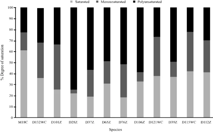
Higher concentrations of saturated and monounsaturated fatty acids and generally lower percentages of polyunsaturated fatty acids where found
Figure 1 -Percentage composition by degree of saturation of the microalgae species examined
M18C = Amphidinium carteraeM18C, D132WC =Chlamydomonas sp. D132WC, D101Z = Chlorellacf. minutissimaD101Z, D28Z = Chlorococcum sp. (cf. hypnosporum) D28Z, D37Z = Chlorococcum sp. (cf. hypnosporum) D37Z, D65Z = Chlorococcumsp. (cf. hypnosporum) D65Z, D76Z = Chlorococcumsp. (cf. hypnosporum) D76Z, D106Z = Chlorococcumsp.D106Z, D121WC = Pediastrum tetrasD121WC, D39Z = Planktothrix isothrixD39Z, D115WC = Scenedesmus acuminatusD115WC, D112Z = Synechococcus nidulansD112Z
in the microalgae examined here (Figure 1); only Chlorococcumsp. (cf. hypnosporum) strains D28Z (75%), D37Z (56.9%), and D76Z (51.8%) andChlorococcumsp. D106Z (58.9%) showed high concentrations of PUFA.
In terms of the percentages of the omegas ω-3,
most of those compounds are bio-accumulated through the food chain (in which microalgae have a principal role) (SAWYERet al., 2016). As such, the possibility of using microalgae to obtain EFA appears quite promising.
CONCLUSIONS
1. Interspecific variations seen in the growth characteristics and chemical compositions of 12 microalgae indicated that each displayed a different response to standard culture conditions;
2. The species Chlorella cf. minutissima D101Z, Chlorococcum sp. (cf. hypnosporum) strains D28Z, D37Z, D65Z, and D76Z,Pediastrum tetrasD121WC, Planktothrix isothrix D39Z, and Scenedesmus
acuminatus D115WC demonstrated elevated
percentages of PUFA, including ω-3, ω-6, and ω-9, as well as carotenoids and chlorophylls - nutrients vital to human health - making those species potential alternative sources of metabolites for food industries.
Figure 2 - Percentage of omega ω-3, ω-6, and ω-9 fatty acids based on the total percentages of fatty acids in the microalgae examined
M18C = Amphidinium carteraeM18C, D132WC =Chlamydomonas sp. D132WC, D101Z = Chlorellacf. minutissimaD101Z, D28Z = Chlorococcum sp. (cf. hypnosporum) D28Z, D37Z = Chlorococcum sp. (cf. hypnosporum) D37Z, D65Z = Chlorococcumsp. (cf. hypnosporum) D65Z, D76Z = Chlorococcumsp. (cf. hypnosporum) D76Z, D106Z = Chlorococcumsp.D106Z, D121WC = Pediastrum tetrasD121WC, D39Z = Planktothrix isothrixD39Z, D115WC = Scenedesmus acuminatusD115WC, D112Z = Synechococcus nidulansD112Z
ACKNOWLEDGMENTS
The authors thank the Ministério da Ciência Tecnologia e Inovação (MCTI), Finep (Processo: 2557/09), and the Conselho Nacional de Desenvolvimento Científico e Tecnológico (CNPq) (Processo: 407519/2013-0) for their financial support; Prof. Dr. Nelson R. Antoniosi Filho of the Laboratório de Métodos de Extração e Separação (LAMES/UFG) for his assistance with the analyses of the fatty acids; and LARBIM/UFPB for the use of their installations and providing the microalgae investigated.
REFERENCES
AMBROZOVA, J. V. et al. Influence of extractive solvents
on lipid and fatty acids content of edible freshwater algal and seaweed products, the green microalgaChlorella kessleriand the cyanobacterium Spirulina platensis.Molecules, v. 19, p. 2344-2360, 2014.
ANDRADE, L. M.et al.Chlorella andSpirulina microalgae
supplements: an overview. MOJ Food Processing and Technology, v. 6, n. 1, 2018.
BARREDO, J. L. Microbial carotenoids from bacteria and microalgae. In: Methods and protocols. New York: Humana Press, 2012. p. 5.
BELLOU, S. et al. Microbial oils as food additives: recent
approaches for improving microbial oil production and its polyunsaturated fatty acid content. Current Opinion in Biotechnology, v. 37, p. 24-35, 2016.
BOUMAN, H. A. et al. Photosynthesis-irradiance parameters
of marine phytoplankton: synthesis of a global data set.Earth System Science Data, v. 10, n. 1, p. 251, 2018.
CAMPOS, V. B.; BARBARINO, E.; LOURENÇO, S. O. Crescimento e composição química de dez espécies de microalgas marinhas em cultivos estanques.Ciência Rural, v. 40, n. 2, p. 339-347, 2010.
CANDELA, C. G.; LÓPEZ, L. M. B.; KOHEN, V. L. Importance of a balanced omega 6/omega 3 ratio for the maintenance of health: nutritional recommendations. Nutrición Hospitalaria, v. 26, n. 2, p. 323-329, 2011.
CHEW, K. W. et al. Microalgae biorefinery: high value
products perspectives.Bioresource Technology, v. 229, p. 53-62, 2017.
CHIU, S. Y.et al. Reduction of CO2 by a high-density culture of Chlorella sp. in a semicontinuous photobioreactor.Bioresource Biotechnology, v. 99, p. 3389-3396, 2008.
FOLCH, J.; LEES, M.; STANLEY, G. H. S. A simple method for the isolation and purification of total lipids from animal tissues. Journal of Biological Chemistry, v. 226, p. 497-509, 1957. GUILLARD, R. R. L.; LORENZEN, C. J. Yellow-green algae with chlorophyllide c.Journal of Phycology, v. 8, p. 10-14, 1972.
HAYES, M.et al. Microalgal proteins for feed, food and health. In:Microalgae-Based Biofuels and Bioproducts, p. 347-368, 2018.
HO, S-H. et al. Optimizing biodiesel production in marine
Chlamydomonas sp. JSC4 through metabolic profiling and
an innovative salinity-gradient strategy. Biotechnology for Biofuels, v. 7, p. 97-113, 2014.
JEFFREY, S. W., HUMPHREY, G. F. New spectrophotometric equation for determining chlorophyll a, b, c1 and c2. Biochemistry and Molecular Biology of Plant Hormones, v. 167, p. 194-204, 1975.
KIRAN, B. et al. Growth pattern and biofuel production
potential of newly isolated microalga,Chlorococcum sp.
IM-03 under nitrogen limited conditions. Journal of Chemical Technology and Biotechnology, v. 90, 2015.
KOCHERT, G. Carbohydrate determination by the phenol-sulfuric method.In: HELLEMBUST, J. A., CRAIGIE, J. S. (Ed.).
Handbook of phycological methods. Cambridge: Cambridge University, 1978. p. 95-97.
LEE, J. M.et al. Fatty acid desaturases, polyunsaturated fatty acid regulation, and biotechnological advances.Nutrients, v. 8, n. 1, p. 23, 2016.
LEMAHIEU, C. et al. Impact of feed supplementation with
different omega-3 rich microalgae species on enrichment of eggs of laying hens. Food Chemistry, v. 141, p. 4051-4059, 2013.
LOWRY, O. H. et al. Protein measurement with the folin phenol reagent.The Journal of Biological Chemistry, v. 193, n. 1, p. 265-75, 1951.
MAHAPATRA, D. M.; RAMACHANDRA, T. V. Algal biofuel: bountiful lipid fromChlorococcumsp. proliferating in municipal wastewater.Current Science, v. 105, n. 1, 2013.
MENEZES, R. S. et al. Avaliação da potencialidade de microalgas dulcícolas como fonte de matéria-prima graxa para a produção de biodiesel.Química Nova,v. 36, n. 1, p. 10-15,
2013.
NAKANISHI, A.et al. Development of lipid productivities under different CO2 conditions of marine microalgaeChlamydomonas
sp.Bioresource Technology, v. 152, p. 247–252, 2014. PALIWAL, C.et al. Abiotic stresses as tools for metabolites in
microalgae.Bioresource Technology, v. 244, p. 1216-1226, 2017.
PEMMARAJU, D. et al. Chlorophyll rich biomolecular fraction of A. cadamba loaded into polymeric nanosystem coupled with photothermal therapy: a synergistic approach for cancer theranostics. International Journal of Biological Macromolecules, v. 110, p. 383-391, 2018.
RADMANN, E. M.; COSTA, J. A. V. Conteúdo lipídico e composição de ácidos graxos de microalgas expostas aos gases CO2, SO2 e NO. Química Nova, v. 31, n. 7, p. 1609-1612,
2008.
RINCÓN-CERVERA, M. A. et al. Supplementation with antioxidant-rich extra virgin olive oil prevents hepatic oxidative stress and reduction of desaturation capacity in mice fed a high-fat diet: effects on high-fatty acid composition in liver and extrahepatic tissues.Nutrition, v. 32, n. 11, p. 1254-1267, 2016.
RODOLFI, L. et al. Microalgae for oil: strain, induction of
lipid synthesis and outdoor mass cultivation in a low-cost photobioreactor.Biotechnology Bioengery, v. 102, p. 100–112, 2009.
RUBIO-RODRÍGUEZ, N. et al. Production of omega-3 polyunsaturated fatty acid concentrates: a review.Food Science and Emerging Technologies, v. 11, p. 1-12, 2010.
RYCKEBOSCH, E.et al. Nutritional evaluation of microalgae
oils rich in omega-3 long chain polyunsaturated fatty acid as alternative for fish oil. Food Chemistry, v. 160, p. 393-400, 2014.
SAWYER, J. M. et al. A general model of polyunsaturated fatty acid (PUFA) uptake, loss and transformation in freshwater fish. Ecological Modelling, v. 323, p. 96-105, 2016.
STEIN, J. R. Handbook of phycological methods: culture methods and growth measurements. In: Handbook of phycological methods. Cambridge University Press; p. 448. 1973.
STRICKLAND, J. D. H., PARSONS, T. R.A practical handbook of seawater analysis. 2. ed. Ottawa: Fisheries Research Board of Canada Bull, 1968. 293 p.
SUBASH-BABU, P.; ALSHATWI, A. A. Effects of increasing ratios of dietary omega-6/omega-3 fatty acids on human
monocyte immunomodulation linked with atherosclerosis. Journal of Functional Foods, v. 41, p. 258-267, 2018. WALNE, P. R. Experiments in the large scale culture of the larvae ofOstrea edulis.Journal Fishery Investiment, v. 25, n. 4, p. 1-53, 1966.
WARNER, D. et al. ω3-PUFA supplementation reduced ALT levels In mice exposed to a low linoleic acid diet with or without ethanol in a drinking in the dark binge model. The FASEB Journal, v. 31, n. 1, p. 894.4-894.4, 2017.
ZARROUK, C. Contribution à l’étude d’une cyanophycée: influence de divers facteurs physiques et chimiques sur la croissance et la photosynthèse de Spirulina maxima (Setch et Gardner) Geitler. 1966. Theises (Ph. D.) - Faculty of Science, Université des Paris, Paris,1966.