Gallus gallus domesticus: immune system and its potential
for generationof immunobiologics
Gallus gallus domesticus: sistema imunológico e seu potencial para geração de imunobiológicos
Álvaro Ferreira Júnior
1*Jandra Pacheco dos Santos
2Iara de Oliveira Sousa
3Ian Martin
3Endrigo Gabellini Leonel Alves
3Isabel Rodrigues Rosado
3ISSNe 1678-4596
INTRODUCTION
Chicken (Gallus gallus domesticus) has
made valuable contributions to our understanding of
immunology. However, the “chicken is not a mouse
with feathers” (by JIM KaufMaN (KaISeR,
2012)), and “the hens’ immune system differs from
mammals’ in various ways” (SCHade et al., 2001).
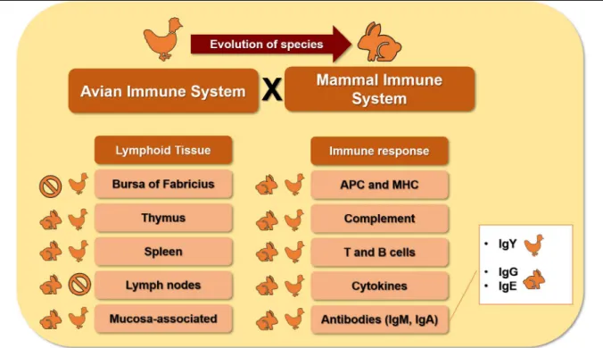
In this context, important differences exist, especially
in the diversity of the lymphoid tissue. for instance,
the bursa of fabricius is present in hens but not in
mammals. The major blood antibody class present
in hens is immunoglobulin Y (IgY), whereas that
in mammals is IgG. Ige antibodies are absent in the
hens’ immune system. additionally, the transference
of maternal antibodies in the hen occurs by egg yolk
absorption, and by transplacental passage in mammals.
One aspect that hens and mammals have
in common, is the presence of both, the innate as
well as the acquired immune response. These
animal groups possess immune cells and molecules.
among these immune cells are the dendritic
cells (dC), macrophages, and lymphocytes.
With regard to the hens’ immunity, the crucial
function of these molecules as signaling proteins
has been demonstrated. They are also known as
cytokines. Besides, a lytic protein system named the
complement system protects the host by both, innate
and acquired immune response mechanisms.
Interestingly, according to phylogenetic
analysis, hens developed before mammals. In
addition, the avian immune system is genetically
simpler than that of the mammalian immune
system. The former can mount a robust immune
response against a wide range of antigenic targets.
Corroborating this robustness, the avian repertoire
of antibodies has an elevated number of antigen
binding combinations. These have been achieved
1escola de Veterinária e Zootecnia, Medicina Veterinária Preventiva, universidade federal de Goiás (ufG), 74.690-900, Goiânia, GO, Brasil.
e-mail: alvaro.ferreira@ufg.br. *Corresponding author.
2Programa de Pós-graduação em Ciências Veterinárias, universidade federal de uberlândia (ufu), uberlândia, MG, Brasil.
3Programa de Pós-Graduação em Sanidade e Produção animal nos Trópicos, universidade de uberaba (uNIuBe), uberaba, MG, Brasil.
ABSTRACT: Gallus gallus domesticus’ immune system is a promising tool for generation of antibody-based immunobiologics. Immunoglobulin Y (IgY) is extracted from egg yolk and has equivalent functions to mammal’s IgG antibody. Avian immune system can be stimulated to produce a high-quality antibody repertoire. In this review, we present an overview of avian immune system emphasizing IgY and its applications as an immunobiologic.
Key words: avian lymphoid tissues; avian immune response; IgY antibody.
RESUMO: O sistema imunológico de Gallus gallus domesticus é uma ferramenta promissora para a geração de imunobiológico a partir de anticorpos. a imunoglobulina Y (IgY) é extraída da gema do ovo e apresenta funções equivalentes ao anticorpo IgG dos mamíferos. O sistema imune aviário pode ser estimulado para produzir um repertório de anticorpos de alta qualidade. Nesta revisão apresentamos aspectos gerais do sistema imune aviário enfatizando o IgY e suas aplicações como um imunobiológico
Palavras-chave: tecidos linfoides aviários; resposta imune aviária; anticorpo IgY.
by the antibodies’ gene recombination and gene
conversion. In conclusion, the efficacy of the hens’
immune system has been proven in its elaborate
defenses against aggressors by different mechanisms.
The IgY antibodies are a valuable tool in the hens’
immune system, and, a promising immunobiological
reagent. Hence, these can be tapped as an alternative
for the mammals’ IgG antibodies.
Lymphoid Tissues
Chicken has made valuable contributions to
our understanding of immunology (KaISeR, 2012).
The avian and mammal immune systems are organized
into groups of immune cells, such as the T cells and B
cells, and are homed into organized lymphoid tissues,
which are strategically positioned to protect the
host (BOeHM et al., 2012; ROSTaMI et al., 2018).
Functionally, the lymphoid tissue has been classified
into the primary lymphoid tissue, such as the thymus
and bursa of fabricius, and the secondary lymphoid
tissue, such as the spleen (figure 1) (MadeJ
et al.,
2015; SuN et al., 2016; IfRaH et al., 2017).
The hens’ primary lymphoid tissue
includes the thymus and bursa of fabricius (BOeHM
et al., 2012). The thymus is located at the ventral neck
region and the bursa of fabricius is reported at the top
of the cloacal region (SuN et al., 2016; IfRaH et al.,
2017). Primary lymphoid tissue works by selecting
lymphocytes such as the T cells (thymus-dependent
cells) and the B cells (bursa of fabricius-selected
cells) for an appropriate immune response and
avoiding autoimmunity (SuN et al., 2016; IfRaH et
al., 2017). The T and B cell precursors are generated
by the lymphoid stem cells in the bone marrow
(BOeHM et al., 2012).
The bursa of fabricius is a lymphoid tissue
that is absent in the immune system of mammals, as
a consequence of the species’ evolution (figure 1)
(BOeHM et al., 2012). after puberty, in the hens and
mammals, the primary lymphoid tissue is involuted
by the effects of the sex hormones.
The selected T and B cells, leaving the
primary lymphoid tissue, move forward to their
defense position in the secondary lymphoid tissue,
such as the spleen and mucosa-associated lymphoid
tissue (MaLT) (LaNNING & KNIGHT, 2015;
MadeJ
et al., 2015; SePaHI & SaLINaS, 2016).
also, they are present in parenchyma, the bursa of
fabricius, and the bone marrow tissue (MadeJ
et al., 2015). Peripheral lymphoid tissue has been
established as the site for the generation of an immune
response following contact with a pathogen. The
spleen is a capsulated tissue reported in abdominal
cavity, close to the stomach (ZHaNG
et al., 2015),
while the MaLT is a lymphoid tissue scattered
throughout the body, on surfaces, such as the mucosa
of the digestive system and the eyes (Harderian
glands) (VaN GINKeL et al., 2012; GuRJaR et al.,
2013), the respiratory system, and skin (SMIaLeK et
al., 2011; LaNNING & KNIGHT, 2015; SePaHI &
SaLINaS, 2016). Some peripheral lymphoid tissue
is absent in the avian immune system, such as the
lymph nodes, (figure 1), however, there are lymphoid
aggregates, such as Meckel’s diverticulum and cecal
tonsils (BOeHM et al., 2012; HeIdaRI et al., 2015).
The apparent absence of lymphotoxin genes might
explain the lack of lymph nodes in hens, because
these genes are crucial to lymph node formation in
mammals (KaISeR, 2012).
The health of the reproductive
tract is important for the formation and production
of high quality and hygienic eggs (YOSHIMuRa
& BaRua, 2017). The hen ovary and oviduct
have lymphoid tissue that contains populations of
immunocompetent cells such as macrophages and
lymphocytes. Influx of immune cells increases
with hen maturity and decreases with aging. The
ovary’s parenchyma and the oviduct’s lamina
propria express TLR (Toll-like receptor) molecules,
triggering the production of pro-inflammatory
cytokines and chemokines, and defensin molecules
(YOSHIMuRa & BaRua, 2017).
Regarding the lymphocyte position in the
secondary lymphoid tissue, the T cells are reported
close to the B cells, in a location called the germinal
center. following contact with pathogens, the spleen
is enlarged in size by hyperplasia tissue, in a process
called splenomegaly. Lymphocyte hyperplasia has
been found in MaLT as well. The germinal center is
the site that effectively produces the avian antibodies.
Avian Immune System
avian immune response is divided in two
arms, the innate immune response and the acquired
immune response (JeuRISSeN
et al., 2000; KaISeR,
2012). The former involves the quick activation of
immune mechanisms, such as the acute inflammatory
reaction, which includes cells and molecules such
as macrophages and the complement system (GuO
et al., 2008)2008. Conversely, the acquired immune
response is delayed and characterized by antibody
production (figure 2) and immune memory (PeI
& COLLISON, 2005; SINGH
et al., 2010). It has
been emphasized that the innate immunity does not
develop an immune memory like that observed in
acquired immunity (GuO et al., 2008)2008. Recently,
it has been demonstrated that some differences occur
in populations of immunocompetent cells between
various hens breeds (BÍLKOVÁ et al., 2017).
The innate immune response starts when
the sentinel cells, such as the dendritic cells and
macrophages, trap non-self-compounds (antigens)
(QuReSHI et al., 2000; de GeuS & VeRVeLde,
2013; NaGY
et al., 2016). These sentinel cells
recognize the pathogen-associated molecular
patterns (PaMPS) by their pathogen recognition
receptors (PRR) such as the Toll-like receptor (TLR),
after which they trigger an acute inflammatory
reaction (QuReSHI
et al., 2000; NaNG
et al.,
2011; GRueBeR
et al., 2014). Pathogens are
classified according to their growth environment,
as extracellular pathogens, such as certain bacteria,
or intracellular pathogens, like viruses. Neutrophils
and eosinophils are absent in hens and the latter is
replaced by heterophils in the avian immune system
(KaISeR, 2012; MuKHeRJee et al., 2016).
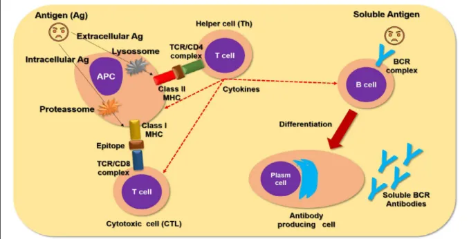
following the pathogen trapping, the
sentinel cells must process the protein antigens in
the antigenicity determinant regions, also called the
epitopes (figure 2) (WaNG et al., 2016). There are
different antigen processing pathways, such as the
lysosomal pathway for extracellular pathogens, and
the proteasome pathway for intracellular pathogens
(figure 2) (BLuM
et al., 2013). although both
antigen processing pathways produce epitopes,
the antigen processing by the lysosome enzymes
generate a larger peptide sequence than the
enzymatic proteasome pathway (HaSSeLGReN
& fISCHeR, 1997).
certain pathogens (KaufMaN, 2000). In this
context, there is a higher chance for this compact
and simple avian MHC does not present a give
protective epitope during the antigen presentation
to T (KaufMaN, 2000).
antigen presenting cells (aPC), such as
the dendritic cells (dC) and macrophages, present
the epitope-MHC to the lymphocyte cells, such as T
lymphocytes (figure 2) (BeCKeR, 2003). The T cells,
using their antigen receptor complex (TCR), bind
to the epitopes and recognize the MHC molecules.
This recognition is carried out by complementary
receptors, such as Cd8 and Cd4, which recognize
the self-class I MHC and class II MHC molecules,
respectively. Therefore the T cells are named the
MHC-restricted lymphocytes (figure 2) (XIaO
et
al., 2017). Intracellular specialized T Cd8 cells are
called the cytotoxic lymphocyte cells (CTL), and
cytokines producing T Cd4 cells are called helper T
cells (Th) (SHaRMa & TIZaRd, 1984; KOGuT,
2000; MeLIef, 2003; aRuN et al., 2011).
T-helper cells produce signaling proteins
named cytokines (figure 2) that orchestrate the
acquired immunity (KOGuT, 2000; NaNG
et
al., 2011; QuINTeIRO-fILHO
et al., 2017).
Produced cytokines are classified into profiles
according to the major kind of cytokine, which
is guided by the antigen nature, for instance,
if they are from an extracellular pathogen
or from an intracellular pathogen (KaISeR,
2010). In general, intracellular pathogens elicit
higher production of interferon gamma (IFN-γ)
(GuRJaR
et al., 2013) and interleukin-2 (IL-2),
and this profile is named Th1 (SaNTHaKuMaR
et al., 2017). after an infection by an extracellular
pathogen, the cytokine polarization is featured by
the production of IL-4 and IL-5 cytokines, this is
named Th2 (deGeN et al., 2005).
The other crucial lymphocyte population is
the B cells, which are featured as aPC and antibody
producing cell (figure 2) (XIaO et al., 2017). The
B cells are not MHC-restricted lymphocytes and
hence are able to capture soluble antigens. following
the entrapment of the antigen, the B cells begin a
clonal expansion. These cells are then differentiated
into plasma cells which are the “antibody factories”
(figure 2) (TaeBIPOuR et al., 2017).
antibodies are antigen binding proteins
that are highly specific and sensitive to the antigenic
target (aRNOLd & CHuNG, 2018). avian immune
response has been described to possess three antibody
classes; immunoglobulin M (IgM), Iga, and IgY
(figure 1) (SMIaLeK
et al., 2011; ZHaNG et al.,
2017), while the mammals’ immune system has five
antibody classes; IgM, Iga, IgG, Ige, and Igd.
IgY antibody shares structural similarities
with mammalian IgG, like the antigen binding
fragment (fab) with complementarity determining
regions (CdR) and crystallizable fragments (fc).
However, IgY antibody lacks a hinge region and
has a longer heavy chain. additionally, IgY does not
binding to mammal’s fc receptor, rheumatoid factor
or proteins of complement (C1q and C3). Together,
these features are able preventing the occurrence
of false positive findings in diagnostic platforms
and make IgY a suitable innovation as an
immune-reagent (Lee et al., 2017).
according to the type of antigen
(t-dependent or t-independent) that immune system
reacts, the predominant immunocompetent cells
and the antibody class, the immune response can
be classified as a primary or secondary immune
response (GuRJaR
et al., 2013). The primary
immune response is characterized by predominately
IgM producing cells rather than IgY/IgG
with
an incipient immune memory. Conversely, the
secondary immune response has a higher production
of IgY/IgG and the development of a solid immune
memory (MeuNIeR et al., 2017; Ou et al., 2017).
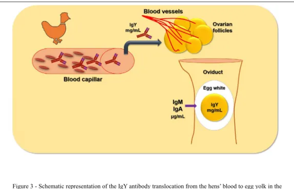
Maternal IgY or IgG
antibodies-based newborn protection is crucial for avian and
mammal species, respectively (LeaNdRO
et al.,
2011). However, the transference of these maternal
antibodies has been established by different pathway
comparing hens and mammals. Transference of IgY
antibodies occurs by their translocation into the egg
yolk, while mammal’s transplacental passage has
been demonstrated for IgG antibodies (LeaNdRO
et al., 2011; MeRRILL & GRINdSTaff, 2014;
BeRNaRdINI et al., 2017).
The egg yolk is concentrated daily into
the hens’ ovarian follicle by the translocation of
compounds from hens’ blood molecules. among these
are proteins such as the IgY antibodies (figure 3). The
egg yolk IgY deposition follows a circadian rhythm
with five day intervals between the passage of higher
and lower IgY concentrations (He et al., 2014).
The IgY antibodies are easily extracted
from the egg yolk and the process does not require a
bleeding procedure on hens. also, among the many
advantages of IgY is their antigen binding repertoire,
which is achieved by gene conversion using the
insertion of segments from pseudogenes (KaISeR,
2012); their avidity maturation; the propensity to
avoid false positive results in the mammalian model
of immunoassay platforms; enzyme and fluorescence
antibody conjugation; immune-gold beads antibody
labeling, and the production of monoclonal antibodies,
such as single chain fragment variable (scfv) by
cloning the fragment antigen binding (fab) coding
genes from the hen B cell (feRReIRa JÚNIOR et
al., 2012; NIe et al., 2014; ZHaNG et al., 2016; da
ROCHa et al., 2017; BORGeS et al., 2018).
CONCLUSION
The avian immune system has been
demonstrated to be highly competent, with a robust
innate and acquired immune response against different
kinds of pathogens. In this context, IgY antibodies are a
crucial character in the hens’ immune response due to
their specific antigen-binding properties. Hence, it has a
wide range application as an immunobiological reagent.
ACKNOwLEDGEMENTS
The authors wish to thank the Coordenação de aperfeiçoamento de Pessoal de Nível Superior (CaPeS, Brasil), fundação de amparo à Pesquisa de Minas Gerais (faPeMIG) e Instituto de estudos avançados em Medicina Veterinária “José
Caetano Borges” (UNIUBE) for granting a finnancial support to the present review in the field of IgY-technology.
DECLARATION OF CONFLICTING OF
INTERESTS
The authors declare no conflict of interest. The
founding sponsors had no role in the design of the study; in the collection, analyses, or interpretation of data; in the writing of the manuscript, and in the decision to publish the results.
REFERENCES
aRNOLd, K. B.; CHuNG, a. W. Prospects from systems serol-ogy research. Immunolserol-ogy, v. 153, n. 3, p.279-289, Mar 2018. ISSN 1365-2567. available from: <https://www.ncbi.nlm.nih.gov/ pubmed/29139548>.
aRuN, K. V.; TaLWaR, a.; KuMaR, T. S. T-helper cells in the etiopathogenesis of periodontal disease: a mini review. J Indian Soc Periodontol, v. 15, n. 1, p. 4-10, Jan 2011. ISSN 0975-1580. avail-able from: <https://www.ncbi.nlm.nih.gov/pubmed/21772714>.
BeCKeR, Y. Immunological and regulatory functions of unin-fected and virus inunin-fected immature and mature subtypes of den-dritic cells--a review. Virus Genes, v. 26, n. 2, p. 119-30, 2003. ISSN 0920-8569. available from: <https://www.ncbi.nlm.nih.gov/ pubmed/12803463>.
BeRNaRdINI, R. et al. Neonatal protection and preterm birth reduction following maternal group B streptococcus vaccination in a mouse model. J Matern Fetal Neonatal Med, v. 30, n. 23, p.
2844-2850, dec 2017. ISSN 1476-4954. available from: <https:// www.ncbi.nlm.nih.gov/pubmed/27973991>.
BLuM, J. S.; WeaRSCH, P. a.; CReSSWeLL, P. Pathways of antigen processing. Annu Rev Immunol, v. 31, p. 443-73, 2013. ISSN 1545-3278. available from: <https://www.ncbi.nlm.nih.gov/ pubmed/23298205>.
BOeHM, T.; HeSS, I.; SWaNN, J. B. evolution of lymphoid tissues. Trends Immunol, v. 33, n. 6, p. 315-21, Jun 2012. ISSN 1471-4981. available from: <https://www.ncbi.nlm.nih.gov/pubmed/22483556>.
BORGeS, I. P. et al. antiparasitic effects induced by polyclonal IgY antibodies anti-phospholipase a. Int J Biol Macromol, v. 112, p. 333-342, Jan 2018. ISSN 1879-0003. available from: <https://www.ncbi. nlm.nih.gov/pubmed/29391226>.
BÍLKOVÁ, B. et al. different breeds, different blood: Cytometric anal-ysis of whole blood cellular composition in chicken breeds. Vet Im-munol Immunopathol, v. 188, p. 71-77, Jun 2017. ISSN 1873-2534. available from: <https://www.ncbi.nlm.nih.gov/pubmed/28615130>.
da ROCHa, d. G. et al. The complementarity-determining region sequences in IgY antivenom hypervariable regions. Data Brief, v. 13, p. 717-722, aug 2017. ISSN 2352-3409. available from: <https:// www.ncbi.nlm.nih.gov/pubmed/28748206>.
de GeuS, e. d.; VeRVeLde, L. Regulation of macrophage and dendritic cell function by pathogens and through immunomodulation in the avian mucosa. Dev Comp Immunol, v. 41, n. 3, p. 341-51, Nov 2013. ISSN 1879-0089. available from: <http://www.ncbi.nlm. nih.gov/pubmed/23542704>.
deGeN, W. G. et al. Th1/Th2 polarization by viral and helminth in-fection in birds. Vet Microbiol, v. 105, n. 3-4, p. 163-7, feb 2005. ISSN 0378-1135. available from: <https://www.ncbi.nlm.nih.gov/ pubmed/15708812>.
feRReIRa JÚNIOR, Á. et al. Production, characterization and
applications for Toxoplasma gondii-specific polyclonal chicken
egg yolk immunoglobulins. PLoS One, v. 7, n. 7, p. e40391, 2012. ISSN 1932-6203. available from: <http://www.ncbi.nlm. nih.gov/pubmed/22808150>.
GRueBeR, C. e.; WaLLIS, G. P.; JaMIeSON, I. G. episodic positive selection in the evolution of avian toll-like receptor in-nate immunity genes. PLoS One, v. 9, n. 3, p. e89632, 2014. ISSN 1932-6203. available from: <https://www.ncbi.nlm.nih. gov/pubmed/24595315>.
GuO, X. et al. Molecular mechanisms of primary and second-ary mucosal immunity using avian infectious bronchitis virus as a model system. Vet Immunol Immunopathol, v. 121, n. 3-4, p. 332-43, feb 2008. ISSN 0165-2427. available from: <https:// www.ncbi.nlm.nih.gov/pubmed/17983666>.
GuRJaR, R. S.; GuLLeY, S. L.; VaN GINKeL, f. W. Cell-mediated immune responses in the head-associated lymphoid tissues induced to a live attenuated avian coronavirus vaccine. Dev Comp Immunol, v. 41, n. 4, p. 715-22, dec 2013. ISSN 1879-0089. available from: <https://www.ncbi.nlm.nih.gov/ pubmed/23948147>.
He, J. X. et al. Chronobiological studies of chicken IgY: monitor-ing of infradian, circadian and ultradian rhythms of IgY in blood and yolk of chickens. Vet Immunol Immunopathol, v. 160, n. 3-4, p. 266-72, aug 2014. ISSN 1873-2534. available from: <https:// www.ncbi.nlm.nih.gov/pubmed/24998020>.
HeIdaRI, M.; fITZGeRaLd, S. d.; ZHaNG, H. Immune Responses in Cecal Tonsils of Marek’s disease Virus-Infect-ed Chickens. Avian Dis, v. 59, n. 2, p. 213-26, Jun 2015. ISSN 0005-2086. available from: <https://www.ncbi.nlm.nih.gov/ pubmed/26473671>.
IfRaH, M. e. et al. The role of the bursa of fabricius in the mune response to vaccinal antigens and the development of im-mune tolerance in chicks (Gallus domesticus) vaccinated at a very young age. Poult Sci, v. 96, n. 1, p. 51-57, Jan 2017. ISSN 1525-3171. available from: <https://www.ncbi.nlm.nih.gov/ pubmed/27418658>.
JeuRISSeN, S. H. et al. defence mechanisms against viral infec-tion in poultry: a review. Vet Q, v. 22, n. 4, p. 204-8, Oct 2000. ISSN 0165-2176. available from: <https://www.ncbi.nlm.nih.gov/ pubmed/11087131>.
KaISeR, P. advances in avian immunology--prospects for dis-ease control: a review. Avian Pathol, v. 39, n. 5, p. 309-24, Oct 2010. ISSN 1465-3338. available from: <https://www.ncbi.nlm. nih.gov/pubmed/20954007>.
KaISeR, P. The long view: a bright past, a brighter future? for-ty years of chicken immunology pre- and post-genome. Avian Pathol, v. 41, n. 6, p. 511-8, dec 2012. ISSN 1465-3338. available from: <https://www.ncbi.nlm.nih.gov/pubmed/23237363>.
KaufMaN, J. The simple chicken major histocompatibility com-plex: life and death in the face of pathogens and vaccines. Philos Trans R Soc Lond B Biol Sci, v. 355, n. 1400, p. 1077-84, aug 2000. ISSN 0962-8436. available from: <https://www.ncbi.nlm. nih.gov/pubmed/11186309>.
KaufMaN, J. Co-evolution with chicken class I genes. Immunol Rev, v. 267, n. 1, p. 56-71, Sep 2015. ISSN 1600-065X. available from: <https://www.ncbi.nlm.nih.gov/pubmed/26284471>.
KOGuT, M. H. Cytokines and prevention of infectious diseases in poultry: a review. Avian Pathol, v. 29, n. 5, p. 395-404, Oct 2000. ISSN 1465-3338. available from: <https://www.ncbi.nlm.nih.gov/ pubmed/19184830>.
LANNING, D. K.; KNIGHT, K. L. Diversification of the Primary
antibody Repertoire by aId-Mediated Gene Conversion. Results Probl Cell Differ, v. 57, p. 279-93, 2015. ISSN 0080-1844. avail-able from: <https://www.ncbi.nlm.nih.gov/pubmed/26537386>.
LeaNdRO, N. M. et al. Maternal antibody transfer to broiler progeny varies among strains and is affected by grain source and cage density. Poult Sci, v. 90, n. 12, p. 2730-9, dec 2011. ISSN 0032-5791. available from: <https://www.ncbi.nlm.nih.gov/ pubmed/22080011>.
Lee, W. et al. Insights into the chicken IgY with emphasis on the generation and applications of chicken recombinant monoclo-nal antibodies. J Immunol Methods, v. 447, p. 71-85, 08 2017. ISSN 1872-7905. available from: <https://www.ncbi.nlm.nih.gov/ pubmed/28502720>.
LIVeRSIdGe, J.; fORReSTeR, J. V. antigen processing and pre-sentation in the eye: a review. Curr Eye Res, v. 11 Suppl, p. 49-58,
1992. ISSN 0271-3683. available from: <https://www.ncbi.nlm. nih.gov/pubmed/1424751>.
MadeJ, J. P.; STefaNIaK, T.; BedNaRCZYK, M. effect of in ovo-delivered prebiotics and synbiotics on lymphoid-organs’ mor-phology in chickens. Poult Sci, v. 94, n. 6, p. 1209-19, Jun 2015. ISSN 0032-5791. available from: <https://www.ncbi.nlm.nih.gov/ pubmed/25877410>.
MeLIef, C. J. Mini-review: Regulation of cytotoxic T lymphocyte responses by dendritic cells: peaceful coexistence of cross-priming and direct priming? Eur J Immunol, v. 33, n. 10, p. 2645-54, Oct 2003. ISSN 0014-2980. available from: <https://www.ncbi.nlm. nih.gov/pubmed/14515248>.
MeRRILL, L.; GRINdSTaff, J. L. Maternal antibody transfer can lead to suppression of humoral immunity in developing zebra
finches (Taeniopygia guttata). Physiol Biochem Zool, v. 87, n. 5, p. 740-51, 2014 Sep-Oct 2014. ISSN 1537-5293. available from: <https://www.ncbi.nlm.nih.gov/pubmed/25244385>.
MeuNIeR, M. et al. Promising new vaccine candidates against Campylobacter in broilers. PLoS One, v. 12, n. 11, p. e0188472, 2017. ISSN 1932-6203. available from: <https://www.ncbi.nlm. nih.gov/pubmed/29176789>.
MILLeR, M. M.; TaYLOR, R. L. Brief review of the chicken Major Histocompatibility Complex: the genes, their distribution on chromosome 16, and their contributions to disease resistance. Poult Sci, v. 95, n. 2, p. 375-92, feb 2016. ISSN 0032-5791. avail-able from: <https://www.ncbi.nlm.nih.gov/pubmed/26740135>.
MuKHeRJee, S.; KaRMaKaR, S.; BaBu, S. P. TLR2 and TLR4 mediated host immune responses in major infectious dis-eases: a review. Braz J Infect Dis, v. 20, n. 2, p. 193-204, 2016 Mar-apr 2016. ISSN 1678-4391. available from <https://www. ncbi.nlm.nih.gov/pubmed/26775799>.
NaGY, N.; BÓdI, I.; OLÁH, I. avian dendritic cells: Phenotype and ontogeny in lymphoid organs. Dev Comp Immunol, v. 58, p. 47-59, May 2016. ISSN 1879-0089. available from: <https://www. ncbi.nlm.nih.gov/pubmed/26751596>.
NANG, N. T. et al. Induction of inflammatory cytokines and Toll-like receptors in chickens infected with avian H9N2 influenza vi -rus. Vet Res, v. 42, p. 64, May 2011. ISSN 1297-9716. available from: <https://www.ncbi.nlm.nih.gov/pubmed/21592354>.
NIe, G. et al. detection of Clonorchis sinensis circulating an-tigen in sera from Chinese patients by immunomagnetic bead eLISa based on IgY. PLoS One, v. 9, n. 12, p. e113208, 2014. ISSN 1932-6203. available from: <https://www.ncbi.nlm.nih.gov/ pubmed/25474577>.
Ou, H. et al. Longevity of protective immune responses induced
by a split influenza A (H7N9) vaccine mixed with MF59 adjuvant
in BaLB/c mice. Oncotarget, v. 8, n. 54, p. 91828-91840, Nov 2017. ISSN 1949-2553. available from: <https://www.ncbi.nlm. nih.gov/pubmed/29190879>.
PaRKeR, a.; KaufMaN, J. What chickens might tell us about the MHC class II system. Curr Opin Immunol, v. 46, p. 23-29, Jun 2017. ISSN 1879-0372. available from: <https://www.ncbi. nlm.nih.gov/pubmed/28433952>.
PEI, J.; COLLISSON, E. W. Specific antibody secreting cells
corona-virus. Dev Comp Immunol, v. 29, n. 2, p. 153-60, 2005. ISSN 0145-305X. available from: <https://www.ncbi.nlm.nih.gov/ pubmed/15450755>.
QuINTeIRO-fILHO, W. M. et al. Heat stress decreases
expres-sion of the cytokines, avian β-defensins 4 and 6 and Toll-like re -ceptor 2 in broiler chickens infected with Salmonella enteritidis. Vet Immunol Immunopathol, v. 186, p. 19-28, apr 2017. ISSN 1873-2534. available from: <https://www.ncbi.nlm.nih.gov/ pubmed/28413046>.
QuReSHI, M. a.; HeGGeN, C. L.; HuSSaIN, I. avian mac-rophage: effector functions in health and disease. Dev Comp Immunol, v. 24, n. 2-3, p. 103-19, 2000 Mar-apr 2000. ISSN 0145-305X. available from: <https://www.ncbi.nlm.nih.gov/ pubmed/10717282>.
ROSTaMI, H. et al. Supplementing dietary rosemary (Rosmarinus
officinalis L.) powder and vitamin E in broiler chickens: evaluation
of humoral immune response, lymphoid organs, and blood proteins. Environ Sci Pollut Res Int, Jan 2018. ISSN 1614-7499. available from: <https://www.ncbi.nlm.nih.gov/pubmed/29330815>.
SaNTHaKuMaR, d. et al. avian Interferons and Their antiviral effectors. Front Immunol, v. 8, p. 49, 2017. ISSN 1664-3224. avail-able from: <https://www.ncbi.nlm.nih.gov/pubmed/28197148>.
SCHade, R. et al. Chicken egg yolk antibodies, production and application. IgY-Technology. . 1st. 2001. 255.
SePaHI, a.; SaLINaS, I. The evolution of nasal immune systems in vertebrates. Mol Immunol, v. 69, p. 131-8, Jan 2016. ISSN 1872-9142. available from: <https://www.ncbi.nlm.nih.gov/pubmed/26391349>.
SHaRMa, J. M.; TIZaRd, I. avian cellular immune effector mech-anisms--a review. Avian Pathol, v. 13, n. 3, p. 357-76, Jul 1984. ISSN 0307-9457. available from: <https://www.ncbi.nlm.nih.gov/ pubmed/18766854>.
SINGH, S. et al. Avian influenza viral nucleocapsid and hemaggluti -nin proteins induce chicken Cd8+ memory T lymphocytes. Virology, v. 399, n. 2, p. 231-8, apr 2010. ISSN 1096-0341. available from: <https://www.ncbi.nlm.nih.gov/pubmed/20116819>.
SMIAŁEK, M. et al. Local immunity of the respiratory mucosal sys -tem in chickens and turkeys. Pol J Vet Sci, v. 14, n. 2, p. 291-7, 2011. ISSN 1505-1773. available from: <https://www.ncbi.nlm.nih.gov/ pubmed/21721419>.
SuN, H. et al. Thymus transcriptome reveals novel pathways in response to avian pathogenic escherichia coli infection. Poult Sci, v. 95, n. 12, p. 2803-2814, dec 2016. ISSN 1525-3171. available from: <https://www.ncbi.nlm.nih.gov/pubmed/27466434>.
TaeBIPOuR, M. J. et al. evaluation of blood monocyte and lym-phocyte population in broiler chicken after vaccination and experi-mental challenge with Newcastle disease virus. Vet Immunol Im-munopathol, v. 190, p. 31-38, aug 2017. ISSN 1873-2534. avail-able from: <https://www.ncbi.nlm.nih.gov/pubmed/28778320>.
VaN GINKeL, f. W. et al. Conjunctiva-associated lymphoid tis-sue in avian mucosal immunity. Dev Comp Immunol, v. 36, n. 2, p. 289-97, feb 2012. ISSN 1879-0089. available from: <https:// www.ncbi.nlm.nih.gov/pubmed/21641931>.
WANG, Y. et al. Review on the identification and role of Toxo -plasma gondii antigenic epitopes. Parasitol Res, v. 115, n. 2, p. 459-68, feb 2016. ISSN 1432-1955. available from: <https:// www.ncbi.nlm.nih.gov/pubmed/26581372>.
XIAO, J. et al. Conserved peptides enhance immune efficiency of inactive vaccines against emerging avian influenza viruses in
chicken. Sci China Life Sci, v. 60, n. 12, p. 1340-1347, dec 2017. ISSN 1869-1889. available from: <https://www.ncbi.nlm.nih.gov/ pubmed/29230639>.
YOSHIMuRa, Y.; BaRua, a. female Reproductive System and Immunology. Adv Exp Med Biol, v. 1001, p. 33-57, 2017. ISSN 0065-2598. available from: <https://www.ncbi.nlm.nih.gov/ pubmed/28980228>.
ZHANG, Q. et al. Identification and structural composition of the
blood-spleen barrier in chickens. Vet J, v. 204, n. 1, p. 110-6, apr 2015. ISSN 1532-2971. available from: <https://www.ncbi.nlm. nih.gov/pubmed/25779339>.
ZHaNG, X. et al. IgY: a key isotype in antibody evolution. Biol Rev Camb Philos Soc, v. 92, n. 4, p. 2144-2156, Nov 2017. ISSN 1469-185X. available from: <https://www.ncbi.nlm.nih.gov/ pubmed/28299878>.