Normal skin of HIV-infected individuals
contains increased numbers of dermal
CD8 T cells and normal numbers of
Langerhans cells
1Instituto de Pesquisa Clínica Evandro Chagas, Fundação Oswaldo Cruz,
Rio de Janeiro, RJ, Brasil
2Departamento de Patologia, Universidade Estadual do Rio de Janeiro,
Rio de Janeiro, RJ, Brasil
3Serviço de Patologia, Hospital Estadual Colônia Curupaiti, and
4Departamento de Medicina Preventiva, Núcleo de Estudos em Saúde Coletiva,
Universidade Federal do Rio de Janeiro, Rio de Janeiro, RJ, Brasil Laboratórios de 5AIDS e Imunologia Molecular, and 6Hanseníase,
Instituto Oswaldo Cruz, Fundação Oswaldo Cruz, Rio de Janeiro, RJ, Brasil
7Serviço de Dermatologia, Hospital Universitário Clementino Fraga Filho,
Universidade Federal do Rio de Janeiro, Rio de Janeiro, RJ, Brasil M.C.G. Galhardo1,
F.F. Alvarenga2,
G. Schueler3,
M. Perez4,
M.G. Morgado5,
H. Ferreira6,
L.M.S. Azevedo7,
E.P. Sampaio6
and E.N. Sarno6
Abstract
Dysregulation of the skin immune system (SIS) could explain the high prevalence of skin disorders in HIV+ individuals. The present study was carried out to determine whether alterations in the cell population of SIS and epidermal immunoactivation occur in the normal skin of HIV+ individuals. Forty-five biopsies were taken from the normal upper arm skin of 45 HIV+ patients and of 15 healthy controls. HIV+ individuals were divided into three categories according to their CD4 cell blood count (<200, 200-499 and ≥500/µl). Hematoxylin-eosin was used to stain tissue sections for morphological analysis and immunohistochemistry was used for the evaluation of the frequency of macrophages, Langerhans cells, and CD lymphocyte subsets. In addi-tion, semiquantitative analysis of LFA-1, ICAM-1 and HLA-DR was determined in epidermal cells. Macrophages, Langerhans cells, and CD lymphocyte subsets did not differ significantly between any of the patient categories and the control group. When all HIV+ individuals were compared as a group to the control group, a significant increase in dermal CD8+ T lymphocytes (P < 0.01) and lower CD4-CD8 ratios (P < 0.01) were observed in the HIV+ individuals. Epidermal ICAM-1 and HLA-DR expression was negative in both HIV+ and normal skin biopsies. No evidence of a depletion of the SIS population or of epidermal immunoactivation in normal skin from HIV+ individuals was demonstrable, suggesting that alterations in the central immune system are not necessarily reflected in the SIS of HIV-infected pa-tients.
Correspondence
M.C.G. Galhardo Instituto de Pesquisa Clínica Evandro Chagas, FIOCRUZ Avenida Brasil, 4365 22045-900 Rio de Janeiro, RJ Brasil
Fax: +55-21-2590-9988 E-mail: [email protected]
Received May 27, 2003 Accepted December 2, 2003
Key words
•HIV
•Skin immune system
•CD8 T cells
•CD4-CD8 ratio
Introduction
During HIV infection, the skin is an or-gan of major morbidity. Increasingly severe cutaneous disorders most commonly occur during the course of the disease and some-times they provide the earliest clue to the existence of HIV or of its progression (1-3). Suggestive of both local permissiveness and responsiveness to the presence of HIV, a change in the cutaneous environment also appears to take place. As such, an dysregula-tion of the skin immune system (SIS) could be the prime reason for the increasing preva-lence of skin disorders in HIV+ individuals (4-7). However, few studies have been con-ducted on the SIS of HIV+ patients, in whom, similar to what occurs in other specialized lymphoid systems, changes in the immune cells associated with the SIS would be pre-dictable(8-10).
Most of the studies on SIS in HIV infec-tion have focused on the role of Langerhans cells (Lc), most probably because of their important function as antigen-presenting cells and because of their expression of CD4 cells. Although results continue to be contradic-tory, the hypothesis that the Lc population may suffer alterations during infection has been the subject of numerous studies (11-16). Few data are available regarding the role of T cells and the other components. Skin mast cell density has been shown to remain unchanged in HIV+ individuals (17). Even though keratinocytes do not become infected with HIV, they are capable of se-creting a variety of immunomodulatory cy-tokines (interleukin (IL)-1, IL-6 and tumor necrosis factor-α) which may enhance HIV replication and the spreading of the virus in the skin (7,18). In addition, as can be seen in a wide variety of dermatoses, keratinocytes may become immunoactivated by means of the induction of expression molecules, such as the intercellular adhesion molecule 1 (ICAM-1) and human leukocyte antigen (HLA)-DR, responsible for the increase in
lymphocyte traffic in the epidermis and its susceptibility to inflammatory reactions (19). The skrelated mechanisms in HIV in-fection are unknown, although SIS dysregu-lation has been consistently thought to play a role in triggering dermatoses. Using histo-logical and immunohistochemical tech-niques, we attempted to determine whether the SIS becomes impaired in the clinically normal-appearing skin of HIV+ patients at different stages of infection. The number of skin immune cells (Lc, dermal dendrite cells, T cell subsets and macrophages), the extent of perivascular infiltrates, and the presence of immunoactivated cell components (HLA-DR, ICAM-1 and leukocyte function-associ-ated antigen-1 (LFA-1)) were determined in situ.
Material and Methods
Subjects
inhibi-tors saquinavir and ritonavir) in a variety of combinations (Table 1). The control group consisted of 15 healthy individuals (8 males and 7 females; mean age, 35.4 ± 7.4; range, 20-42 years).
Skin biopsies
Punch biopsies (6 mm) were obtained from clinically normal skin on the sun-pro-tected inner side of the upper arm from HIV patients and control subjects. Biopsy samples were cut into two parts and processed for histological and immunohistochemical evalu-ation as described below.
Histopathological analysis
The skin fragment was fixed in 10% neu-tral-buffered formalin overnight, embedded in paraffin, and stained with hematoxylin-eosin. The material was submitted to the following morphological determinations: 1) count of the number of layers in the epider-mis and analysis of the keratinocytes in each layer; 2) evaluation of the dermal and subcu-taneous adnexa and blood vessels, and 3) measurement of the extension of the inflam-matory infiltrate. Samples were examined by two independent observers and morpho-logical features were recorded. Quantitative measurements (mean %) of the infiltrated area were made during microscopic exami-nation (100X magnification) of histological sections using a ProLite image analyzer, which permitted the viewing of fields with a Nikon E 450 microscope. For each sample, three different fields of the upper dermis were scanned (20X). The most extended infiltrates (four of them) were identified and measured in each field (40X), and the mean of each sample was calculated.
Immunohistochemistry
Biopsies were snap-frozen and kept in liquid nitrogen until the time for use. Frozen
sections were cut with a cryostat, air dried, and fixed in cold acetone for 10 min. The sections were incubated with the primary antibody for 60 min, washed in PBS, and then incubated with the biotinylated second-ary antibody (Dako Corporation, Carpinte-ria, CA, USA), followed by the avidin-bi-otin-peroxidase complex (Vectastain ABC kit; Vector Laboratories, Inc., Burlingame, CA, USA), and processed according to manu-facturer instructions. Finally, sections were developed with aminoethylcarbazole as the chromogenic substrate. In control specimens, the primary monoclonal antibody was omit-ted or isotype control antibodies of irrel-evant specificity were used. The sections were analyzed by light microscopy and pho-tomicrographs were taken with a Nikon Microphot system.
Antibodies
Mouse anti-human monoclonal antibodies were used for the identification of specific cell types, proteins, and surface molecules. Anti-CD1 (Lc) antibody, anti-CD3 (pan T cell marker), anti-CD4, anti-CD8 T cell subsets,
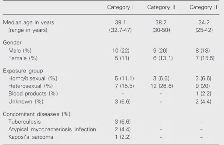
Table 1. Clinical data of the HIV+ individuals enrolled in the present study.
Category I Category II Category III
Median age in years 39.1 38.2 34.2 (range in years) (32.7-47) (30-50) (25-42)
Gender
Male (%) 10 (22) 9 (20) 8 (18) Female (%) 5 (11) 6 (13.1) 7 (15.5)
Exposure group
Homo/bisexual (%) 5 (11.1) 3 (6.6) 3 (6.6) Heterosexual (%) 7 (15.5) 12 (26.6) 9 (20) Blood products (%) - - 1 (2.2) Unknown (%) 3 (6.6) - 2 (4.4)
Concomitant diseases (%)
Tuberculosis 3 (6.6) - -Atypical mycobacteriosis infection 2 (4.4) - -Kaposi’s sarcoma 1 (2.2) -
anti-CD45RO (previous activated T cells), and anti-CD45RA (naive T cells) were obtained from Becton & Dickinson (San Jose, CA, USA) and were diluted 1:25 in PBS. Anti-CD68 (macrophage marker; Dako Corpora-tion) and anti-HLA-DR (Becton & Dickinson) were diluted 1:100. Anti-ICAM-1 and anti-LFA-1 were obtained from Immunotech
(Marseille, France) and were used at 1:50 dilution. The antibody concentration used was found to be optimal, providing maximum spe-cific staining with the lowest background sig-nal. Positive cells were identified by reddish-brown staining and compared with the control slides from which the primary antibody had been omitted. Staining for CD1 cells was con-sidered to be positive when the cell body was seen to have one or more dendritic processes. The total number of CD1-positive epidermal cells per field was determined by using a 40X objective for the length of the epidermis. The result was the ratio between the number of positive cells and the number of fields cover-ing the length of the epidermis. For quantifica-tion of CD3, CD4, CD8, CD45RO, CD45RA, and macrophages in the dermal infiltrate, count-ing was performed by focuscount-ing on the largest infiltrates in five fields in which the total number of positive cells per field was counted with 40X and 100X objectives. For HLA-DR, ICAM-1 and LFA-1 expression, semiquanti-tative analysis was performed as follows: for HLA-DR, (0) = only Lcs were stained, (+) = epidermal cells were stained not including all the layers or the total length of the epidermis, and (++) = all positive epidermal cells were stained. For ICAM-1, (0) = when the epider-mis was negative, (+) = only one area stained positive, and (++) = more than one area stained positive. For LFA-1, (0) = absent staining, (+) = sparse staining, (++) = moderate stain-ing, and (+++) = intense staining.
Viral load and T lymphocyte subsets. Blood samples were collected by venipunc-ture from all participants at the time of bi-opsy into Vacutainer tubes containing EDTA and heparin for the determination of viral load and T lymphocyte subsets, respectively. HIV-1 RNA was determined by quantita-tive nucleic acid sequence-based amplifica-tion (Organon, Teknika, Boxtel, The Nether-lands), as recommended by the manufac-turer. The detection limit of the assay was <400 copies/ml. CD3, CD4 and CD8 lym-phocyte counts were determined by
four-A
B
color staining (CD45, CD3, CD4, CD8) us-ing standard flow cytometry.
Statistical analysis
Results are reported as mean ± SD. A simple description of variables was elaborated in order to compare the different HIV catego-ries and controls with respect to clinical and laboratory data. ANOVA (Scheffé) and the chi-square test (Fischer) were used to measure the effect of HIV staging associated to inde-pendent variables. In addition, the Pearson-product-moment correlation coefficient (r) was calculated. The level of significance was set at P < 0.05 in all analyses.
Results
Histological analysis
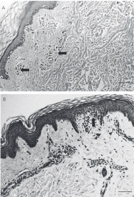
Hematoxylin-eosin staining showed a similar histological pattern and a nonspe-cific dermis infiltrate in HIV patients and control subjects. No major differences in mean percent area occupied by the infiltrate were noted between the patient categories and the control group, with values of 702.8 ± 621.6 for category I, 448.8 ± 274.8 for cat-egory II, 626.4 ± 408.6 for catcat-egory III, and 749.5 ± 462.3 for controls. Similarly, there was no statistically significant difference between HIV+ individuals (592.6 ± 760.1) and controls (749.5 ± 462.3; Figure 1A,B).
Immunohistological evaluation
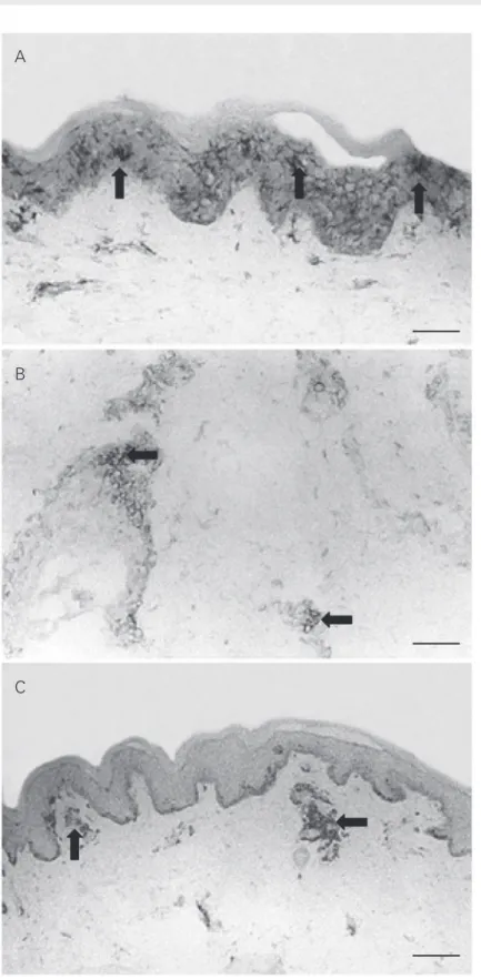
Immunohistological staining of frozen biopsies for T cell subsets showed that the numbers of CD3, CD4 and CD8 T lympho-cytes and the CD4-CD8 lymphocyte ratio in the perivascular dermis did not differ signifi-cantly among patient categories or between groups of HIV patients and controls (Table 2 and Figure 2B,C). However, a significant increase in CD8 T lymphocytes was de-tected in HIV+ individuals (5.3 ± 7.3) in the
Figure 2. Normal skin biopsies obtained from HIV+ patients. A, There is a diffuse distribu-tion of Langerhans cells (arrows) in the epidermis. Few Langerhans cells are present in the dermis (anti-CD1 staining, 200X). B, CD4 T lymphocytes (arrows) are present in the dermal cellular infiltrate (anti-CD8 staining, 200X). C, CD8 T lymphocytes (arrows) are shown in the dermal cellular infiltrate (anti-CD4 T staining, Original magnification, 200X. Bars = 80 µm.
A
B
22.5 ± 14.4 and 1.0 ± 1.4 for the control group (Figure 2A). Likewise, the mean mac-rophage cell density in the dermis was not significantly different between the HIV+ categories and the control group or among the different patient categories (data not shown). Epidermal HLA-DR and ICAM-1 expression was negative in the keratinocytes (0+) and LFA-1 and ICAM-1 were expressed constitutively in the blood vessels and in dermal lymphocytes.
Correlation between CD8 T lymphocytes in the perivascular dermis infiltrate and T lymphocyte subsets in the blood
The number of CD3-, CD4- and CD8-positive cells in blood was estimated in all the individuals enrolled in the study. As expected, CD4 cell averages were signifi-cantly different between the various catego-ries (except category III, CD4 ≥500/µl) and the control group, and individuals with AIDS presented the lowest averages (Table 2). When data for all HIV+ individuals were pooled, lower mean CD4-CD8 ratios were detected (0.44 ± 0.45) compared to the con-trol group (2.12 ± 1.24; P < 0.001). More-over, evaluation of the cellular infiltrate showed a positive correlation between CD8
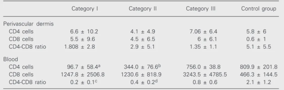
Table 2. Numbers of CD4 and CD8 T cells in the perivascular dermis and blood.
Category I Category II Category III Control group
Perivascular dermis
CD4 cells 6.6 ± 10.2 4.1 ± 4.9 7.06 ± 6.4 5.8 ± 6 CD8 cells 5.5 ± 9.6 4.5 ± 6.5 6 ± 6.1 0.6 ± 1 CD4-CD8 ratio 1.808 ± 2.8 2.9 ± 5.1 1.35 ± 1.1 5.1 ± 5.5
Blood
CD4 cells 96.7 ± 58.4a 344.0 ± 76.6b 756.0 ± 38.8 809.9 ± 201.8 CD8 cells 1247.8 ± 2506.8 1230.6 ± 818.9 3243.5 ± 4785.5 466.3 ± 144.5 CD4-CD8 ratio 0.2 ± 0.1c 0.4 ± 0.2d 0.8 ± 0.6 2.1 ± 1.2
Data are reported as means ± SD. Categories: I = CD4 <200/µl; II = CD4 200-499/µl; III = CD4 ≥500/µl. aAIDS patients had significantly lower numbers of CD4 lymphocytes in blood compared with category II (P < 0.02), category III (P < 0.001), and control (P < 0.001). bIn category II, CD4 numbers were also significantly lower compared to category III (P < 0.001), and to control (P < 0.001). cThe CD4-CD8 T lymphocyte ratio was significantly lower for the AIDS group compared to control (P < 0.001), and for dcategory II compared to control (P < 0.001). ANOVA was used for all comparisons.
perivascular dermis compared to control (0.6 ± 1; P < 0.01). In addition, these patients presented a lower CD4-CD8 ratio (2 ± 3.4) compared to control (5.1 ± 5.6; P < 0.01). A positive correlation was also observed be-tween the number of CD3 and CD8 T lym-phocytes (r = 0.29; P < 0.028) and between the number of CD4 lymphocytes and the CD4-CD8 ratio in the perivascular dermis (r = 0.80; P < 0.001) in all individuals. Staining for the CD45RO and CD45RA isoforms showed no differences among the various groups (data not shown).
Langerhans cells
perivascular T lymphocyte counts and CD8 counts in blood (r = 0.29; P < 0.02) and a negative correlation between perivascular CD8 T cells and the CD4-CD8 ratio in blood (r = 0.31; P < 0.01).
With respect to viral load, 37 patients showed detectable levels of HIV-1 RNA in plasma with no significant difference among the 3 groups (category I: 4.72 ± 0.69, cat-egory II: 3.9 ± 0.85, and catcat-egory III: 4.4 ± 0.85). No correlations were detected between cell components and viral load.
Discussion
In view of the great importance of skin morbidity in HIV infection and the scarcity of studies on the possible participation of the SIS in the development of the disease, the original purpose of this study was to ascer-tain the morphological aspects and to char-acterize the cell population of the immune system in the normal skin of HIV+ individu-als.
Most of the patients in this study were receiving antiretroviral therapy, a fact that might probably have had some impact on the results. Generally, the introduction of highly active antiretroviral therapy is associated with significantly lower cutaneous morbidity in HIV patients (20-23). However, despite the antiretroviral therapy, it was possible to di-vide the population studied into categories according to the variations observed in their CD4 T cell counts, which indicated sharply different degrees of immunodeficiency (P < 0.05), especially considering individuals with AIDS (category I), who were severely im-munodepressed (CD4 = 96/µl).
In the present study, surprisingly, no re-duction in CD4 counts was found in the perivascular infiltrate of the dermis, nor did we observe any propensity toward the loss of CD45RO cells, normally described to occur in the earlier stages of HIV infection (24). This finding appears to indicate that the CD4 cells are preserved in the skin in contrast to
the decreased number in peripheral blood. In addition, the dynamic process of replication/ destruction of CD4+ T cells seems to be occurring in most patients, as indicated by the presence of HIV-1 RNA in plasma.
In contrast, among HIV+ individuals, an increase of CD8 T cells was noted in the perivascular dermis. Cases of skin CD8 + T-cell infiltrate associated with advanced HIV infection have been described (25-30). Func-tional studies with CD8 T cells obtained from the HIV skin infiltrates have shown a cytotoxic immune response to HIV protein. It has been suggested that, in addition to their known role in controlling the retroviral in-fection, these cytotoxic T lymphocytes may also be involved in the pathogenesis of the cutaneous inflammatory disorders occurring during the course of the disease (26,27). In this respect, a cutaneous lymphoma express-ing a CD8 T cell phenotype has been re-ported in HIV patients (31).
Furthermore, the reversed CD4-CD8 T cell ratio reported for blood (described in the present study), as well as in other specialized lymphoid systems and clearly observed dur-ing HIV infection, was not seen in the dermis (Table 2) (9,10,32). Nevertheless, the perivas-cular CD4-CD8 ratio was significantly lower in HIV-infected patients compared to the control group, a fact that could probably be attributed to the increased number of CD8+ T cells.
As reported in other recent studies in-volving a larger number of patients, there was no detectable reduction in Lc density in the normal-appearing skin (14-16). Although Lc is the sole cell of the epidermis that can be infected by HIV (33,34), simultaneously al-lowing the efficient replication of the virus (35,36), the frequency of infected Lc is low (37). It would appear then that the involve-ment of Lc in HIV infection is a great deal more qualitative than quantitative in nature (38,39).
patients or between them and the control group. Previous studies have also failed to detect any HIV viral particles or viruses in such cells (40). On the other hand, in other sites such as the lung, where HIV can be isolated from 50% of the macrophages in bronchoalveolar lavage fluids, numerical, phenotypical, and functional changes in mac-rophages have been reported to occur (3). Similarly, no increase in HLA-DR or ICAM-1 expression was observed in keratinocytes, suggesting that there is no inflammatory re-sponse in the normal skin of HIV+ individu-als.
The present study has shown the preser-vation of immune cells such as dermal CD4+ T cells, dermal dendritic cells, macrophages, and Lc in the normal skin of HIV patients. It can now be convincingly argued that the skin is not a major site of HIV replication and that the SIS in HIV infection acts differently
from other local immune systems where HIV-related changes take place. Conversely, it would seem that subtle interactions do oc-cur, as indicated mainly by the increased number of CD8+ T cells in the skin. Al-though in the present study we did not assess the activation of CD8 cells, the presence of an increased number of cells in the perivas-cular normal skin may somehow lead to tissue damage, and, thereby actually contrib-uting to the pathogenesis of skin disease in HIV infection.
Acknowledgments
The authors wish to thank Dr. Sérgio Luiz Gomes Antunes for helpful advice and the Laboratório de Produção e Tratamento de Imagens, Instituto Oswaldo Cruz, Rio de Janeiro, RJ, Brazil, for the figures.
References
1. Coldiron BM & Bergstresser PR (1989). Prevalence and clinical spectrum of skin disease in patient infected with human immuno-deficiency virus. Archives of Dermatology, 125: 357-361.
2. Uthayakumar S, Nanwani R, Drinkwater T, Nayagam AT & Darley CR (1997). The prevalence of skin disease in HIV infection and its relationship to the degree of immunosuppression. British Medical Journal, 137: 595-598.
3. Spira R, Mignard M, Doutre MS, Morlat P & Dabis F (1998). Preva-lence of cutaneous disorders in a population of HIV-infected pa-tients. Archives of Dermatology, 34: 1208-1212.
4. Duvic M (1995). Human immunodeficiency virus and the skin: se-lected controversies. Journal of Investigative Dermatology, 105 (Suppl): 117S-121S.
5. Tschachler E, Bergstresser PR & Stingl G (1996). HIV-related skin diseases. Lancet, 348: 659-663.
6. Henry M & Tschachler E (1996). The skin immune system in the course of HIV-1 infection. Photochemistry and Photobiology, 64: 275-279.
7. Blauvelt A & Katz SI (1995). The skin as target, vector, and effector organ in human immunodeficiency virus disease. Journal of Investi-gative Dermatology, 105: 122S-126S.
8. Bray DH, Squire SB, Bagdades E, Mulvenna PM, Johnson MA & Poulter LW (1992). Alveolar macrophage populations are distorted in immunocompromised patients with pneumonitis. European Res-piratory Journal, 5: 545-552.
9. Lim SG, Condez A, Lee CA, Johnson MA, Elias C & Poulter L (1993). Loss of mucosal CD4 lymphocytes is an early feature of HIV infec-tion. Clinical and Experimental Immunology, 92: 448-454.
10. Olaitan A, Johnson MA, MacLean A & Poulter LW (1996). The distribution of immunocompetent cells in the genital tract of HIV-positive women. AIDS, 10: 759-764.
11. Belsito DV, Sanchez MR, Baer RL, Valentine F & Thorbecke J (1984). Reduced Langerhans’ cell Ia antigen and ATPase activity in patients with the acquired immunodeficiency syndrome. New Eng-land Journal of Medicine, 310: 1279-1282.
12. Dréno B, Milpied B, Bignon JD, Stalder JF & Litoux P (1988). Prognostic value of Langerhans cells in the epidermis of HIV pa-tients. British Journal of Dermatology, 118: 481-486.
13. Kanitakis J, Marchand C, Su H, Thivolet J, Zambruno G, Schmitt D & Gazzolo L (1989). Immunohistochemical study of normal skin of HIV-1 infected patients shows no evidence of infection of epidermal Langerhans cells by HIV. AIDS Research and Human Retroviruses, 5: 293-302.
14. Kalter DC, Greenhouse JJ, Orenstein JM, Schnittman SM, Gendelman HE & Meltzer MS (1991). Epidermal Langerhans cells are not principal reservoirs of virus in HIV disease. Journal of Immu-nology, 146: 3396-3404.
15. Nandwani R, Gazzard BG, Barton SE, Hawkins DA, Zemelman V & Staughton RC (1996). Does HIV disease progression influence epi-dermal Langerhans cell density? British Journal of Dermatology, 134: 1087-1092.
16. Comptom CC, Kupper TS & Nadire KB (1996). HIV-1 infected Lan-gerhans cells constitute a significant proportion of the epidermal Langerhans cell population throughout the course of HIV disease. Journal of Investigative Dermatology, 107: 822-826.
Geog I, Ferreira H & Sarno EN (2000). Search for evidence of a Th2 profile in HIV+ patients. International Journal of Dermatology, 39: 109-115.
18. Oxholm A, Oxholm P & Permin H (1989). Epidermal tumour necro-sis factor α and interleukin 6-like activities in AIDS-related Kaposi’s sarcoma. Acta Pathologica et Microbiologica Scandinavica, 97: 533-538.
19. Wantzing GL, Ralfkiaer E, Lisby S & Rothlein R (1989). The role of intercellular adhesion molecules in inflammatory skin reactions. British Journal of Dermatology, 119: 141-145.
20. Mirmirani P, Maurer TA, Berger TG, Sands LP & Chren MM (2002). Skin-related quality of life in HIV-infected patients on highly active antiretroviral therapy. Journal of Cutaneous Medicine and Surgery, 6: 10-18.
21. Johnson RA (1999). Human immunodeficiency virus disease in the era of HAART: a reevaluation of the mucocutaneous manifesta-tions. Current Clinical Topics in Infectious Diseases, 19: 252-286. 22. Costner M & Cockerell CJ (1998). The changing spectrum of
cuta-neous manifestations of HIV infection. Archives of Dermatology, 134: 1290-1292.
23. Calista D, Morri M, Stagno A & Boschini A (2002). Changing morbid-ity of cutaneous diseases in patients with HIV after the introduction of antiretroviral therapy including a protease inhibitor. American Journal of Clinical Dermatology, 3: 359-362.
24. Gruters RA, Terpstra FG, De Goede RE, Mulder JW, De Wolf F, Schellekens PT, Van Lier RA, Tersmette M & Miedema F (1991). Immunological and virological markers in individuals progressing from seroconversion to AIDS. AIDS, 5: 837-844.
25. Janier M, Katlama C, Flageul B, Valensi F, Moulonguet I, Sigaux F, Dompmartin D & Civatte J (1989). The pseudo-Sezary-syndrome with CD8 phenotype in a patient with the acquired immunodeficien-cy syndrome (AIDS). Annals of Internal Medicine, 110: 738-740. 26. Bachelez H, Hadida F & Gorochov G (1996). Massive infiltration of
the skin by human immunodeficiency virus-specific cytotoxic CD8 T cells (Letter). New England Journal of Medicine, 335: 61-62. 27. Bachelez H, Hadida F, Parizot C, Flageul B, Kemula M, Dubertret L,
Debree P & Gorochov G (1998). Oligoclonal expansion of immuno-deficiency virus-specific cytotoxic CD8 T cells in the skin of human immunodeficiency virus-1 infected patients with pseudolymphoma. Journal of Clinical Investigation, 101: 2506-2516.
28. Friedler S, Parisi MT, Waldo E, Wieczorek R, Sidhu G & Rico MJ (1999). Atypical cutaneous lymphoproliferative disorder in patients with HIV infection. International Journal of Dermatology, 38: 111-118.
29. Guitart J, Variakojis D, Kuzel T & Rosen S (1999). Cutaneous CD8+ T cell infiltrates in advanced HIV infection. Journal of the American
Academy of Dermatology, 41: 722-727.
30. Pirovano S, Signorini L, Facchetti F, Santoro A, Albertin A & Imberti L (2001). Polyclonal T-cell expansion in a HIV+ patient with atypical cutaneous lymphoproliferative disorder, large granular lymphocyte proliferation and SENV infection. Haematologica, 86: 881-882. 31. Burns MK & Cooper KD (1993). Cutaneous T-cell lymphoma
associ-ated with HIV infection. Journal of the American Academy of Der-matology, 29: 394-399.
32. Bofill M, Janossy G, Lee CA, MacDonald-Burns D, Phillips AN, Sabin C, Timms A, Johnson MA & Kernof PBA (1992). Laboratory control values for CD4 and CD8 T lymphocytes. Implications for HIV-1 diagnosis. Clinical and Experimental Immunology, 88: 243-252.
33. Tschachler E, Groh V, Popovic M, Mann DL, Konrad K, Safai B, Eron L, Veronese FM, Wolff K & Sting G (1987). Epidermal Langerhans cells - a target for HTLV-III/LAV infection. Journal of Investigative Dermatology, 88: 233-237.
34. Giannetti A, Zambruno G, Cimarelli A, Marconi A, Negroni M, Girolomoni G & Bertazzoni U (1993). Direct detection of HIV-1 in epidermis Langerhans cells of HIV-1 infected patients. Journal of Acquired Immune Deficiency Syndromes, 6: 329-333.
35. Kawamura T, Cohen SS, Borris DL, Aquilino EA, Glushakova S, Margolis LB, Orenstein JM, Offord RE, Neurath AR & Blauvelt A (2000). Candidate microbicides block HIV-1 infection of human im-mature Langerhans cells within epithelial tissue explants. Journal of Experimental Medicine, 192: 1491-1500.
36. Reece JC, Handley AJ, Anstee EJ, Morrison WA, Crowe SM & Cameron PU (1998). HIV-1 selection by epidermal dendritic cells during transmission across human skin. Journal of Experimental Medicine, 187: 1623-1631.
37. Cimarelli A, Zambruno G, Marconi A, Giralomoni G, Bertazzoni U & Giannetti A (1994). Quantitation by competitive PCR of HIV-1 provi-ral DNA in epidermal Langerhans cells of HIV-1 infected patients. Journal of Acquired Immune Deficiency Syndromes, 7: 230-235. 38. Blauvelt A, Clerici M, Lucey DR, Steinberg SM, Yarchoan R, Walker
R, Shearer GM & Katz SI (1995). Functional studies of epidermal Langerhans cells and blood monocytes in HIV-1 infected persons. Journal of Immunology, 154: 3506-3515.
39. Blauvelt A, Chougnet C, Shearer GM & Katz SI (1996). Modulation of T cell responses to recall antigens presented by Langerhans cells in HIV-discordant identical twins by anti-interleukin (IL)-10 antibodies and IL-12. Journal of Clinical Investigation, 97: 1550-1555. 40. Kalter DC, Gendelman HE & Meltzer MS (1991). Monocytes,