Wild mushrooms and their mycelia as sources of bioactive
compounds: antioxidant, anti-inflammatory and cytotoxic
properties
SOUILEM FEDIA
Dissertation submitted to Escola Superior Agrária de Bragança to obtain the Degree of Master in Biotechnological Engineering
Supervised by
Dr. Anabela R.L. Martins
Dr. Isabel C.F.R. Ferreira
Dr. Fathia Skhiri
Dissertation made under the agreement of Double Diploma between the Escola Superior Agrária de Bragança|IPB and the
ACKNOWLEDGEMENTS
First of all, I want to acknowledge my supervisors, Dr. Anabela Martins, Dr. Isabel C.F.R. Ferreira and Dr. Fethia Skhiri, for their generosity and infinite support, great help in laboratory procedures, continuous encouragement and support in the writing of this thesis.
My special thanks Dr. Lillian Barros for her practical guidance and encouragement and to Dr. João Barreira for his collaboration in statistical analyses.
Many thanks to Dr. Ângela Fernandes and Dr. Ricardo Calhelha for their excellent support in the laboratorial experiments.
My profound gratitude to all the people of BioChemCore. I really appreciate all your efforts and I am really happy to be part of your research team. Also, to Mountain Research Centre (CIMO) for all the support.
Lastly, I would like also to express my gratitude to all of my friends and my family especially my father Mohamed, my mother Monia, my sister Fida and my brothers Lassaad and Houcem who have helped and given moral support during the happy and sad times; I do not know what I would do without you.
TABLE OF CONTENT
INDEX OF FIGURES ... v
INDEX OF TABLES ... vii
ABBREVIATIONS LIST ... viii
ABSTRACT ... xi
RESUMO ... xii
CHAPTER 1. FRAMEWORK ... 1
CHAPTER 2. INTRODUCTION ... 2
1. Studied mushroom species ... 2
1.1 Pleurotus eryngii (DC.) Quél. ... 2
1.2 Suillus bellinii (Inzenga) Watling ... 3
2. Bioactive compound in wild mushrooms ... 4
2.1 Phenolic acids ... 4
2.2 Ergosterol ... 10
3. Bioactivity of wild mushroom extracts ... 11
3.1 Antioxidant activity ... 12
3.2 Anti-inflammatory activity ... 15
3.3 Cytotoxic activity ... 16
4. Production of mushroom mycelia by in vitro culture ... 18
4.1 In vitro culture applied to mushrooms ... 18
4.2 Case-studies with wild mushrooms ... 19
5. Objectives ... 21
CHAPTER 3. MATERIALS AND METHODS ... 22
1. Wild samples and in vitro production of mycelia ... 22
2. Standards and reagents ... 22
3. Preparation of the extracts ... 23
4.1 Analysis of phenolic acids ... 23
4.2 Analysis of ergosterol ... 24
5. Evaluation of bioactive properties ... 25
5.1 Antioxidant activity ... 25
5.1.1 DPPH radical scavenging activity ... 25
5.1.2 Reducing power ... 26
5.1.3 Inhibition of 𝛽-carotene bleaching ... 27
5.1.4 Thiobarbituric acid reactive substances (TBARS) assay ... 28
5.2 Anti-inflammatory activity ... 28
5.2.1 Cells treatment ... 29
5.2.2 Nitric oxide determination ... 29
5.3 Cytotoxic activity ... 30
5.3.1 In human tumor cell lines ... 30
5.3.2 In non-tumor cells ... 31
6. Statistical ... 32
CHAPTER 4. RESULTS AND DISCUSSION ... 33
1. Mycelia growth ... 33
2. Chemical characterization of the extracts ... 35
3. Antioxidant activity ... 38
4. Anti-inflammatory activity ... 40
5. Cytotoxic activity ... 41
CHAPTER 5. CONCLUDING REMARKS AND FUTURE PERSPECTIVES ... 44
INDEX OF FIGURES
Figure 1. Fruiting body of Pleurotus eryngii (DC.) Quél. ... 2 Figure 2. Fruiting body of Suillus bellinii (Inzenga) Watling ... 4 Figure 3. Chemical structures of benzoic and cinnamic acid derivatives usually found in
mushrooms (Heleno et al., 2014). ... 6
Figure 4. General representation of the phenolic acids biosynthesis derived from the shikimic
acid pathway. (Heleno et al., 2014). The numbers find the correspondence in the entries of Table 2. ... 7
Figure 5. Steroid nucleus. ... 10 Figure 6. Chemical structure of ergosterol. ... 10 Figure 7. Major causes for over production of free radical (oxidative stress), possible cellular
targets and conditions that were associated to oxidative stress (Ferreira et al., 2009). ... 12
Figure 8. Natural antioxidants separated in classes. Green words represent exogenous
antioxidants, while yellow ones represent endogenous antioxidants (Carocho & Ferreira, 2013a). ... 13
Figure 9. Overview of the main reactions involving reactive Oxygen species (ROS) / reactive
Nitrogen species (RNS), and major endogenous enzymatic and non-enzymatic antioxidant defenses in the cell. The most representative endogenous sources (traced rectangles) of ROS/RNS are presented and include: Mitochondrial ETS (Electron transport system), NADPH oxidases, Xanthine oxidase for ROS and NO synthases for RNS. The main antioxidant defenses are presented in shaded rectangles and the enzymes involved are presented in italic. Molecular Oxygen (O2), superoxide anion(O2•), hydrogen peroxide (H2O2),
hydroxyl radical (HO•), hydroxide ion (HO-), membrane lipids (LH), lipid radical (L•), peroxyl radical (LOO•), hydroperoxide lipid (LOOH), lipid alkoxyl radical (LO•), nitric oxide (NO•), radicals (R•), non-radicals (R), alcohols (LOH), glutathione (GSH), glutathione disulphide (GS-SG), α-tocopherol or vitamin E (vit. E), vitamin E radical (vit. E•), vitamin C (vit. C), vitamin C radical (vit. C•), S-nitrosoglutathione (GSNO), nicotinamide adenine dinucleotide phosphate: oxidized (NADP+), reduced (NADPH). Enzymes: superoxide
dismutase (SOD), catalase (CAT), glutathione peroxidase (GPx), glutathione redutase (Gred), glutathione- S-transferases (GST), Mitochondrial ETS (electron transport system), nitric oxide synthase (NOS) (Ferreira et al., 2009). ... 14
Figure 10. Low-molecular-weight (LMW) and high-molecular-weight (HMW) compounds
Figure 11. Step of solvent removal in the preparation of the extracts. ... 23
Figure 12. HPLC-DAD equipment used in the analysis of phenolic acids. ... 24
Figure 13. HPLC-UV system used in the analysis of ergosterol. ... 25
Figure 14. Microplate used in the evaluation of DPPH radical-scavenging activity. ... 26
Figure 15. Microplate used in the evaluation of reducing power. ... 27
Figure 16.Test tubes used in the β-carotene/linoleate assay. ... 27
Figure 17. Test tubes used in the TBARS assay. ... 28
Figure 18. Microplate showing the NO assay with methanolic extracts. ... 30
Figure 19. Microplate used in the evaluation of the methanolic extracts cytotoxicity. ... 31
Figure 20. Pleurotus eryngii (a) and Suillus bellinii (b) mycelia cultivated in vitro, in both liquid (PDB and iMMN) and solid medium (PDA and iMMN). ... 33
Figure 21. Growth in diameter of the mycelia of P. eryngii cultivated in different culture media throughout time. ... 34
Figure 22. Growth in diameter of the mycelia of S. bellinii cultivated in different culture media throughout time. ... 34
INDEX OF TABLES
Table 1. Classification through number of carbons and basic structure of molecules within the
phenolic compounds family (Carocho & Ferreira, 2013). ... 5
Table 2. Main enzymes involved in the biosynthesis of phenolic acids through shikimate
pathway from L- phenylalanine or L- tyrosine. ... 8
Table 3. Antioxidant activity of methanolic extracts prepared from fruiting bodies and
mycelia of the species studied within this work. ... 15
Table 4. Anti-inflammatory activity of extracts prepared from fruiting bodies of the species
studied within this work. ... 16
Table 5. Cytotoxic activity of extracts prepared from fruiting bodies of the species studied
within this work. ... 18
Table 6. Ergosterol content (mg/g extract) and phenolic acids composition (μg/g extract) in
the mycelia and culture media of P. eryngii and S. bellinii. The values corresponding to the
fruiting body of both mushrooms (wild samples) are also presented. Values are given as mean±standard deviation. ... 36
Table 7. Antioxidant activity (EC50 values, mg/mL extract) of the mycelia and culture media
of P. eryngii and S. bellinii. The values corresponding to the fruiting body of both mushrooms
(wild samples) are also presented. Values are given as mean±standard deviation ... 39
Table 8. Anti-proliferative (GI50 values, μg/mL extract) and anti-inflammatory activity (EC50
values, μg/mL extract) of the mycelia and culture media of P. eryngii and S. bellinii. The
ABBREVIATIONS LIST
5-LOX: 5-Lipooxygenase
ANOVA: One vary analysis of variance CAT: Catalase
COMT: Caffeic/5-hydroxyferulic acid-O-transferase
COX: Cyclooxygenase COX-2: Cyclooxygenase-2 DAD: Diode array detector
DMEM: Dulbecco’s modified Eagle’s minimum essential medium DNA: Deoxyribonucleic acid
DPPH: 2,2-Diphenyl-1-picrylhydrazyl radical Dw: Dry weight
EC50:Extract concentration corresponding to 50% of antioxidant activity ECACC: European collection of animal cell culture
ETS: Electron transport system ECM: Ectomycorrhizal
FBS: Fetal bovine serum Fw: Fresh weight
GAE: Gallic acid equivalents
GI50: Extract concentration that inhibited 50% of the net cell growth GPx: Glutathione peroxidase
Gred: Glutathione reductase GSH: Glutathione
GSNO: S-nitrosoglutathione GS-SG: Glutathione disulphide GST: Glutathione-S-transferases HBSS: Hank’s balanced salt solution HCT15: Colon carcinoma cell line
HeLa: Henrietta Lacks human cervical carcinoma cell line HepG2: Hepatocellular carcinoma cell line
HMW: High molecular weight
HPLC: High performance liquid chromatography
HPLC-UV: High performance liquid chromatography coupled to an ultraviolet detector
HSD:Tukey’s honestly significant difference
HO•: Hydroxyl radical HO-:Hydroxyl ion H2O2: Hydrogen peroxide
ICAM-1: Intercellular adhesion molecule-1 IL-1 β: Interleukin 1β
IL-6: Interleukin 6 IL-8: Interleukin 8
iMMN: Incomplete melin-norkans medium iNOS: Inducible nitric oxide synthase L•: Lipidic radical
LH: Membrane lipids
LMW: Low molecular weight LO•:Lipid alkoxyl radical LOH: Alcohols
LOO•: Peroxyl radical LOOH: Hydroperoxide lipid LPS: Lipopolysaccharide
MCF-7: Breast adenocarcinoma cell line MDA: Malondialdehyde
MMN: Melin–norkans medium
NADPH: Nicotinamide adenine dinucleotide reduced
NADP+:Nicotidamide adenine dinucleotide phosphate: oxidized NCI-H460: Human lung cancinoma cell line
NF-KB: Nuclear factor kappa B
NED: N-(1-napthyl) ethylenediamine hydrochloride NOS: Nitric oxide synthase
NO: Nitric oxide
NO˙: Nitric oxide radical
NSAIDs: Nonsteroidal anti-inflammatory drugs PAL: Phenylalanine ammonialyase
PLP2: Non- tumor primary culture of porcine liver cells PGE2: Prostaglandin E2
R: Non-radicals R•:Radicals
RNS: Nitrogen reactive species ROS: Oxygen reactive species RSA: Radical scavenging activity SRB: Sulphorodamine B
SOD: Superoxide dismutase TAL: Tyrosine ammonia lyase
TBARS: Thiobarbituric acid reactive substances TBA: Thiobarbituric acid
TCA: Trichloroacetic acid
TNF- α: Tumour necrosis factor α O2•: Superoxide anion
Vit. C: Vitamin C
Vit. C•: Vitamin C radical Vit. E: Vitamin E
Vit. E•:Vitamin E radical UV: Ultraviolet
ABSTRACT
Mushrooms are an important source of natural compounds with acknowledged bioactivity.
Pleurotus eryngii (DC.) Quél., in particular, is widely recognized for its organoleptic quality
and favorable health effects, being commercially produced in great extent. On the other hand,
Suillus bellinii (Inzenga) Watling is an ectomycorrhizal symbiont, whose main properties
were only reported in a scarce number of publications. Some current trends point toward using the mycelia and the culture media as potential sources of bioactive compounds, in addition to the fruiting bodies. Accordingly, P. eryngii and S. bellinii were studied for their
composition in phenolic acids and sterols, antioxidant capacity (scavenging DPPH radicals,
reducing power, β-carotene bleaching inhibition and TBARS formation inhibition),
inflammatory effect (by down-regulating LPS-stimulated NO in RAW264.7 cells) and anti-proliferative activity (using MCF-7, NCI-H460, HeLa, HepG2 and PLP2 cell lines). Overall,
S. bellinii mycelia showed higher contents of ergosterol and phenolic compounds (which were
also detected in higher quantity in its fruiting body) and stronger antioxidant activity than P.
eryngii. On the other hand, P. eryngii mycelia showed anti-inflammatory (absent in S. bellinii
mycelia) and a cytotoxicity similar (sometimes superior) to its fruiting bodies, in opposition to
S. bellinii, whose mycelia presented a decreased anti-proliferative activity. Furthermore, the
assayed species showed differences in the growth rate and yielded biomass of their mycelia, which should also be considered in further applications.
Keywords: Pleurotus eryngii (DC.) Quél., Suillus bellinii (Inzenga) Watling, ergosterol,
RESUMO
Os cogumelos são uma importante fonte de compostos naturais com reconhecida bioatividade.
Pleurotus eryngii (DC.) Quél., em particular, é largamente reconhecido pela sua qualidade
organolética e benefícios para a saúde, sendo comercialmente produzido em larga escala. Por outro lado, Suillus bellinii (Inzenga) Watling é um fungo simbionte ectomicorrízico, cujas
propriedades estão pouco documentadas. A produção de micélio através da cultura in vitro
revela-se como uma fonte potencial de obtenção de compostos bioativos, para além da produção pelos carpóforos. Assim sendo, P. eryngii e S. bellinii foram estudados na sua
composição em ácidos fenólicos e esteróis, capacidade antioxidante (capacidade de captação de radicais de DPPH; poder redutor, inibição do branqueamento do β-caroteno e Inibição da formação de TBARS), capacidade anti-inflamatória (através da regulação negativa da NO estimulada pela LPS na linha celular RAW264.7) e a atividade anti-proliferativa (nas linhas celulares MCF-7, NCI-H460, HeLa, HepG2 e PLP2). O micélio de S. bellinii mostrou
maiores conteúdos de ergosteral e de compostos fenólicos (que também foram detetados em maiores quantidades).
Por outro lado, o micélio de P. eryngii apresentou capacidade anti-inflamatória e
citotoxicidade similares (e por vezes superiores) ao corpo frutífero, ao contrário do que acontece com S. bellinii, cujo micélio apresenta uma atividade anti-proliferativa diminuída.
Além disso, as espécies estudadas apresentam diferenças nas taxas de crescimento e de produção de biomassa dos seus micélios, que tem que ser considerada em futuras aplicações.
Palavras-chave: Pleurotus eryngii (DC.) Quél., Suillus bellinii (Inzenga) Watling, ergosterol,
CHAPTER 1. FRAMEWORK
The number of mushroom species on earth is evaluated to be 140.000, proposing that only 10% are already known (Ferreira et al., 2010).
For several centuries, wild mushrooms have been part of the normal human diet, and also extensively consumed due to their organoleptic properties including distinctive flavor, texture, and taste. Edible wild mushrooms have been described as source of chemical and nutritional properties. They are rich in minerals, water (80 to 90%), protein, fiber and vitamins (thiamine, riboflavin, ascorbic acid, vitamin D2, and carbohydrates). They are low caloric foods due to low lipids level (free fatty acids, mono-, di- and triglycerides, sterols, and phospholipids) (Heleno et al., 2009; Kalac, 2009, 2012). Beyond the nutritional characteristics, several medicinal properties have been reported, mainly due to their richness in bioactive compounds, secondary metabolites, including phenolic compounds (Carocho & Ferreira, 2013), polysaccharides (Ren et al., 2016), and lipids (Heleno et al., 2009), that present antioxidant (Ferreira et al., 2009), antitumor (Ferreira el al., 2010), anti-inflammatory (Taofiq et al., 2015), antibacterial (Alves et al., 2012) and antifungal (Alves et al., 2013) properties, among other bioactivities.
Not just fruiting bodies, also their mycelium has been recognized as a source of natural new drugs. In this context, mushroom mycelium is produced through novel biotechnologies in order to produce bioactive compounds using as pharmaceutical agent or functional food (Reis et al., 2011).
CHAPTER 2. INTRODUCTION
1. Studied mushroom species
1.1 Pleurotus eryngii (DC.) Quél.
The genus Pleurotus (Fries) Kummer was defined by Paul Kummer in 1871 and belongs to
the phylum Basidiomycota and the order Agaricales. Many of the species of this genus produce mushroom that usually live in wood and grow in clusters. They have a range of spores varying from white to pink, brown, and nearly black (Trudell & Ammirati, 2009).
Figure 1. Fruiting body of Pleurotus eryngii (DC.) Quél.
This group of mushrooms can be recognized by its rather soft and fleshy fruiting body (Arora, 1986). It exhibits a short-term growth compared to other mushrooms (Liu et al., 2016), it has a high nutritional value and several therapeutic properties, being also used in different biotechnological and environmental applications (Knop et al., 2015;Corrêa et al., 2016).
Pleurotus eryngii (DC.) Quél. is a widespread species in the Mediterranean, central Europe,
central Asia, and North Africa. Ithas a convex cap and very decurrent whitish gills (Figure
1). It is a weak parasite, and can live on the root or stem base of plants. P. eryngii taxon is
described as ‘complex’ due to the significant variations in morphology, biochemical and
genetic makeup so it has a wide geographical distribution. It is also used in biotechnological processes including food production, bioremediation of soil and industrial waters, biotransformation of raw plant materials to feed, among others (Stajic et al., 2009; Reis et al., 2014).
reported by Reis et al., (2014) was as follows: moisture (82.59±0.36 g/100 g fw), ash (14.95±0.91 g/100 g dw),proteins (2.09±0.01 g/100 g dw) and energy (362.00±2.06 kcal/100 g dw); its free sugars were mannitol (1.40±0.09 g/100 g dw) andtrehalose (14.21±0.23 g/100 g dw), and tocopherols included α-tocopherol (6.79±1.80 μg/100 g dw), β-tocopherol (48.24±9.53 μg/100 g dw) and γ-tocopherol (31.55±7.56 μg/100 g dw). It presented low levels of fatty acids being the major ones palmitic acid (17.44±0.21%), stearic acid (4.77±0.08%), oleic acid (47.52±0.07%) and linoleic acid (24.71±0.28%). The percentages of saturated, monounsaturated and polyunsaturated fatty acids were 25.79±0.18%, 49.05±0.24% and 25.17±0.24%, respectively (Reis et al., 2014). The main organic acids found in P. eryngii
were oxalic acid (2.02±0.03 mg/g dw), malic acid (18.48±0.07 mg/g dw), citric acid (28.73±0.57 mg/g dw) and fumaric acid (2.50±0.05 mg/g dw) (Barros et al., 2013).
P. eryngii is esteemed primarily for its nutritional value, but it can also have bioactive
properties through different active substances such as polysaccharides, lipids, peptides, sterols, dietary fibers, etc. Furthermore, several recent studies have indicated that P. eryngii
polysaccharides have several functions, including hepatoprotector, immunity enhancer and antihyperlipidemia, and can inhibit the growth of several types of tumor cell lines (Ren et al., 2016).
1.2 Suillus bellinii (Inzenga) Watling
The genus Suillus (Inzenga) Watling is equivalent to the Boletaceae (Hawksworth et al.,
1995); it is a genus from the phylum Basidiomycota and order Agaricales (Arora, 1986).
The fruiting bodies are yellow, brown and reddish in many species of the genus (Trudell & Ammirati, 2009). In addition, Arora, (1986) illustrated that the genus Suillus has a “slippery”
cap, the pores are radially elongated in some species, having a veil or a glandular-dotted stem or both, and being mycorrhizal almost exclusively with conifers. Furthermore, Suillus spp. is
considered C-demanding species able to produce large amounts of biomass and produce large amounts of exudates (Izumi et al., 2013).
Suilus bellinii (Inzenga) Watling is known as an ectomycorrhizal symbiont associated with a
Figure 2. Fruiting body of Suillus bellinii (Inzenga) Watling
Suillus bellinii has different organic acids such as malic acid, quinic acid (31.2±1.37 g/kg
dw); 42% of nonaromatic acids; citric acid (11.2±0.27 g/kg dw); 20% of nonaromatic acids and also fatty acids, mainly palmitic acid (160.702 mg/kg dw) (Ribeiro et al., 2009).
S. bellinii is also rich in alcohols, which are considered to be the main odorants of the
mushroom like aroma (Guedes De Pinho et al., 2008).
2. Bioactive compound in wild mushrooms
Mushrooms have become attractive as functional foods and as a source of physiologically beneficial bioactive compounds. Herein, we will give emphasis to phenolic acids and ergosterol.
2.1 Phenolic acids
Phenolic compounds represent about 8000 different phenolic structures. They have one or more aromatic rings with one or more hydroxyl groups, including different subclasses such as flavonoids, phenolic acids, stilbenes, lignans, tannins, and oxidized polyphenols (Crozier et al., 2006; Barros et al., 2009; Fraga et al., 2010; Carocho & Ferreira, 2013). Table 1 displays
their structure as well as the number of carbons and representative compounds.
potential of the mushroom extracts (Chan et al., 2011; Bertalanic et al., 2012; Skotti et al., 2014).
Table1. Classification through number of carbons and basic structure of molecules within the phenolic
compounds family (Carocho & Ferreira, 2013).
Number of carbons Classification Example Basic structure
7 Hydroxybenzoic acids Gallic acid
9 Hydroxycinnamic acids p-Coumaric acid
9 Coumarins Esculetin
13 Xanthons Mangiferin
14 Stilbenes Resveratrol
15 Flavonoids Naringenin
Several research studies have reported the bioactive potential of phenolic acids as the main phenolic compounds found in mushroom, as well their quantity was commonly determined by HPLC (High performance Liquid Chromatograhy) (Ferreira et al., 2009; Heleno et al., 2015).
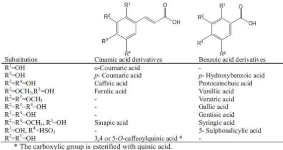
Phenolic acids can be splitted into two major categories, hydroxybenzoic acids and hydroxycinnamic acids, which are derived from non-phenolic molecules of benzoic and cinnamic acid, respectively. These compounds have at least one aromatic ring in which at least one hydrogen is substituted by a hydroxyl group (Figure 3) (Heleno et al., 2014).
sugars or organic acids in plant foods. Hydroxycinnamic acid derivatives are present in the bound form, linked to cell-wall structural components, including cellulose, lignin, and protein, as well as linked to organic acids (Ferreira et al., 2009).
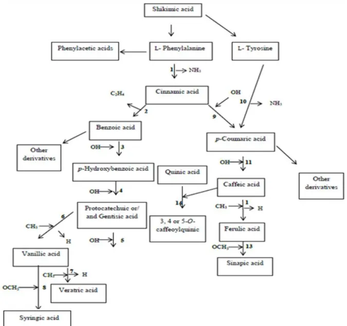
Phenolic acids are synthesized from the shikimate pathway from L-phenylalanine or L- tyrosine (Rice-Evans et al., 1996) (Figure 4). These amino acids are the common precursors
of the majority of the phenolic natural products.
Figure 3. Chemical structures of benzoic and cinnamic acid derivatives usually found in mushrooms (Heleno et
al., 2014).
Phenolic acids are abundant in a balanced daily diet, showing different bioactivities such as antioxidant (Rice-Evans et al., 1996, Ferreira et al., 2009), antitumor (Carocho & Ferreira, 2013), and antimicrobial (Alves et al., 2013a) properties. Cinnamic acid has strong capacity to inhibit the growth of human tumor cell lines such as non-small lung carcinoma (NCI-H460) (Vaz et al., 2012), colon carcinoma (HCT15) and cervical carcinoma (HeLa) cell lines (Heleno et al., 2014a, 2015).
Mushrooms have been extensively studied due to their bioactive properties (Mau et al., 2002; Murcia et al., 2002; Yang et al., 2002; Ferreira et al., 2009) assigned to several molecules including phenolic acids (Valentão et al., 2005; Puttaraju et al., 2006; Ribeiro et al., 2007; Kim et al., 2008; Barros et al., 2009). Nevertheless, in vivo, these compounds are metabolized
The main phenolic acids reported in P. eryngii are p-hydroxybenzoic acid (3.81±0.41 mg/100
g dw) (Reis et al., 2014), gallic acid andprotocatechuic acid (Ferreira et al., 2009; Heleno et al., 2015).
Figure 4. General representation of the phenolic acids biosynthesis derived from the shikimic acid pathway.
Table 2. Main enzymes involved in the biosynthesis of phenolic acids through shikimate pathway from L-
phenylalanine or L- tyrosine.
Entry Starting molecule Enzyme Final compound
1
Phenylalanine
Phenylalanine ammonialyase (PAL)
Cinnamic acid
2
Cinnamic acid
Oxidase (presumed b-oxidation)
Benzoic acid
3
Benzoic acid
Benzoic acid 4-hydroxylase
p-Hydroxybenzoic acid
4
p-Hydroxybenzoic acid
p-Hydroxybenzoic acid 3-hydroxylase
Gentisic acid
5
Protocatechuic acid
Protocatechuic acid 5-hydroxylase
Gallic acid
6
Protocatechuic acid
Protocatechuic acid 3-O-methyltransferase
7
Vanillic acid
Vanillic acid 4-O-methyltransferase
Veratric acid
8
Vanillic acid
Vanillic acid 5 hydroxylase and vanillic acid 5-O-methyltransferase
Syringic acid
9
Cinnamic acid
Cinnamic acid 4-hydroxylase
p-Coumaric acid
10
L- Tyrosine
Tyrosine ammonia lyase (TAL)
p-Coumaric acid
11
p-Coumaric acid
p-Coumaric acid 3-hydroxylase
Caffeic acid
12
Caffeic acid
Caffeic acid 3-O-methyltransferase
Ferulic acid
13
Ferulic acid
Ferulic acid 5-hydroxylase and caffeic/5-hydroxyferulic acid O-methyltransferase (COMT)
2.2 Ergosterol
The presence of sterols in animals, plants and fungi has been reported by Barreira & Ferreira, (2015). They are organic compounds with a tetracyclic structure of four rings linked together, of three to six carbons and other ring with five carbons (steroid nucleus) (Figure 5) with a
hydroxyl group at theC3 position and an aliphatic chain linked to the steroid nucleus (Fahy et al., 2005).
Figure 5 . Steroid nucleus.
Ergosterol is evidently the main sterol found in mushrooms, being a precursor of vitamin D2 (Figure 6) (Mattila et al., 2002; Barreira & Ferreira, 2015).
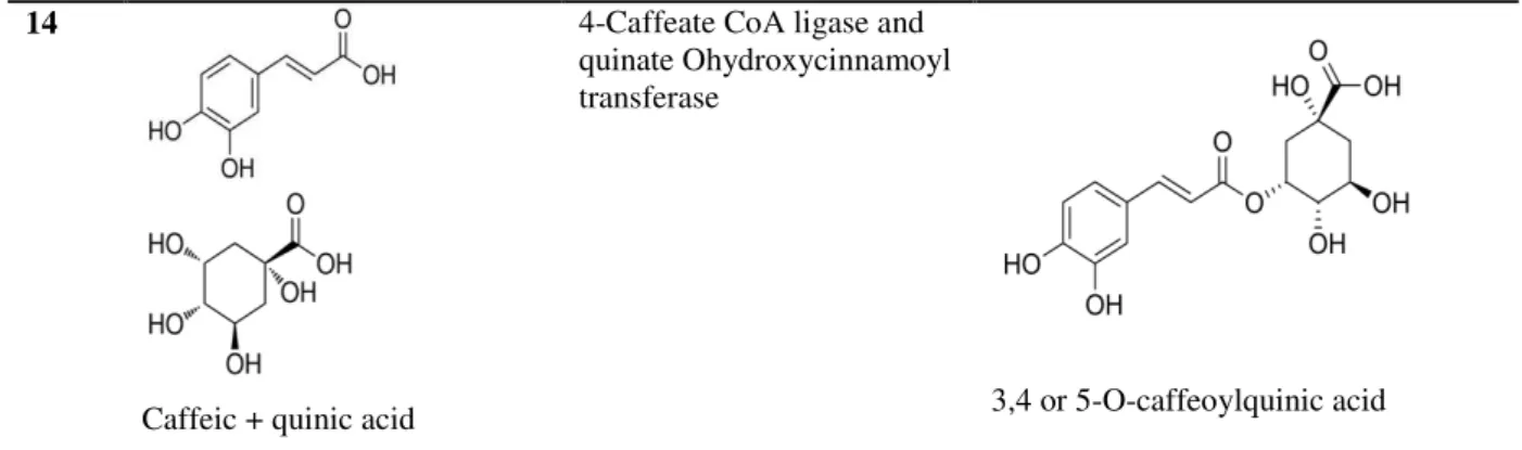
Figure 6. Chemical structure of ergosterol. 14
Caffeic + quinic acid
4-Caffeate CoA ligase and quinate Ohydroxycinnamoyl transferase
Ergosterol is a solid compound, crystalline, colourless with the scientific name 5,7,22-ergostatrien-3β-ol, the empirical formula C28H44O and a molecular weight of 396.36 g/mol.
Its melting point is in the order of 161-166ºC.In addition, ergosterol supports the temperature of 250 °C without decomposition. It has a side chain with a double bond at C22 and two double bonds at the level of positions C5 and C7 (ring B), giving a maximum absorption in the UV in a range of 240 to 300 nm (Barreira et al., 2014). Ergosterol is practically insoluble in water; however, it is soluble in organic solvents, such as chloroform. Exposure to ultraviolet light causes a photochemical reaction that converts ergosterol to ergocalciferol (vitamin D2) (Villares et al., 2012).
According to Barreira et al., (2014), ergosterol can be determined by different analytical methods such as high performance liquid chromatography coupled to ultraviolet detection. Savón et al., (2002) and Jasinghe & Perera, (2005) reported that the concentration of ergosterol in mushrooms, depends on the part of the mushroom tissue, developmental stage, growth conditions, and environmental factors.
Sterols, in general, have been reported to play several biological functions that include antioxidant (Shao et al., 2010), anti-inflammatory (Kuo et al., 2011) and antitumoral activity (Villares et al., 2012; Barreira & Ferreira, 2015), having also the ability to activate the expression of specific defense genes (Lochman & Mikes, 2005). Furthermore, these compounds permit a reduction in pain related inflammation, reduce cardiovascular disease and inhibit cyclooxygenase (COX) enzymes (Yasukawa et al., 1994; Zhang et al., 2002; Ravi & Abplanalp, 2003).
The ergosterol content in Pleurotus eryngii obtained after Soxhlet extraction with hexane and
a saponification step was 187±1mg/100 g dw(Barreira et al., 2014).
3. Bioactivity of wild mushroom extracts
Mushrooms grow in darkness and dampness in highly competitive environments and protect themselves from microbes by excretion of natural substances; this explains their richness in bioactive compounds (Ferreira et al., 2010).
immunomodulator (Borchers et al., 2008), antiatherogenic (Mori et al., 2008), hypoglycemic (Hu et al., 2006) and anti-inflammatory (Taofiq et al., 2016) activities.
3.1 Antioxidant activity
Oxidation is essential to many organisms, which can produce energy to fuel biological processes (Huanga et al., 2015).
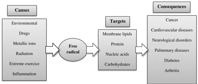
A free radical is an atom or molecule possessing unpaired electrons in the outer orbit, generally unstable and very reactive (Halliwell & Gutteridge, 1999; Gutteridge & Halliwell, 2000; Carocho & Ferreira, 2013a). Free radicals and non-radical species are produced in the normal metabolism of aerobic cells, mostly in the form of oxygen or/and nitrogen reactive species (ROS or/and RNS) (Valko et al., 2007;Ferreira et al., 2009).They can be produced in the mitochondria, through xanthine oxidase, peroxisomes, inflammation processes, and phagocytosis, among other. Nevertheless, some external factors also help to promote the production of free radicals such as smoking, environmental pollutants, radiation, drugs, pesticides, industrial solvents and ozone (Figure 7) (Ferreira et al., 2009).
Figure 7. Major causes for over production of free radical (oxidative stress), possible cellular targets and
conditions that were associated to oxidative stress (Ferreira et al., 2009).
Free radicals are neutralized by cellular antioxidant defenses such as enzymes and non-enzymatic molecules. Maintaining the equilibrium between free radicals production and antioxidant defenses is an essential condition for normal organism functioning; this equilibrium might be displaced either by the overproduction of ROS or by the loss of the cell antioxidant defenses (Figure 8) (Ferreira et al., 2009; Lobo et al., 2010; Carocho & Ferreira,
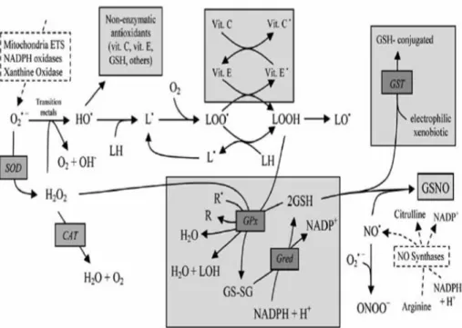
leads to oxidation and damage of cellular lipids, proteins and DNA, and causing several diseases including cancer, cardiovascular diseases, neurological disorders, renal disorders, liver disorders, hypertension, among others (Valko et al., 2007; Ferreira et al., 2009; Lobo et al., 2010, Carocho & Ferreira, 2013a). Several ROS production pathways and the main endogenous antioxidant defenses of the cell are described in Figure 9 (Ferreira et al., 2009).
Moreover, the synthetic antioxidants most commonly used in industrial processing may be cytotoxic. Thus, it is necessary to enhance natural and nontoxic antioxidants (Yating et al., 2015), that can be found in mushrooms.
Figure 9. Overview of the main reactions involving reactive Oxygen species (ROS) / reactive Nitrogen species
(RNS), and major endogenous enzymatic and non-enzymatic antioxidant defenses in the cell. The most representative endogenous sources (traced rectangles) of ROS/RNS are presented and include: Mitochondrial ETS (Electron transport system), NADPH oxidases, Xanthine oxidase for ROS and NO synthases for RNS. The main antioxidant defenses are presented in shaded rectangles and the enzymes involved are presented in italic. Molecular Oxygen (O2), superoxide anion(O2•), hydrogen peroxide (H2O2), hydroxyl radical (HO•), hydroxide
ion (HO-), membrane lipids (LH), lipid radical (L•), peroxyl radical (LOO•), hydroperoxide lipid (LOOH), lipid
alkoxyl radical (LO•), nitric oxide (NO•), radicals (R•), non-radicals (R), alcohols (LOH), glutathione (GSH),
glutathione disulphide (GS-SG), α-tocopherol or vitamin E (vit. E), vitamin E radical (vit. E•), vitamin C (vit. C),
vitamin C radical (vit. C•), S-nitrosoglutathione (GSNO), nicotinamide adenine dinucleotide phosphate: oxidized
(NADP+), reduced (NADPH). Enzymes: superoxide dismutase (SOD), catalase (CAT), glutathione peroxidase (GPx), glutathione redutase (Gred), glutathione- S-transferases (GST), Mitochondrial ETS (electron transport system), nitric oxide synthase (NOS) (Ferreira et al., 2009).
In this context, mushrooms contain many different compounds with powerful antioxidant activities, such as phenolic compounds and vitamins (Ferreira et al., 2009) that help the organism in the combat against oxidative stress. As an example, Table 3 shows antioxidant
effects in extracts prepared from the fruiting bodies of the species studied within the present work.
Particularly, P. eyngii showed high reducing power and free radical-scavenging activity (Reis
Table 3. Antioxidant activity of methanolic extracts prepared from fruiting bodies and mycelia of the species
studied within this work.
DPPH: 2,2-Diphenyl-1-picrylhydrazyl; TBARS: thiobarbituric acid reactive substances; EC50: Extract concentration corresponding to 50% of antioxidant activity or 0.5 of absorbance for the Ferricyanide/Prussian blue assay. GAE: Gallic acid equivalents.
3.2 Anti-inflammatory activity
Inflammation is a biological response to eliminate the initial cause of cell injury or harmful toxins stimuli such as pathogens, damaged cells, or irritation. However, inflammation may lead to many diseases including atherosclerosis, obesity, metabolic syndrome, diabetes and cancer (Moro et al., 2012; Taofiq et al., 2016). Macrophages are important components of the immune system, and they play a crucial role in the inflammatory response by supplying an immediate defense against foreign agents and release of excess pro-inflammatory mediators such as interleukins (IL-1β, IL-6, IL-8), tumor necrosis factor (TNF-α), nuclear factor-κB
(NF-κB), intercellular adhesion molecule-1 (ICAM-1), inducible type cyclooxygenase-2
(COX-2), prostaglandin E2 (PGE2), 5-lipoxygenase (5-LOX), and inducible nitric oxide synthase (iNOS) that leads to the production of reactive nitrogen species such as nitric oxide (NO). Overproduction of these inflammatory mediators leads to different kinds of cell damage (Joo et al., 2014; Taofiq et al., 2015).
Therefore, pharmacologic treatment with anti-inflammatories generally begins with nonsteroidal anti-inflammatory drugs (NSAIDs). In contrast, the long-term administration of NSAIDs is associated with a poor safety profile on gastrointestinal, cardiovascular, renal, also high blood pressure and acute tubular necrosis (Taofiq et al., 2015; Rovati et al., 2016).
Species Part Antioxidant assay
Pleurotus eryngii
(Reis et al., 2012)
Fruiting body
Mycelium (MMN) Folin Ciocalteu assay mg (GAE/g extract) 7.14 ± 2.01 9.11 ± 0.23 Fruiting body
Mycelium (MMN) Ferricyanide/Prussian blue assay; EC50 (mg/mL) 3.72 ± 0.09 3.81 ± 0.02 Fruiting body
Mycelium (MMN) DPPH scavenging activity assay; EC50 (mg/mL) 8.67 ± 0.12 25.40 ± 0.33 Fruiting body
Mycelium (MMN) -carotene/linoleate assay; EC50 (mg/mL) 4.68 ± 0.60 1.43 ± 0.60 Fruiting body
Mycelium (MMN) TBARS assay; EC50 (mg/mL) 3.95 ± 0.58 21.03 ± 0.45
Suillus bellinii
(Ribeiro et al., 2006)
In this case, discovering of natural bioactive compounds with anti-inflammatory properties and without or lower toxic impact is essential. Mushrooms have also demonstrated some anti-inflammatory potential based on their ability to reduce the production of anti-inflammatory mediators (Taofiq et al., 2016).
Previous research studies have been carried out on several mushroom species, mainly in methanolic and ethanolic extracts (Table 4).
Table 4. Anti-inflammatory activity of extracts prepared from fruiting bodies of the species studied within this work.
Species Extract NO production inhibition
Pleurotus eryngii Ethanolic EC50 value: 388 ± 17 μg/mL (Taofiq et al., 2015).
Suillus bellinii Not available in literature.
EC50: Extract concentration corresponding to 50% of NO production inhibition.
3.3 Cytotoxic activity
Carcinogenesis is one of the most life-threatening diseases and causes severe health problems in both developed and developing countries. It is a process which normally takes several years during which progressive genetic changes occurs leading to malignant transformations (Ferreira et al., 2010; Jemal et al., 2011; Carocho & Ferreira, 2013; Rashed et al., 2014).
Several endogenous causes are known to increase the risk of cancer. They include external factors such as smoking, dietary factors, infections, exposure to radiation, lack of physical activity, obesity, and environmental pollutants (Anand et al., 2008), and internal factors such as chromosomal abnormalities, somatic mutations, and immunological surveillance (Carocho & Ferreira, 2013a).
Furthermore, literature suggests that there is a link between cancer and oxidative stress. Therefore, the consumption of foods with antioxidant activity may reduce oxidative stress associated to these diseases (Vaz et al., 2010).
In this context, mushrooms are a vast source of powerful potential pharmaceutical drugs. They have been established to inhibit the growth of different tumor cell lines (Poucheret et al., 2006). Moreover, both cellular components and secondary metabolites of a large number of mushrooms have anticarcinogenic actions including, low-molecular-weight (LMW), and high-molecular-weight compounds (HMW) (Figure 10) (Wasser & Weis, 1999; Ferreira et
al., 2010).
Mushrooms have powerful capacities against cancers of the stomach, esophagus, and lungs, among others and are known in China, Japan, Korea, Russia, United States and Canada (Ferreira et al., 2010).
The most important molecules found in mushrooms with antitumor potential are polysaccharides and phenolic compounds or derivatives (Vaz et al., 2012).
Several recent studies have indicated that P. eryngii polysaccharides can inhibit the growth of
several types of cancer (Ren et al., 2016) (Table 5).
Table 5. Cytotoxic activity of extracts prepared from fruiting bodies of the species studied within this work.
Species Extract Type of activity
Pleurotus eryngii Polysaccharidic Inhibited HepG-2 proliferation, induced apoptosis, cell cycle arrest at
the S-phase and intracellular ROS production (Ren et al., 2016).
Suillus bellinii Not available in literature.
4. Production of mushroom mycelia by in vitro culture
4.1 In vitro culture applied to mushrooms
The filamentous fungi are characterized by being non-mobile organisms composed of an organized mass of threadlike cells called hyphae (the assembly of hyphae are called mycelium), having a life cycle with sexual and asexual reproduction that results in spores production, usually from a common haploid "stem" resulting from zygotic meiosis and heterotrophic nutrition (Griffin, 1994; Alexopoulos et al., 1996).
Leiva et al., (2015) pointed out that mycelium growth is affected by the in vitro conditions,
such as size of the inoculum, the period of incubation, pH, temperature, composition of media, thus, cultures on agar and in liquid media are excellent models and are responsible for mycelium development. PDA (Potato Dextrose Agar) and MMN (Melin-Norkrans medium)
have been the successfully media for the cultivation of many wild mushrooms as demonstrated by Pinto et al., (2013).
Stamets and Chilton, (1983) explain the methodology of culture in solid medium: “Cut a piece of the interior cap tissue or the upper region of the "stem" from mushroom and put the fragment at the center of a Petri dish. This technique takes place in a sterile area to avoid contamination (yeast, bacteria), that would stop the growth of mycelia”.
In vitro culture methodology (for mycelia production) is used as alternative products of
fruiting bodies, containing numerous advantages including the shorter cultivation time, storage of mycelia for a long period without genetic alterations, contamination can be easily observed and monitored on the flat of media, so it is easy to know and retain pure culture, it is also important for the preservation of endangered or rare species (Stamets & Chilton, 1983; Ferreira et al., 2009; Zilly et al., 2011; Heleno et al., 2012;Pinto et al., 2013). In addition to these advantages, in vitro culture allows the scaling up of biomass production for
as polysaccharides (Chen et al., 2013), phenolic compounds (Reis et al., 2012) and some other
from mycelium it’s easy, and could be isolated for medicinal purposes (Pinto et al., 2013),
including antioxidant, anti-inflammatory, antitumor, antimicrobial, antiviral, antidiabetic, immunomodulatory, cardiovascular, liver protective, and antifibrotic properties (Borchers et al., 2004;Gonçalves et al., 2011). In this context, Reis et al., (2012) was the first publication comparing in vivo and in vitro culture in terms of bioactivity.
Different media influence growth rates and mycelia biomass production, since liquid MMN media contains available nutrients and oxygen leading to higher mycelia absorption of nutrients, thus a faster growth and higher mycelia biomass (Heleno et al., 2012). On other hand, culture media also may influence the bioactivity of mycelium. Heleno et al., (2012) demonstrated that PDA is the most indicated medium to increase the antioxidant activity of G.
lucidum mycelium.
Corrêa et al., (2016) in a recent review reported that Pleurotus spp mycelium has high
nutritional profile that determines a precious aroma, characteristic flavor, and pharmacological value being a source of important bioactive compounds such as peptides, glycoproteins, polysaccharides, lipids and hydrolytic and oxidative enzymes with antioxidant, antimicrobial, anti-inflammatory, antitumor and immunomodulatory effects.
4.2 Case-studies with wild mushrooms
The antioxidant properties of wild mushrooms with their content in antioxidant compounds including tocopherols, can detoxify damaging forms of activated oxygen. A comparative study of tocopherols composition and antioxidant properties of in vivo (fruiting bodies) and in
vitro (mycelia) ectomycorrhizal fungi: Paxillus involutus and Pisolithus arhizus was carried
out by Reis et al., (2011). Mycelia showed higher levels of total tocopherols than fruiting
bodies, and particularly P. arhizus mycelium demonstrated to be an efficient source of
-tocopherol.
A systematic study was performed in order to compare the antioxidant activity of phenolic and polysaccharidic extracts from fruiting body, spores and mycelium, obtained in three different culture media, of Ganoderma lucidum. The highest levels of total polysaccharides
Another study examines a comparison of the antioxidant activities and phenolic profile of cultivated mushrooms and their mycelia: Agaricus bisporus, Pleurotus ostreatus, Pleurotus
eryngii and Lentinula edodes. L. edodes mycelia have the highest reducing power. Generally, in vivo samples detected higher antioxidant activities than their mycelia obtained by in vitro
culture. On the other hand, phenolic compounds were revealed both in mushrooms and mycelia without any particular abundance. Results demonstrated that there is no correlation between mushrooms and their mycelia obtained in vitro, thus in vitro production may be used
5. Objectives
The main goals of the present work were to produce, chemically characterize and evaluate the bioactivity of mycelia of two mushrooms species, Pleurotus eryngii and Suillus bellinii,
obtained by in vitro culture, using different solid and liquid culture media: iMMN (incomplete
Melin-Norkans medium) and PDA/PDB (Potato Dextrose Agar/ Potato Dextrose broth media).
Furthermore, the mycelia produced, their fruiting body and the culture media were:
i) Chemically characterized in terms of phenolic acids and ergosterol by HPLC-DAD and HPLC-UV, respectively;
CHAPTER 3. MATERIALS AND METHODS
1. Wild samples and in vitro production of mycelia
Two species of wild mushrooms (Pleurotus eryngii (DC.) Quél. and Suillus bellinii (Inzenga)
Watling) were collected in Bragança (Northeast Portugal) during November 2015. Mycelium was isolated from sporocarps of each sample on different solid: i) potato dextrose agar medium (PDA) (Biolab); ii) Melin-Norkans incomplete medium (without micronutrients, casaminoacids and malt extract) (iMMN solid), and liquid: i) potato dextrose broth (PDB); ii) Melin-Norkans incomplete (without micronutrients, casaminoacids and malt extract) (iMMN liquid) culture media (Marx, 1969).
Mycelia were grown in Petri dishes with 10 mL of solid media and flasks with 20 mL of liquid media. Petri dishes and flasks were placed at 22 °C in the dark until mycelium covered most of the medium: 21 days for P. eryngii and 42 days for S. bellinii, approximately. Radial
growth measurements were registered every week from the inoculation time until the full growth of the mycelium (covering all available area). The mycelia were further recovered from the medium.
Fruiting bodies, mycelia and culture media were lyophilized (FreeZone 4.5, Labconco, MO, USA), ground to a fine powder (20 mesh) and weighted to obtain the dry biomass (dw).
2. Standards and reagents
The solvents acetonitrile 99.9% and methanol were of high-performance liquid chromatography (HPLC) grade from Lab-Scan (Lisbon, Portugal). Ergosterol, phenolic compounds (gallic, protocatechuic; p-hydroxybenzoic, p-coumaric, and cinnamic acids) and
trolox (6-hydroxy-2,5,7,8- tetramethylchroman-2-carboxylic acid) standards were purchased from Sigma (St. Louis, MO, USA). 2,2-Diphenyl-1-picrylhydrazyl (DPPH) was obtained from Alfa Aesar (Ward Hill, MA, USA). Dulbecco’s modified Eagle’s medium, hank’s balanced salt solution (HBSS), fetal bovine serum (FBS), L-glutamine, trypsin-EDTA, penicillin/streptomycin solution (100 U/mL and 100 mg/ mL, respectively) were purchased from Gibco Invitrogen Life Technologies (Paisley, UK). Sulforhodamine B, trypan blue, trichloroacetic acid (TCA) and Tris were purchased from Sigma Chemical Co. (Saint Louis,
MO, USA). RAW264.7 cells were purchased from ECACC (“European Collection of Animal
Cell Culture”) (Salisburg, UK), lipopolysaccharide (LPS) from Sigma and DMEM medium
dexamethasone from Sigma. Thiamine, casamino acids, malt extract and agar were obtained from Panreac AppliChem (Barcelona, Spain). PDA and PDB were acquired from Oxoid microbiology products (Hampshire, United Kingdom). All other reagents and solvents were of analytical grade and obtained from common sources. Water was treated in a Milli-Q water purification system (TGI Pure Water Systems, Greenville, SC, USA).
3. Preparation of the extracts
The extraction was carried out by stirring the samples (≈2 g) with methanol (30 mL) at 25°C and 150 rpm, for 1 h. The extract was separated from the residue by filtration through Whatman No. 4 paper to a round flask. The residue was re-extracted once more and the filtrates were combined and concentrated under vacuum (rotary evaporator, Büchi, Flawil Switzerland) (figure 11) (Reis et al., 2012).
Figure 11. Step of solvent removal in the preparation of the extracts.
4. Chemical characterization of the extracts
4.1 Analysis of phenolic acids
The final extracts were dissolved in methanol:water 20:80 (v/v) at 20 mg/mL and filtered
through a 0.22 μmnylon disposable filter. The analysis was performed using a Shimadzu 20A
series ultra-fast liquid chromatograph (UFLC, Shimadzu Coperation, Kyoto, Japan) as previously described by Reis et al., (2012) (figure 12). Separation was achieved on a Waters
Spherisorb S3 ODS2 C18 column (3 µm, 150 mm x 4.6 mm) thermostatted at 35ºC. The
The phenolic acids were quantified by comparison of the area of their peaks recorded at 280 nm with calibration curves (5-100 µg/mL) obtained from commercial standards of each compound:
Protocatechuic acid (y = 164741x, R2=0.9996), p-hydroxybenzoic acid (y = 113523x,
R2=0.9993), p-coumaric acid (y = 433521x, R2=0.9981) and cinnamic acid (y = 583527x,
R2=0.9961). The results were expressed as µg per g of extract.
Figure 12. HPLC-DAD equipment used in the analysis of phenolic acids.
4.2 Analysis of ergosterol
The final extracts were dissolved in methanol at 20 mg/mL and filtered through a 0.22 μm nylon disposable filter. Ergosterol analysis was performed by high performance liquid chromatography coupled to an ultraviolet detector (HPLC-UV) as previously described by Barreira et al., (2014) (Figure 13). The components of the HPLC-UV integrated system
include a pump (Knauer, Smartline system1000, Berlin, Germany), an UV detector (Knauer Smartline 2500), a degasser system (Smartline manager 5000) and an injector (autosampler) (AS-2057 Jasco, Easton, MD, USA). Chromatographic separation was performed with an Inertsil 100A ODS-3 reversed-phase column (4.6×150 mm, 5 μm BGB Analytik AG, Boeckten, Switzerland) at 35 °C (7971R Grace oven). The mobile phase was acetonitrile/methanol (70:30, v/v), at a flow rate of 1 mL/min, and the injection volume was
20 μL. The detection was performed at 285 nm and data were analysed using Clarity 2.4
Figure 13. HPLC-UV system used in the analysis of ergosterol.
5. Evaluation of bioactive properties
5.1 Antioxidant activity
The final extracts were dissolved in methanol at appropriate concentrations (10-80 mg/mL) and several dilutions were obtained from the stock solutions: 0.005-50 mg/mL, depending on the assay.
The in vitro antioxidant activity of the extracts was evaluated by four different assays: DPPH
radical-scavenging activity, reducing power, β-carotene bleaching inhibition and thiobarbituric acid reactive substances (TBARS) assay (Heleno et al., 2010). Trolox was used as positive control.
5.1.1 DPPH radical scavenging activity
In this assay, the deep violet chromogen DPPH radical is reduced to slight yellow color in the presence of hydrogen donating antioxidants leading to the formation of non-radical form. The reaction mixture consisted of one of the different concentrations of the diluted methanolic
extracts (30 μL) and methanolic solution (270 μL) containing DPPH radicals (6×10⁻⁵ mol/L).
The microplate was stored in the dark at 25 ºC for 1h. Absorbances were recorded using ELX800 microplate Reader (Bio-Tek Instruments, Inc.; Winooski, VT, USA) at 515 nm
(Figure 14).
The results were expressed in mg/mL as the extract concentration responsible for 50% of
radical scavenging activity (RSA) percentage against extract concentration and expressed in
mg/mL of extract.
%RSA = [(ADPPH-AS)/ADPPH] ×100, (AS- absorbance of the solution when the sample extract
has been added at a particular level and ADPPH- absorbance of the DPPH solution).
Figure 14. Microplate used in the evaluation of DPPH radical-scavenging activity.
5.1.2 Reducing power
This methodology is based on the ability to reduce yellow ferric form (Fe3+) to blue ferrous
form (Fe2+) by the action of electron-donating antioxidants (Benzie et al., 1999).
Different concentrations of the methanolic extracts (0.5 mL), sodium phosphate buffer (0.5
mL, 200 mmol/L, pH 6.6) and potassium ferricyanide (0.5 mL, 1% w/v) were mixed in
eppendorf tubes. The tubes were incubated at room temperature (50 °C) for 20 min. After the
addition of trichloroacetic acid (0.5 mL, 10% w/v), the mixture (0.8 mL) was transferred into
48 well microplates as also deionized water (0.8 mL) and ferric chloride (0.16 mL, 0.1% w/v).
Absorbance was then read at 690 nm in the microplate reader mentioned above (Figure 15).
The extract concentration providing 0.5 of absorbance (EC50) was calculated from the graph
of absorbance against extract concentration and expressed in mg/mL of extract.
Figure 15. Microplate used in the evaluation of reducing power.
5.1.3 Inhibition of 𝛽-carotene bleaching
This assay is based on the capacity of antioxidants to neutralize the linoleate free radical. This neutralization is detected by the discoloration of the yellowish color of 𝛽-carotene.
The reagent is prepared by dissolving 2 mg of 𝛽-carotene in 10 mL of chloroform in a round-bottom flask. This solution was concentrated in a rotary evaporator to remove the chloroform, and then 400 mg of Tween 80 emulsifier, 2 drops of linoleic acid and 100 mL of distilled water were added to the flask with vigorous agitation. About 4.8 mL of this mixture were added to 0.2 mL of each dilution distributed in test tubes. After reading zero time absorbance at 470 nm, the tubes were incubated in a water bath at 50 °C with agitation (100 rpm) for 2h, and the absorbances were measured for the second time at 470 nm (UV–Vis spectrophotometer SPECORD 200, Analytik Jena AG, Jena, Germany) (Figure 16). 𝛽
-carotene bleaching inhibition was calculated through this equation: (absorbance after 2 h of assay/initial absorbance) × 100 and further converted to EC50 (extract concentration
responsible for 50% of 𝛽-carotene bleaching inhibition), expressed in mg/mL of extract.
5.1.4 Thiobarbituric acid reactive substances (TBARS) assay
TBARS is a colorimetric assay in which lipid peroxidation produces malondialdehyde (MDA) as secondary breakdown product, and reacts with the thiobarbituric acid (TBA) to form MDA-TBA complex with the production of a pink pigment (Ndhlala et al., 2010).
The inhibition of lipid peroxidation in porcine (Sus scrofa) brain homogenates in the presence
of antioxidant is detected by measuring the absorbance of MDA-TBA complex at 532 nm.
Porcine brains were purchased froma local slaughter house, dissected and dissolved in Tris-HCl buffer (20 mmol/L, pH 7.4) in a proportion of 1:2 w/v brain tissue homogenate which
was centrifuged at 3000 g for 10 min. Each dilution of the sample solutions (200 μL) was
pipetted into test tubes, adding also 100 μL of ascorbic acid (0.1 mmol/L), 100 μL of FeSO4
(10 mmol/L) and 100 μL of the supernatant of brain tissue homogenate. The tubes were
incubated at 37 °C for 1h. Trichloroacetic acid (500 μL, 28% w/v) was added to stop reaction,
together with 380 μL thiobarbituric acid (TBA, 2% w/v), and the mixture was then incubated
at 80 °C for 20 min. The mixtures were centrifuged at 3500 rpm for 5 min to eliminate the precipitated protein; the absorbances of the supernant samples were measured at 532 nm
(Figure 17). The percentage of inhibition was calculated through this equation: inhibition
ratio (%) = [(A−B)/A] × 100; (A- absorbance of the control and B- absorbance of the sample
solution) and converted to EC50 values (extract concentration responsible for 50% of lipid
peroxidation inhibition) expressed in mg/mL of extract.
Figure 17. Test tubes used in the TBARS assay.
5.2Anti-inflammatory activity
5.2.1 Cells treatment
The mouse macrophage-like cell line RAW264.7 was cultured in DMEM medium supplemented with 10% heat-inactivated foetal bovine serum, 100 U/mL penicillin and 100 mg/mL streptomycin and were incubated at 37 ºC in a humidified atmosphere containing 5% CO2. For each experiment, cells were detached with a cell scraper. Under our experiment cell
density (5 x 10⁵ cells/mL), the proportion of dead cells was less than 1%, according to Trypan
blue dye exclusion tests.
Cells were seeded in 96-well plates at 150.000 cells/well and allowed to attach to the plate overnight. Then, cells were treated with the different concentrations of each of the extracts for 1h. Dexamethasone (50 µM) was used as a positive control for the experiment. The following step was stimulation with LPS (1 µg/mL) for 18h. The effect of all the tested samples in the absence of LPS was also evaluated, in order to observe if they induced changes in NO basal levels. In negative controls, no LPS was added. Both extracts and LPS were dissolved in supplemented DMEM.
5.2.2 Nitric oxide determination
For the determination of nitric oxide, Griess Reagent System kit (Promega) was used, which contains sulfanilamide, N-(1-napthyl) ethylenediamine hydrochloride (NED) and nitrite solutions. A reference curve of the nitrite (sodium nitrite 100 µM to 1.6 µM; y=0.0064x+0.1311, R2=0.9981) was prepared in a 96-well plate. One hundred microliters of
the cell culture supernatant was transferred to the plate in duplicate and mixed with sulfanilamide and NED solutions, 5-10 minutes each, at room temperature.
The nitric oxide produced was determined by measuring the absorbance at 540 nm using microplate reader described above (Figure 18), and by comparison with the standard
calibration curve. The percentage of inhibition of the NO production was calculated, for each sample concentration, by the equation: [(Substrate concentration - Basal cells)/LPS - Basal cells] x100.
For an easier comparison of the results, EC50 values were calculated based on 50% of
Figure 18. Microplate showing the NO assay with methanolic extracts.
5.3 Cytotoxic activity
The final extracts were dissolved in water at 8 mg/mL and several dilutions were obtained from the stock solutions: 0.125 to 0.4 mg/mL. Ellipticine was used as positive control.
5.3.1 In human tumor cell lines
Four human tumor cell lines were used: MCF-7 (breast adenocarcinoma), NCI-H460 (non-small cell lung carcinoma), HeLa (cervical carcinoma) and HepG2 (hepatocellular carcinoma).
The cell lines were maintained with RPMI-1640 medium supplemented with 10% heat-inactivated fetal bovine serum (FBS), glutamine (1%, 2 mM) and antibiotic (1%) at 37 ºC, in a humidified air incubator containing 5% CO2 for 24h. The medium was changed every 2
days. The cell concentrations were determined by counting in a hematocytometer chamber under a microscope using trypan blue solution.
For the determination of cell density, sulforhodamine B assay was used (Guimaraes et al., 2013). In brief, the cell lines were cultured at an appropriate density (1×10⁴ cells/mL per well
in 96-well plates) and then treated with the different extract solution at 37°C in a humidified incubator with 5% CO2 for 48h. After incubation, cells were fixed with 100𝜇l of 10% cold
trichloroacetic acid (TCA) and maintained at 4∘C for 1h. The plates were then washed with slow-running tap water and dried. Sulforhodamine B solution (0.1% in 1% acetic acid, 100
solubilized in 200 𝜇l of 10 mM Tris base solution for the absorbance determination at 540 nm using the microplate reader mentioned above (Figure 19).
Figure 19. Microplate used in the evaluation of the methanolic extracts cytotoxicity.
The extract concentration that inhibited 50% of the net cell growth (GI50) was calculated from
the graph of sample concentration against percentage of growth inhibition and expressed in
μg/mL extract.
5.3.2 In non-tumor cells
The effect of the extracts on the growth of porcine liver primary cells (PLP2), established by the group, was evaluated by the sulforhodamine B colorimetric assay with some modifications as described by Abreu et al., (2011). Briefly, theliver tissues were rinsed in Hank’s balanced salt solution containing 100 U/mL penicillin and 100 µg/mL streptomycin and divided into 1×1 mm3 explants. Some of these explants were placed in 25 cm3 tissue flasks in DMEM
supplemented with 10% fetal bovine serum, 2 mM nonessential amino acids and 100 U/mL
penicillin, 100 mg/mL streptomycin and incubated at 37 ºC with a humidified atmosphere
containing 5% CO2. The medium was changed every 2 days. Cultivation of the cells was
continued with direct monitoring every 2-3 days using a phase contrast microscope. Before
confluence, cells were sub-cultured and plated in 96-well plates at a density of 1.0×10⁴
cells/well, and cultivated in DMEM medium with 10% FBS, 100 U/mL penicillin and 100 µg/mL streptomycin. Cells were treated for 48 h with the different diluted sample solutions and the SRB assay was performed. The results were expressed in GI50 values (sample
6. Statistical
For each culture component, fruiting body and fungal species, three independent samples were analysed. Data were expressed as mean ± standard deviation. All statistical tests were performed at a 5% significance level using IBM SPSS Statistics for Windows, version 22.0. (IBM Corp., USA).
The fulfilment of the one-way ANOVA requirements, specifically the normal distribution of the residuals (data not shown) and the homogeneity of variance, was tested by means of the
Shapiro Wilk’s and the Levene’s tests, respectively. All dependent variables were compared
using Tukey’s honestly significant difference (HSD) or Tamhane’s T2 multiple comparison
CHAPTER 4. RESULTS AND DISCUSSION
1. Mycelia growth
Edible mushrooms, in general, are esteemed primarily for their nutritional value and bioactive properties mostly provided by different active substances, such as polysaccharides, lipids, peptides, sterols, or fiber (Ren et al., 2016). The great majority of the studies reporting the previous features are conducted on the fruiting body, but the mycelia, as well as the culture media utilized in different stages of mushroom production, might also represent a good source of valuable compounds.
Besides the differences in bioactive compounds and corresponding activities, each strain of mushroom produces a special type of mycelium and this range of characteristics varies in form, color and growth rate. P. eryngii presented depigmented and cottony mycelia, whereas
S. bellinii had pigmented and rhizomorphic mycelia (Figure 20).
Figure 20. Pleurotus eryngii (a) and Suillus bellinii (b) mycelia cultivated in vitro, in both liquid (PDB and iMMN) and solid medium (PDA and iMMN).
The growth rate and yielded biomass of mycelia are of paramount importance, since these parameters might define the industrial interest of each species. Accordingly, both indicators are presented in Figures 21, 22 and 23, to allow a proper assessment of their true
applicability. P. eryngii and S. bellinii mycelia started to grow after 3 days of culture. For P.
eryngii, the mycelium grown in PDA media showed the fastest radial growth and for S. bellinii, the mycelium grown in iMMN solid media showed the fastest radial growth. These
results are in agreement with the already known better growth of mycorrhizal fungi in MMN medium when compared with PDA medium (Marx, 1969). The opposite is true for nonmycorrhizal species like P.eryngii.