Development of nutraceutical formulations based on
mycelium of
Pleurotus ostreatus
and
Agaricus bisporus
ROSSANA VEVIANA CENTEIO CARDOSO
Dissertation presented to the Escola Superior Agrária de Bragança
to obtain the Degree of Master in Biotechnological Engineering
Supervised by
Anabela Rodrigues Lourenço Martins (PhD)
Isabel Cristina Fernandes Rodrigues Ferreira (PhD)
Bragança
ACKNOWLEDGEMENTS
"Every victory the recognition due of my God, for only He is worthy of all honor, glory and praise" Lord, thank you for the end of more this step.
To my supervisors, PhD. Anabela Martins and PhD. Isabel Ferreira, for the great contributions during the thesis writing, and understanding and patience that have accompanied me during the research time. For your generosity and infinite support, great help in laboratory procedures, continuous encouragement and support in the writing of this thesis. I want to recognize the advices and encouragement. You have been really helpful and gave me all necessary information for the successful completion of this project. Thank you for being wonderful persons and mentors to me.
To my boyfriend Artur Cardoso, for your love, for always being by my side helping and supporting me, and for all that we share with affection.
To my great family, for love and support always. My parents António Cardoso and Maria Elda Cardoso, my brothers Edson Martins and Katia Brandão, and my aunt Maria Elvira Monteiro.
My profound gratitude also goes to my coordinator of course biotecnology Engineering, PhD. Anabela Martins and for all the members of this scientific committee, Phd. Paula Rodrigues and PhD. Antonio Peres. Also for all the teaching and non-teaching staff of the Polytechnic Institute of Braganca. This is an amazing opportunity for me and I am really glad.
My special thanks to PhD. Lillian Barros for her practical guidance and encouragement, for her collaboration in statistical analyses, for her support and for being there for me. I appreciate your persistent help. Also many thanks to PhD. Ângela Fernandes and PhD. Ricardo Calhelha for theirs excellent support in the laboratorial experiments. To Maria Isabel Afonso for her help in the laboratory. I am forever grateful for taking out time to fully support me towards completion of this project. I am delighted to have worked with you.
I would like also to express my gratitude to all of my friends who have helped and gave me moral support during the happy and sad times. For theirs friendship, advice, encouragement and fellowship shared during that course time that helped me on this journey.
To the Polytechnic Institute of Bragança for the opportunity of realization of this master Course.
To all, that in a way were made present and contributed to the realization of this work.
TABLE OF CONTENT
LIST OF TABLES ... V
LIST OF FIGURES ... VI
LIST OF ABREVIATIONS ...VIII
ABSTRACT ... XI
CHAPTER 1. ... 1
1.INTRODUTION ... 1
1.1. Description of the species to be studied ... 2
1.1.1. Agaricus bisporus L. ... 3
1.1.2. Pleurotus ostreatus (Jacq. Ex Fr.) P. Kumm. ... 5
1.2. The mushrooms as a source of bioactive compounds ... 6
1.2.1. Phenolic acids ... 7
1.2.2. Ergosterol ... 9
1.3. Bioactivity of extracts rich in phenolic acids and ergosterol ... 11
1.3.1. Antioxidant activity ... 11
1.3.2. Anti-inflammatory activity ... 13
1.3.3. Cytotoxic activity ... 15
1.4. In vitro culture as a tool to improve the production of bioactive compounds ... 17
1.4.1. Advantages and disadvantages of in vitro culture ... 17
1.4.2. Mycelium production ... 19
1.5 Objectives ... 20
CHAPTER 2. ... 22
2.MATERIALANDMETHODOS ... 22
2.1. Standards and reagents ... 22
2.2. Samples and mycelium production ... 22
2.3. Preparation of the extracts ... 24
2.4. Chemical characterization ... 25
2.4.1. In phenolic acids ... 25
2.4.2. In ergosterol ... 26
2.5. Evaluation of bioactive properties ... 27
2.5.1. Antioxidant activity ... 27
2.5.1.1. DPPH radical scavenging activity ... 27
2.5.1.2. Reducing power ... 28
2.5.1.3. Inhibition of discoloration of β-carotene ... 29
2.5.1.4. Inhibition of lipid peroxidation in the presence of thiobarbituric reactive substances (TBARS) ... 30
2.5.2. Anti-inflammatory activity ... 31
2.5.2.1. Cells treatment ... 31
2.5.2.2. Nitric oxide determination ... 32
2.5.3. Cytotoxic activity ... 32
2.5.3.1. For human tumor cell lines ... 32
2.5.3.2. For non-tumor cell lines ... 33
2.5.4. Statistical analysis ... 34
3.RESULTSANDDISCUSSION ... 35
3.1. Mycelia prodution ... 36
3.2. Chemical characterization of the extracts ... 40
3.3. Antioxidant activity ... 46
3.4. Anti-inflammatory activity ... 50
3.5. Cytotoxic activity ... 51
CHAPTER 4. ... 55
4.CONCLUDINGREMARKSANDFUTUREPERSPECTIVES ... 55
LIST OF TABLES
Table 1: Taxonomic classification of Agaricus bisporus (Braga et al., 1998). ... 5
Table 2: Taxonomic classification of Pleurotus ostreatus (Adapted of Alexopoulos et al., 1996). ... 6
Table 3: Phenolic acids in mushrooms reported in the literature (A - Barros et al., 2009; B - Muszyńska et al., 2013; C - Reis et al., 2012). ... 7
Table 4: Ergosterol contents in mushrooms reported in the literature (a -Barreira et al., 2014 and b- Villares et al., 2014). ... 7
Table 5: Chemical structure of the benzoic acid derivatives found in mushrooms (Ferreira
et al., 2009). ... 9
Table 6: Chemical structure of the cinnamic acid derivatives found in mushrooms
(Ferreira et al., 2009). ... 9
Table 7: Reducing power, scavenging activity and lipid peroxidation inhibition of the
studied edible mushrooms according with Reis et al. (2012b)... 13
Table 8: Some previous studies on anti-inflammatory activity of the two studied
mushroom species. ... 15 Table 9: Compounds with antitumor potential found in the studied mushroom species
(Ferreira et al., 2010) and cytotoxic activity of extracts obtained from the same speices against the two carcinoma cell lines (Younis et al., 2014). ... 17
Table 10: Ergosterol (mg/g extract) and phenolic acids (μg/g extract) content in the
mycelia and culture media of A. bisporus and P. ostreatus. The values corresponding to the fruiting body of both mushrooms (edible samples) are also presented. Values are given as mean±standard deviation. ... 45
Table 11: Antioxidant activity (EC50 values, mg/mL) of the mycelia and culture media of A. bisporus and P. ostreatus. The values corresponding to the fruiting body of both mushrooms (edible samples) are also presented. Values are given as mean±standard deviation. ... 49
LIST OF FIGURES
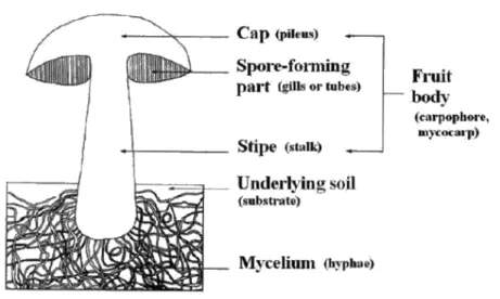
Figure 1: Schematic representation of mushrooms (Kalac, 2000). ... 3
Figure 2: Agaricus bisporus. ... 4
Figure 3:Pleurotus ostreatus. ... 5
Figure 4: Steroid nucleus (Barreira & Ferreira, 2015). ... 10
Figure 5: Pleurotus ostreatus in bales. ... 23
Figure 6: Illustration of the mycelium production. ... 24
Figure 7: Illustration of mycelia separation from the medium. ... 24
Figure 8: Illustration of the extraction procedure. ... 25
Figure 9: HPLC-DAD equipment used in the analysis of phenolic acids. ... 26
Figure 10: HPLC- UV system used in the analysis of ergosterol... 27
Figure 11: Microplate used in the evaluation of DPPH radical-scavenging activity. ... 28
Figure 12: Microplate used in the evaluation of reducing power. ... 29
Figure 13: Test tubes used in the β-carotene/linoleate assay. ... 30
Figure 14: Test tubes used in the TBARS assay. ... 31
Figure 15: Microplate used in the evaluation of the NO assay. ... 32
Figure 16: Microplate used in the evaluation of the cytotoxicity using human tumor cell lines. ... 33
Figure 17: Microplate used in the evaluation of the cytotoxicity in non-tumor cell lines. 34 Figure 18: Pleurotus ostreatus mycelia cultivated in vitro, in both liquid (A-PDB and B -iMMN) and solid medium (C and E- PDA and D- iMMN). ... 37
Figure 19:Agaricus bisborus mycelia cultivated in vitro, in both liquid (F- PDB and G -iMMN) and solid medium (H- PDA and I- iMMN). ... 38
Figure 20: Radial growth of A. bisporus (A) and P. ostreatus (B) mycelia cultivated in different culture media throughout time. ... 39
Figure 21: Total biomass of A. bisporus (A)and P. ostreatus (B) mycelia. ... 40
Figure 22: Chromatographic profiles of phenolic compounds of A. bispous (Mycelium: 1- p-hydroxybenzoic acid, 2- p-coumaric acid, 3- cinnamic acid) and P. ostreatus (Fruiting body: 1- protocatechuic acid, 2- p-hydroxybenzoic acid, 3- p-coumaric acid, 4- cinnamic acid). ... 43
LIST OF ABREVIATIONS
5-LOX: 5-Lipooxygenase
ANOVA: One vary analysis of variance
B2: Riboflavin B3: Niacin
B5: Pantothenic acid
BHA: Butylated hydroxyanisole BHT: Butylated hydroxytoluene
COX-2: Cyclooxygenase-2
DMEM: Dulbecco’s modified Eagle’s minimum essential medium
DNA: Deoxyribonucleic acid
DPPH: 2,2-Diphenyl-1-picrylhydrazyl radical Dw: Dry weight
EC50: Extract concentration corresponding to 50% of antioxidant activity or 0.5 of
absorbance in reducing power assay
ECACC: European collection of animal cell culture FAO: Food and Agriculture Organization
FBS: Fetal bovine serum Fw: Fresh weight
GAE: Gallic acid equivalents
GI50: Extract concentration that inhibited 50% of the net cell growth HBSS: Hank’s balanced salt solution
HBV: Hepatitis B virus HCV: Hepatitis C virus
HeLa: Henrietta Lacks human cervical carcinoma cell line HepG2: Hepatocellular carcinoma cell line
HIV: Human immunodeficiency virus
HPLC: High performance liquid chromatography
HPLC-DAD: Diode array detector
HPLC-UV: Ultraviolet detector
HPV: Human papillomavirus
IAEA: International Atomic Energy Agency
IC50: Extract concentration corresponding to 50% of cytotoxic activity ICAM-1: Intercellular adhesion molecule-1
IFN-δ: Interferon gamma IL-1 β: Interleukin 1β IL-6: Interleukin 6 IL-8: Interleukin 8 IL-12: Interleukin 12
iMMN: Incomplete melin-norkans medium
iNOS: Inducible nitric oxide synthase LPS: Lipopolysaccharide
MA: Massachusetts
MCF-7: Breast adenocarcinoma cell line MD: Maryland
MDA: Malondialdehyde
MMN: Melin–norkans medium
NCI-H460: Human lung cancinoma cell line
NF-KB: Nuclear factor kappa B
NED: N-(1-napthyl) ethylenediamine hydrochloride NK-cell: Natural killer cell
NSAIDs: Nonsteroidal anti-inflammatory drugs PDA: Potato dextrose agar medium
PDB: Potato dextrose broth
PLP2: Non-tumor primary culture of porcine liver cells PG: Propyl gallate
PGE2: Prostaglandin E2
RAW 264.7: Mouse leukaemic monocyte macrophage cell line
RNA: Ribonucleic acid
ROS: Oxygen reactive species RSA: Radical scavenging activity
SRB: Sulphorodamine B
TBARS: Thiobarbituric acid reactive substances TBA: Thiobarbituric acid
TCA: Trichloroacetic acid
TNF-α: Tumor necrosis factor alpha
μl: Microliter
USA: United States of America UK: United Kingdom
UV: Ultraviolet
UFLC: Ultra-fast liquid chromatography VT: Vermont
v/v: Volume/Volume
w/v: Weight/Volume
ABSTRACT
The importance of improving people's quality of life has aroused the interest of the food industry to develop new products with functional characteristics that may be associated with possible beneficial effects on human health, either preventive or therapeutic aspects. Functional foods and nutraceuticals present in its composition bioactive substances that provide medical benefits and, therefore, have aroused the interest of many consumers around the world. Mushrooms are a source of proteins, vitamins, minerals, and especially bioactive compounds. These compounds, including ergosterol, phenolic compounds, tocopherols, ascorbic acid and carotenoids, have been associated with antioxidant, anti-inflammatory, antimicrobial and cytotoxic properties, among others attributed to mushrooms and associated to its promoting health effects. Following this approach, the present work aimed to develope nutraceutical formulations based on mycelium of Agaricus bisporus L. and Pleurotus ostreatus (Jacq. Ex Fr.) P. Kumm. The present study highlights the potential of in vitro culture as a tool to improve production of bioactive compounds by two different mushroom species. Accordingly, A. bisporus and P. ostreatus were studied for their composition in phenolic acids and sterols, antioxidant activity (DPPH radicals scavenging, reducing power, β-carotene bleaching inhibition and TBARS formation inhibition), anti-inflammatory effect (by down-regulating LPS-stimulated NO in RAW264.7 cells) and cytotoxic activity (using MCF-7, NCI-H460, HeLa, HepG2 and PLP2 cell lines). Therefore, the mycelia of A. bisporus and P. ostreatus was cultured in different solid and liquid media, and further submitted to solid-liquid extraction processes; these assayed species showed differences in the growth rate and yielded biomass. Overall, P. ostreatus mycelia showed higher contents of ergosterol and phenolic compounds (but the mycelia of A. bisporus produced in PDA presented a higher amount of p -hydroxybenzoic acid) and stronger antioxidant activity than the corresponding fruting body. On the other hand, P. ostreatus and A. bisporus did not show anti-inflammatory activity. However, P. ostreatus showed cytotoxicity in human tumor cell lines in opposition to A. bisporus, that didn't present cytotoxic activity. In conclusion, the results show that these mushrooms are a good source of compounds with antioxidant and cytotoxic capacity, with variations among species.
Keywords: Nutraceutical formulations; Mycelium; Phenolic compounds; Ergosterol;
RESUMO
Palavras-chave: Formulações nutracêuticas; Micélio; Compostos fenólicos; Ergosterol;
CHAPTER 1.
1. INTRODUTION
In recent decades the demands of consumers in terms of food production have changed considerably. Consumers increasingly believe that food directly contribute to their health and well-being. Today's foods are not only intended to satisfy hunger and provide the necessary nutrients, but also to prevent disease and improve physical and mental well-being of consumers. In this context, functional foods play a very important role. The growing demand for such foods can be explained by the rapid advances in science and technology, increasing health care costs, changes in effective food laws tam labels and product claims, the steady increase in life tenancy, the desire of older people to improving quality of life in their late years and the increased interest in maintaining good health through diet (Siro et al., 2008).
The subject “nutraceuticals” and “functional foods” is experiencing a broad discussion in
the scientific community, and with the recent improvements and biotechnological discoveries of recent decades, it becomes a recent research hot topic.
According to DeFelice (1995) and subsequent references, “a nutraceutical is any substance that is a food or a part of food and provides medical or health benefits, including the
prevention and treatment of disease” (DeFelice, 1995; Brower, 1998 and Rishi, 2006). Foods, known as “functional foods,” are thought to provide benefits beyond basic nutrition
and may play a role in reducing or minimizing the risk of certain diseases and other health conditions (Cencic & Chingwaru, 2010). Nutraceuticals and functional characteristics of many traditional foods are being discovered and studied, while new food products are being developed to include beneficial components. By knowing which foods can provide specific health benefits, we can make food and beverage choices that allow us to take greater control of our health.
Mushrooms belong to the kingdom Fungi, a group very distinct from plants, animals and bacteria. Fungi depend on other organisms for food, absorbing nutrients from the organic material in which they reside (Adamovic et al., 1998).
of the pharmaceutical and food industries due to its nutritional value and the presence of numerous bioactive substances (Kalac, 2009). These molecules are responsible for the use of mushrooms as important partners in the complementary treatment of many diseases such as cancer, hepatitis, human papillomavirus (HPV) and human immunodeficiency virus (HIV). In addition to these therapeutic effects, mushrooms are also used in the prevention of diseases including cancer, diabetes, stroke, among others (Younis et al., 2014). According to Ferreira and collaborators (2010), in addition to the immunomodulating properties, mushrooms have effective substances for lowering cholesterol, which revert situations of hyperlipidemia, show antithrombotic activity reducing blood pressure, as also hypoglycaemic activity.
The great majority of the studies reporting mushrooms’ properties are conducted with the fruiting bodies, but culture media used in mushroom’s cultivation have also been explored as potential sources of bioactive compounds (Ma et al., 2016). Otherwise, the in vitro culture of mycelia is becoming a promising alternative to obtain sources of bioactive compounds, mainly due to the shorter incubation time and easier culture conditions (less space needed, low probability of contamination and higher biomass yields when compared with fruiting bodies) (Gan et al, 2012 and Zhang et al., 2016).
The fruiting bodies and mycelia of A. bisporus and P. ostreatus were previously studied for their chemical composition, antioxidant, anti-inflammatory and cytotoxic activities, after being harvested at different periods (Barros et al., 2009; Reis et al., 2012; Barreira et al., 2014; Morro et al., 2012; Ferreira et al., 2010; Martins et al., 2012; Taofiq et al., 2015; Younis et al., 2014; Silva, 2015). However, the present study goes further and compares fruiting bodies, mycelia and culture media in terms of bioactive properties and compounds. Thereby, the main objective was to develop nutraceutical formulations based on fruiting body and mycelium of A. bisporus and P. ostreatus, as also the corresponding culture media, highlighting the potential of in vitro culture as a tool to improve production of bioactive compounds by two different mushroom species.
1.1. Description of the species to be studied
Mushrooms (Figure 1) are macrofungi with distinctive and visible fruiting bodies
al., 2010).
Figure 1: Schematic representation of mushrooms (Kalac, 2000).
Agaricus bisporus L., known as champignon, white mushroom or white button mushroom, belongs to the phylum Basidiomycota. This species has several demonstrated and valuable medicinal properties including anti-tumor, anti-aromatase, antimicrobial, immunomodulatory, anti-inflammatory and antioxidant (Stojkovic et al., 2014).
Pleurotus ostreatus (Jacq. Ex Fr.) P. Kumm., better known as oyster mushroom or ‘‘repolgas’’, also belongs to the phylum Basidiomycota. It is the edible mushroom most
produced all over the world. The production and consumption of Pleurotus is increasing, not only for its easily, quickly growing and organoleptic characteristics, but also for its nutritional value and medicinal properties (Ramos et al., 2011). Its medicinal properties include immunologic, antitumor, anti-inflammatory and antimicrobial activities, among others (Fernandes et al., 2015).
1.1.1. Agaricus bisporus L.
The consumption of mushrooms is an ancient practice and literature reports its existence since the beginning of civilization. The mushroom Agaricus bisporus (Figure 2) is the
Figure 2: Agaricus bisporus.
The mushrooms are gaining worldwide recognition as important functional foods and as sources of therapeutical molecules for the treatment of some diseases (Chang, 2008). In addition to these qualities edible mushrooms are very versatile in cooking, as they can be baked, boiled, grilled or sautéed, and even combined with soups, patés, salads and teas (Brill, 2002).
Beelman and collaborators (2003) published a study related with bioactive compounds in A. bisporus, and reported for this species a higer nutritional value but similar bioactive components in comparison with Lentinus edodes and Pleurotus spp.
A. bisporus has a cap of 5 to 12 cm, convex with curved edge, becoming plane with age, white color, smell and mild taste; it has a pink-brown laminae becoming purple-brown and finally dark brown, a stem of 2 to 5 cm with bulbous base, and dark brown spores (Breitenbach & Kranzlin, 1995 and Sanchez, 2008).
A. bisporus is one of the edible cultivated fungi with higher economic importance. It is a microscopic fungus, with distinctive fruiting body, being large enough to be seen with the "naked eye" and be picked up by hand. It has no cholesterol, and insignificant levels of vitamin A or vitamin C, but it is a source of some B vitamins, in particular, riboflavin (B2), niacin (B3) and pantothenic acid (B5), ergosterol, representing 90% of its sterol fraction (when exposed to ultraviolet light is converted to pro-vitamin D), essential minerals (especially selenium, copper and potassium), phenolic compounds, having also a high amount of crude protein (Barros et al., 2008; Chang, 2008 and Barreira et al., 2014).
Table 1: Taxonomic classification of Agaricus bisporus (Braga et al., 1998).
Kingdom: Fungi
Division: Basidiomycota
Class: Homobasidiomycetes
Order: Agaricales
Family: Agaricaceae
Genus: Agaricus
Species A. bisporus
1.1.2. Pleurotus ostreatus (Jacq. Ex Fr.) P. Kumm.
Among the many existing edible mushrooms, only a few species are used as food and commercially cultivated. The three most cultivated species are Agaricus brasiliensis, Lentinula edodes and Pleurotus ostreatus (Figure 3) (Ribas, 2006), being the genus
Pleurotus the third type most produced worldwide (Dias, 2010).
Figure 3: Pleurotus ostreatus.
P. ostreatus presents a convex cap plan-convex, grayish or whitish color and with a diameter from 5 to 15 cm. At the bottom of the hat, the blades are arranged radially, with whitish and rather narrow. This species also has a side foot hair and very short compared to the diameter of the cap. The edible part has a whitish color with pleasant and intense fragrance (Coelho, 2012).
and calories (Manzi et al., 2001 and Bonatti et al., 2004). Moreover, the advances made in the field of biotechnology allowed the identification of important medicinal properties of mushrooms attributed to various bioactive molecules, such as polysaccharides, ergosterol, phenolic compounds, lecithins and some immunomodulatory amino acids including arginine and glutamine (Viegas et al., 2006).
The total phenolic content in P. ostreatus is 12.45 mg GAE/g of extract (Reis et al., 2012b) and its concentration in free ergosterol (determined by HPLC- UV) is 4.40 mg/g dry matter (Jasinghe, 2005).
P. ostreatus is a decomposer mushroom of vegetable matter. This species and other species of mushrooms produce extracellular ligninolytic enzymes such as lignin peroxidase, manganese peroxidase and laccase, which are involved in lignin degradation. This feature gives mushrooms the ability to grow on the trunks of living or dead trees (Martinez et al., 2001 and Coelho, 2012). The taxonomic classification of P. ostreatus species is presented in the following table (Table 2).
Table 2: Taxonomic classification of Pleurotus ostreatus (Adapted of Alexopoulos et al., 1996).
Kingdom: Fungi
Division: Basidiomycota
Class: Agaricomycetes
Order: Agaricales
Family: Pleurotaceae
Genus: Pleurotus
Species P. ostreatus
1.2. The mushrooms as a source of bioactive compounds
The mushrooms are consumed by people of diverse cultures, both for its gastronomic characteristics, as also medicinal features. However, its use as functional foods is more evident in Eastern cultures, in which the use of mushrooms in health maintenance began thousands of years ago with the Chinese (Chang, 1996).
(Table 4), among others, some of them responsible for their nutraceutical potential (Barros
et al., 2008a; Barros et al., 2009 and Ferreira et al., 2009). Nutraceuticals present in mushrooms have been related to their antioxidant, anti-inflammatory and cytotoxic activities (Fernandes et al., 2015).
Table 3: Phenolic acids in mushrooms reported in the literature (A - Barros et al., 2009; B - Muszyńska et al., 2013; C - Reis et al., 2012).
Phenolic acids acid (g/g dw)
Species Sample Gallicaci d Protocatechui c acid p -Hydroxybenzoic acid p-Coumaric acid Cinnamic acid Agaricus bisporus Mushro om
63 ± 13C Nd nd 2.31 ± 0.06 C 0.38 ± 0.02 C
Nd Nd 26 ± 2A nd 8.7 ± 0.7A
Myceliu
m 31 ± 4
C Nd 0.02 ± 0.001 C 3.7 ± 0.2 C 0.76 ± 0.03 C
Pleurotus ostreatus
Mushro om
Nd 0.77 ± 0.02C 1.56 ± 0.06C 0.81 ± 0.03C 0.23 ± 0.02C
Nd 2.52 ±0.03B 3.60 ±0.05B nd 1.09 ±0.01B
Myceliu
m Nd Nd 0.05 ± 0.0001
C nd 9.7 ± 0.9 C
dw- dry weight; n.d. – not detected.
Table 4: Ergosterol content in mushroom fruting bodies reported in the literature (A -Barreira et al., 2014 and B- Villares et al., 2014).
Species Ergosterol (mg/100 g dw)
Agaricus bisporus
352±1 A
6.4±0.2 B
Pleurotus ostreatus
104±1A
3.3 ± 0.2 B
1.2.1. Phenolic acids
Phenolic compounds range from simple molecules up to other with high degree of polymerization (Bravo, 1998 cited by Smith et al., 2002). The presence of these compounds has been highly documented due to its pharmacological and antinutritional activities (Nagem et al., 1992; Gamache et al., 1993; Ivanova et al., 1997; Aziz et al., 1998; Fernandez et al., 1998 and Hollman Katan, 1998 cited by Soares, 2002).
Ferreira and collaborators (2009), phenolic compounds are described as biologically active substances having antioxidant, anti-inflammatory or anti-tumor properties. Phenolic molecules are hydroxylated aromatic compounds having in their basic constitution one or more aromatic rings with one or more hydroxyl groups. They include a large number of subclasses, such as phenolic acids, flavonoids, tannins, stilbenes, among others, which indicate a large diversity of chemical structures. The flavonoids are the most widely distributed sub-class. They are found in higher levels in plants and vegetables but mushrooms also have significant amounts of phenolic compounds (Ferreira et al, 2009 and Palacios et al, 2011).
The major phenolic compounds from mushrooms are phenolic acids. Phenolic acids are characterized by having a benzene ring, a carboxyl group and one or more hydroxyl and/or methoxyl groups in the molecule, imparting antioxidant properties both to food as to the organism after ingestion (Crozier et al., 2006 and Fraga, 2010 cited by Carocho & Ferreira, 2013). Phenolic acids are usually found in vegetables and fruits present in our diet, and some of which are among the most bioactive and therapeutically useful substances (Apak et al., 2007 cited by Ferreira et al., 2009). According to Soares (2002), phenolic acids, in addition to its presence in their natural form, can also bind to each other or other compounds.
Phenolic acids can be divided into two main groups, hydroxybenzoic acids (Table 5) and
hydroxycinnamic acids (Table 6), which are derivatives of the non-phenolic benzoic and
Table 5:Chemical structure of the benzoic acid derivatives found in mushrooms (Ferreira et al., 2009).
* Aldeydes are groups under the corresponding phenolic acid class. Benzoic acid
derivatives
Substitution
X
R1 R2 R3 R4
p-Hydroxibenzoic COOH H H H OH
Protocatechuic COOH H H OH OH
Gallic COOH H OH OH OH
Gentisic COOH OH H H OH
Homogentisic CH2COOH OH H H OH
Vanillic COOH H OCH3 OH H
5-Sulphosalicylic COOH OH H H HSO3
Syringic COOH H OCH3 OH OCH3
Veratric COOH H OCH3 OCH3 H
Vanillin CHO* H OCH3 OH H
Table 6: Chemical structure of the cinnamic acid derivatives found in mushrooms (Ferreira et al., 2009). *The carboxylic group is esterified with quinic acid.
Cinnamic acid derivatives
Substitution
X R1 R2 R3 R4
p-Coumaric H H H OH H
o-Coumaric H OH H H H
Caffeic H H OH OH H
Ferulic H H CH3 O OH H
Sinapic CH3O H CH3O OH CH3O
3-O-caffeoylquinic * H OH OH H
4-O- caffeoylquinic * H OH OH H
5-O- caffeoylquinic * H OH OH H
1.2.2. Ergosterol
Sterols are special forms of steroids that can be found in animals (zoosterols), plants (phytosterols) and fungi (mycosterols) (Barreira & Ferreira, 2015). They consist of a tetracyclic structure of four rings linked together, consisting of three to six carbon atoms and other ring with five carbon atoms (steroid nucleus) (Figure 4) with a hydroxyl group
on the C3 position and an aliphatic chain linked to the steroid nucleus (Fahy et al., 2005 cited by Diz, 2015).
Figure 4: Steroid nucleus (Barreira & Ferreira, 2015).
The ergosterol is abundant in edible mushrooms (Kuo et al., 2011). According Jasinghe and Perera (2005), the nature of ergosterol varies depending on the mushroom species, within the same species, with different varieties of mushrooms and with the maturity stage. These authors indicate that the concentration of sterols in mushrooms, especially ergosterol, depends on the part of the mushroom tissue used, maturity stage and growth conditions. Savón and collaborators (2002) cited by Diz (2015) also reported that, in relation to specific genetic traits, ergosterol content in mushrooms also depends on environmental factors such as light, heat, temperature, humidity and the type of substrate used on the growth of mushrooms.
The ergosterol is present in two basic forms, namely, as free ergosterol and esterified ergosterol. The relative abundances of free and esterified ergosterol are different according with the species. The free ergosterol is most important for the integrity of the cell and contributes to various cellular functions. The ergosterol ester, kept in cytosolic lipid particles, is a fixed storage form of sterol and can serve as an intermediate for the ergosterol functioning (Barreira & Ferreira, 2015).
According to Diz (2015), ergosterol is a molecule with high commercial value and is the main sterol in fungi. For example, it is the most abundant mycosterol in Agaricus bisporus, and is also found in corn oils, cottonseed, groundnut and linseed (Lagarda et al., 2006 cited by Barreira & Ferreira, 2015). Zymosterol, ergosta- 5,7-dienol, 24-methyl cholesterol, and methylene cholesterol are also important examples of sterols in fungi (Mattila et al., 2002 and Christie, 2013 cited by Barreira & Ferreira, 2015).
of 161-166 ºC. In vacuum, it supports the temperature of 250 ºC without decomposition (Barreira et al., 2014 and Ferreira, 1985 cited by Ricardo, 2015).
A metabolite, which is commonly found in mushrooms, is ergosterol peroxide, having become a very promising compound for health and significant pharmacological activities, such as antioxidant, antimicrobial and antitumor. In addition, these compounds reduce the incidence of cardiovascular disease and pain associated with inflammation by inhibiting the cyclooxygenase enzyme (Yuan et al., 2007).
The antitumor activity of ergosterol may be due to its inhibitory capacity of angiogenesis (development of new blood vessels from existing ones) induced by solid tumors (Takaku et al., 2001; Zaidman et al., 2005; Slominski et al., 2005 cited by Ricardo, 2015).
Other studies have indicated that free ergosterol (the main component of the lipid portion) was responsible for the antioxidant activity of the hexane extract of Agaricus bisporus brown mushroom (positive correlation between ergosterol content and antioxidant activity; r2 > 0.89) (Shao et al., 2010). Furthermore, ergosterol may exhibit hypocholesterolemic properties, since it is able to reduce cholesterol levels in the serum (Gil-Ramirez et al., 2013).
1.3. Bioactivity of extracts rich in phenolic acids and ergosterol
Edible mushrooms grow in darkness and dampness in highly competitive environments and protect themselves from microbes by excretion of natural substances; this explains their richness in bioactive compounds. They are appreciated throughout the world not only for its organoleptic characteristics but also for its nutritional and functional properties (Ferreira et al., 2010).
The presence of bioactive compounds, namely ergosterol and phenolic compounds, could explain their antioxidant, anti-inflammatory, cytotoxic and antimicrobial properties (Ricardo, 2013).
1.3.1. Antioxidant activity
In general, the term antioxidant is used for sorting molecules which are present in low quantities in the substrate that is oxidizable and which reacts rapidly in order to suppress or prevent oxidation (Magalhães, 2009).
(enzymes and non-enzymatic molecules) is a prerequisite for the normal functioning of the body. When this equilibrium tends to uncontrolled production of free radicals means that the body is in oxidative stress and in such situations, excess free radicals can oxidize and damage cell lipids, proteins, DNA (deoxyribonucleic acid) and RNA (ribonucleic acid), inhibiting their normal function, leading to various diseases such as atherosclerosis, diabetes, cirrhosis, cataracts, premature aging and cancer (Valko et al., 2007 and Ferreira et al., 2009).
According Ferreira and Abreu (2007), the exposure of organisms to free radicals, led to the development of endogenous defense mechanisms to eliminate them. But despite all organisms possess defense and repair systems that have evolved to protect them against oxidative damage, these systems are often insufficient to prevent completely, the damage induced by oxidative stress (Yang et al., 2001). Nevertheless, the presence of antioxidants in the diet can help the endogenous defense system, reducing oxidative damage (Fang et al., 2002; Liu, 2003 and Barros et al., 2008b).
Currently, antioxidants have a range of applications, from food to pharmaceutical industry, as also plastic’s industry and lubricating oils. The best known synthetic antioxidants are BHA (butylated hydroxyanisole), BHT (butylated hydroxytoluene), TBHQ (tert-butylhydroquinone) and PG (propyl gallate), which are widely used in both food and pharmaceutical industry. However, BHA and BHT have been restricted in their use due to possible toxic and carcinogenic effects (Silva, 2015). Thus, the use of antioxidants from natural sources such as mushrooms, assumes increasing importance.
This antioxidant activity in mushrooms is related mainly to its high content of phenolic compounds, are recognized as excellent antioxidants because of their ability to block free radical by transfer of a single electron and excellent redox properties of the phenolic hydroxyl groups (Cheung, 2008; Kim et al, 2008; Jayakumar et al, 2009 cited by Ricardo 2013).
Agaricus bisporus and Pleurotus ostreatus, as well as other species of cultivated edible mushrooms are a good source of natural antioxidants, presenting in its constitution phenolic compounds, especially phenolic acids, followed by tocopherols (mainly α -tocopherol), ascorbic acid and carotenoids, especially the β-carotene (Barros et al., 2009 and Ferreira et al., 2009). According to Rodriguez-Vaquero and collaborators (2007) and Karaman and collaborators (2010), the strongest antioxidant properties and capability of cell protection against hydrogen peroxide was evident for vanillic acid, and among cinnamic acid derivatives, for caffeic acid. p-Hydroxybenzoic, gallic and protocatechuic acids found in mushrooms are characterized by antioxidant, antibacterial, antiviral, antifungal, anti-inflammatory and gastric secretion-stimulatory actions, documented by in vitro and in vivo studies. However, according to Heleno and collaborators (2015) little is known about the bioactive phenolic forms in vivo.
Studies have shown that ergosterol and its peroxidation products may also contribute to potential health benefits and significant pharmacological activities, namely antioxidant activity (Yuan et al., 2007).
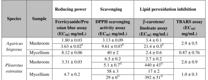
Table 7: Reducing power, scavenging activity and lipid peroxidation inhibition of the studied edible mushrooms according with Reis et al. (2012b).
Species Sample
Reducing power Scavenging Lipid peroxidation inhibition
Ferricyanide/Pru ssian blue assay (EC50; mg/mL)
DPPH scavenging activity assay (EC50; mg/mL)
β-carotene/ linoleate assay (EC50; mg/mL)
TBARS assay (EC50;
mg/mL)
Agaricus bisporus
Mushroom 1.80 ± 0.03 3.13 ± 0.09 3.4 ± 0.1 2.9 ± 0.5
3.63 ± 0.02F 9.61 ± 0.07F 21.4 ± 0.5F
Mycellium 8.12 ± 0.06 40 ± 2 2.4 ± 0.6 0.87 ± 0.76
Pleurotus ostreatus
Mushroom 3.31 ± 0.03 6.5 ± 0.2 2.7 ± 0.2 2.6 ± 0.9
5.1 ± 0.7G 440 ± 43G
Mycellium 4.7 ± 0.2 58 ± 3 17 ± 2 1.0 ± 0.3
29 ± 6G 392 ± 51G
Concerning Folin-Ciocalteu assay, higher values mean higher reducing pawer: for the other assays, the results are presented in EC50 values, what means that higher values correspond to lower reducing power or antioxidant potential. EC50: Extract concentration corresponding to 50% of antioxidant activity or 0.5 of absorbance for the Ferricyanide/Prussian blue assay. F- Martins et al., 2012. G- Silva, 2015.
1.3.2. Anti-inflammatory activity
initiate the process of inflammation (Taofiq et al., 2015).
Among the various inflammatory mediators, the most common are the interleukins (IL-1β, IL-6, IL-8), tumor necrosis factor (TNF-α), nuclear-kB factor (NF-kB), intercellular adhesion molecule 1 (ICAM-1) inducible type of cyclooxygenase-2 (COX-2), prostaglandin E2 (PGE2), 5-lipoxygenase (5-LOX), and the inducible nitric oxide synthase (iNOS), which leads to the production of reactive nitrogen species such as nitric oxide (NO). Over-production of these inflammatory mediators leads to different types of cellular damage (Kanwar et al., 2009 cited by Taofiq et al., 2015).
Nowadays, NSAIDs are generally administered to reduce inflammation in the body. But long-term administration of NSAIDs has significant side effects on the gastrointestinal tract (Smalley et al., 1995 and Sinha et al., 2013), as well as other serious complications such as hypertension and cardiovascular toxicity, etc. (Dugowson & Gnanashanmugam, 2006 and Meek et al., 2010).
Many bioactive compounds present in mushrooms exhibit significant anti-inflammatory properties (Table 8), based on their ability to reduce the production of inflammatory
mediators (Ferreira et al., 2009; Elsayed et al., 2014 and Heleno et al., 2015). Several compounds have been identified as potential natural and safe drugs, without the harmful side effects characteristic of NSAIDs. These compounds are responsible for anti-inflammatory activity, and include mostly beta-glucans, triterpenoids, glycoproteins and phenolic compounds (Taofiq et al., 2015).
According to Yuan and collaborators (2007) the products of ergosterol peroxidation reduce pain associated with inflammation by inhibiting the enzyme cyclooxygenase.
As previously demonstrated, the edible white button mushroom (Agaricus bisporus) enhanced NK cell activity in mice through the increased production of IFN-δ which induced maturation of dendritic cells, and TNF-α, increasing the production of IL-12 (Wu et al., 2007 and Ren et al., 2008).
Table 8: Some previous studies on anti-inflammatory activity of the two studied mushroom species.
Species Inhibition of NO production References
Agaricus bisporus 30% at 0.5 mg/mL
Moro et al., (2012)
Pleurotus ostreatus 15% at 0.5 mg/mL
Extract concentrations responsible for 50% of reduction of NO production (EC50 values) in RAW
264.7 cell line
Species EC50values (μg/mL) References
Agaricus bisporus 190 ± 6
Taofiq et al., (2015)
Pleurotus ostreatus 96 ± 1
1.3.3. Cytotoxic activity
Cancer is a disease characterized by a cell population, which grow and divide without respect to normal limits, invade and destroy adjacent tissues. But, the cells have enzyme systems capable of repairing the majority of such damages. Although these systems are efficient, some injury or even spontaneous errors during DNA replication are not repaired (Kellof & Sigman 2000).
The causes of cancer are diverse, complex, and only partially understood. Cancers are primarily an environmental diseases with 90–95% of cases attributed to environmental factors and 5–10% due to genetics common environmental factors that contribute to cancer death including tobacco (25–30%), diet and obesity (30–35%), infections (15–20%), radiation (both ionizing and non-ionizing, up to 10%), stress, lack of physical activity, and environmental pollutants. Cancer causing viral infections such as HBV/HCV and HPV are responsible for up to 20% of cancer deaths in low and middle-income countries (Younis et al., 2014).
Janardhanan, 2003).
Popovic and collaborators (2013) defines mycotherapy as the study of the use of extracts and compounds derived from mushrooms as medicines or health promoting agents. Mycotherapy of cancer is a scientific field relatively new and promising, which deals with anticancer agents derived from mushrooms.
Identification of active ingredients in the extracts, in other words, the isolation of new anti-tumor mushroom substances has become a matter of great importance, in view of the complexity and distribution of various types of cancer in the world population (Zong et al., 2012).
A wide variety of compounds and complex fractions have been isolated and/or purified from some edible mushrooms, with particular importance in relation to anticancer and cancer preventive activity (Table 9). These activities are attributed mainly to
polysaccharides, sterols, phenolic compounds etc. (Takaku et al., 2001; Liang et al., 2011; Vaz et al., 2012 and Zong et al., 2012).
Some species of the genera Pleurotus, Agaricus, Flammulina, Ganoderma, among others are medicinally valuable due to its anti-tumor activity (Table 9) (Ferreira et al., 2009 and Gunawardena et al., 2013 cited by Younis et al., 2014).
According to Song and collaborators (2009), Ma and collaborators (2013) (cited by Popovic et al., 2013), various derivatives of ergosterol have been isolated from mushrooms, for example, ergosterol peroxide and trametenolic acid, and showed cytotoxicity in prostate cell lines and human breast carcinoma.
According to Takaku and collaborators (2001), oral administration of ergosterol (400 and 800 mg/kg for 20 days) to mice with Sarcoma 180, reduced significantly the tumor growth without any side effects. Other studies showed that ergosterol peroxide induced death of miR-378 transfected cells (Wu et al., 2012), and ergosterol peroxide and trametenolic acid isolated from Inonotus obliquus exerted cytotoxic activity in prostate cell lines and human breast carcinoma (Ma et al., 2013).
Table 9: Compounds with antitumor potential found in the studied mushroom species (Ferreira et al., 2010) and cytotoxic activity of extracts obtained from the same speices against the two carcinoma cell lines
(Younis et al., 2014). Mushroom
Species Antitumor Agents References
Agaricus bisporus
490 Quinone (-L-glutaminyl-4-hydroxy-2,5-benzoquinone) Zaidman et al. (2005)
Selenium Tiffany et al. (1978)
Lectins Wang et al. (1998)
Pleurotus ostreatus
Selenium Zaidman et al. (2005)
Pleuran Bobek & Galbavy (2001)
Mushroom
Species Sample
HepG2 HeLa
Maximum
inhibitory % IC50 (μg/mL)
Maximum
inhibitory % IC50(μg/mL) Agaricus
bisporus
Fruiting bodies 66.6±3.2 30.2±2.2 71.3±1.9 28.8±1.8
Mycelia 41.1±2.1 54.9±2.4 57.4±2.9 38.7±1.9
Pleurotus ostreatus
Fruiting bodies 70.4±2.8 30.4±1.9 70.9±2.7 26.5±1.8
Mycelia 40.8±2.6 56.3±2.5 45.2±1.5 52.2±2.5
IC50: Extract concentration corresponding to 50% of cytotoxic activity.
1.4. In vitro culture as a tool to improve the production of bioactive compounds
The in vitro culture aims to clarify the optimum conditions for fungal growth, regarding culture medium, temperature and time of incubation and this knowledge is a prerequisite for commercial cultivation in formulated substrate (Hatvani, 2001). In vitro culture emerges as a biotechnological tool feasible for the production of bioactive compounds that can be used in several areas, and particularly in order to make additional efforts for the sustainable conservation and rational use of biodiversity (Karuppusamy, 2009 cited by Dias et al., 2016).
1.4.1. Advantages and disadvantages of in vitro culture
In 1994, the United Nations Food and Agriculture Organization (FAO) has authorized the use of in vitro culture techniques as a method for the production of food natural compounds (Anand, 2010 and Roberto & Francesca, 2011). In 2002, FAO published a report in association with the IAEA (International Atomic Energy Agency - Division of Nuclear Techniques in Food and Agriculture) referring to the theme of in vitro culture techniques for the production of bioactive compounds as having a high value, indicating how it can be carried out more economically by researchers and industry (FAO/IAEA, 2002).
The cells of species grown in vitro synthesize, accumulate and sometimes exude many classes of metabolites. The bioactive compounds are of particular interest, and much effort has been devoted to getting some of the most precious and therapeutic properties (Vanisree & Tsay, 2004).
Murthy and collaborators (2015) developed a safety assessment of food ingredients obtained by in vitro culture and proposed some protocols for evaluating the toxicity of these compounds and also its potential bioactivity. The in vitro culture includes handling under aseptic conditions and in case of plant culture, it must be carried out in a medium under controlled conditions of light, humidity and temperature (Smetanka 2008 cited by Dias et al., 2016). This controlled production system allows increased uniformity and standardization of extracts, such as the concentration of the desired compounds, maintaining the same genetic characteristics of the best clones for production (Chaturvedi et al., 2007 Cited by Dias et al., 2016).
The combination of the engineering process, biotechnology and biochemistry led to a significant improvement in production yields, especially for the in vitro culture as the preferred method for the production of bioactive compounds (Zhou & Wu, 2006 and Kolewe et al., 2008 cited by Dias et al., 2016).
However, according to Anand (2010), the most important advantage of in vitro culture is its ability to provide continuous, sustainable, economic and viable production of natural compounds, regardless of the geoclimatic conditions and a highly controlled environment. The in vitro culture, as any other process is sensitive to some problems of environmental or biological order, which affect directly the development of the cultures. Among these problems can be mentioned: low rate of development; it may happen a partial or complete loss of culture. Moreover, if the production on a large scale requires costly equipment, the procedure requires special care and careful observation; there may be error in the identity of the organisms after the culture; risk of infection, if precautions are not taken; risks of genetic variation (Engelmann, 1991; Santos et al., 2001 and Ahmed et al., 2008).
1.4.2. Mycelium production
The mycelium is the mass of hyphae that makes up the vegetative body of a mushroom. Colonies of mushrooms comprised of mycelium are found on the ground and in many other substrates. A single spore germinates in a monokaryotic mycelium, which cannot sexually reproduce; when two compatible monokaryotic mycelia join and form a dikaryotic mycelium, this mycelium can form fruiting bodies such as mushrooms (Leiva et al., 2015).
period of incubation, temperature, pH, size of the inoculum, etc. (Leiva et al., 2015).
The mycelia mushrooms are usually isolated from sporocarps on solid or liquid media (Melin-Norkrans (MMN), PDA medium (Potato Dextrose Agar) or PDB (Potato Dextrose Broth), or others, according to nutrients requirements of the species). The strains are maintained on Petri dishes (9 cm diameter) containing the same media mentioned above at 25°C in the dark and subcultured. After growth, the mycelium is recovered from the medium (Reis et al., 2011 and Carocho et al., 2012).
As mentioned in the previous sections, mushrooms mycelia have demonstrated several bioactive properties, being used in the treatment, but mostly in the prevention of different diseases. Therefore, with the market requirements for these bioactive compounds, in vitro culture arises as the promissing alternative for their production.
1.5 Objectives
As mentioned before, mushrooms have been used for many years by different cultures as
teas and nutritional foods, because of their special taste and texture. Agaricus bisporus and Pleurotus ostreatus are traditionally used as food, and accumulate a large variety of secondary metabolites, including phenolic compounds, polyketides, terpenes and steroids, with great bioactive potential. These species in particular have antioxidant, anti-inflammatory and cytotoxic activities. Although many people appreciate mushrooms there are still some people that do not like its texture and flavor, being interesting to develop nutraceutical formulations that can be used per si or incorporated into other foods, providing bioactive properties. On the other hand, the in vitro production of mycelium can help to overcome difficulties associated with mushroom’s availability issues related to seasonality or adverse climatic conditions, for example.
Therefore, this work had the main objective of developing nutraceutical formulations based on mycelium of Agaricus bisporus and Pleurotus ostreatus, and will include the following steps:
agar; ii) Potato Dextrose Agar (PDA) and Potato Dextrose Broth (PDB) media, after properly monitorization of the growth.
2) Chemical characterization of the mycelia obtained in terms of phenolic acids (HPLC-DAD) and mycosterols (mainly ergosterol, HPLC-UV).
CHAPTER 2.
2. MATERIAL AND METHODOS
2.1. Standards and reagents
The solvents acetonitrile 99.9% and methanol were of high-performance liquid chromatography (HPLC) grade from Lab-Scan (Lisbon, Portugal). The standards of ergosterol, phenolic compounds (gallic, protocatechuic; p-hydroxybenzoic, p-coumaric, and cinnamic acids) and trolox (6-hydroxy-2,5,7,8- tetramethylchroman-2-carboxylic acid) were purchased from Sigma (St. Louis, MO, USA). 2,2-Diphenyl-1-picrylhydrazyl (DPPH) was obtained from Alfa Aesar (Ward Hill, MA, USA). Dulbecco’s modified
Eagle’s medium, hank’s balanced salt solution (HBSS), fetal bovine serum (FBS), L-glutamine, trypsin-EDTA, penicillin/streptomycin solution (100 U/mL and 100 mg/ mL, respectively) were purchased from Gibco Invitrogen Life Technologies (Paisley, UK). Sulforhodamine B, trypan blue, trichloroacetic acid (TCA), lipopolysaccharide (LPS) and Tris were purchased from Sigma Chemical Co. (Saint Louis, MO, USA). RAW264.7 cells were purchased from ECACC (European Collection of Animal Cell Culture”, Salisburg, UK), and DMEM medium from HyClone (GE Healthcare Life Sciences, Logan, UT, USA). The Griess Reagent System Kit was purchased from Promega, and dexamethasone from Sigma. Thiamine, casamino acids, malt extract and agar were obtained from Panreac AppliChem (Barcelona, Spain). PDA and PDB were acquired from Oxoid microbiology products (Hampshire, United Kingdom). All other reagents and solvents were of analytical grade and obtained from common sources. Water was treated in a Milli-Q water purification system (TGI Pure Water Systems, Greenville, SC, USA).
2.2. Samples and mycelium production
Figure 5: Pleurotus ostreatus in bales.
Mycelia from both species were produced in the Biology and Biotechnology Laboratory of School of Agriculture of Bragança.
Mycelia from each of the mushrooms were isolated from sporocarps in two solid and liquid media: 1) modified Melin-Norkrans solid medium (MMN) pH 6.6 (NaCl 0.025 g/L; (NH4)2HPO4 0.25 g/L, KH2PO4 0.509 g/L; FeCl3 0.0050 g/L, CaCl2 0.050 g/L, MgSO4.7H2O 0.15 g/L, thiamine 100.10 g/L, glucose 10 g/L, agar 20 g/L and tap water) (Marx, 1969 cited by Reis et al., 2011 and Carocho et al., 2012); 2) MMN liquid medium (prepared using the above reagents, without agar); 3) Potato Dextrose Agar solid medium (PDA) and 4) Potato Dextrose Broth liquid medium (PDB).
The strains were maintained on Petri dishes (9 cm diameter) containing 10 mL of solid media and flasks with 20 mL of the same medium mentioned above at 25 °C in the dark and subcultured until mycelium covered most of the medium: approximately 21 days for P. ostreatus in solid medium and 42 days in liquid medium for A. bisporus, 56 days in solid and liquid medium. Radial growth measurements were registered every week from the inoculation time until the full growth of the mycelium (covering all available area) was obtained (Reis et al., 2011 and Carocho et al., 2012).
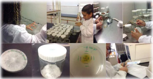
Figure 6: Illustration of the mycelium production.
Afterwards, all the samples (mycelium, culture medium and fruiting body) were placed in beakers and weighed separately to obtain fresh weight (fw) (Figure 7). Subsequently, the
samples were frozen and lyophilized (freeze 4.5 FreeZone model 7750031, Labconco, Kansas, USA) to obtain the corresponding dry weight (dw). Finally, they were reduced to a fine powder (20 mesh).
Figure 7: Illustration of mycelia separation from the medium.
2.3. Preparation of the extracts
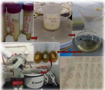
from the residue by filtration through Whatman paper No. 4 into a round flask. The residue was re-extracted once more under the same conditions and the filtrates were combined and concentrated with a rotary evaporated at ≈40°C (rotary evaporator, Büchi, Flawil Switzerland) (Figure 8). The extraction yield was calculated by measuring the extract
weight in relation to the initial mass sample and stock solutions were prepared for the different biological assay measured.
Figure 8: Illustration of the extraction procedure.
2.4. Chemical characterization
2.4.1. In phenolic acids
Each sample was placed in a beaker (≈1g) and was extracted by magnetic stirring with
methanol:water 80:20 (v/v) at 25°C and 150 rpm, for 1 h. The extract was separated from the residue by filtration through Whatman paper No. 4 to a round flask. The residue was re-extracted once more and the filtrate was rotary evaporated at ≈40°C to remove methanol (rotary evaporator, Büchi, Flawil Switzerland) (Reis et al., 2012). The aqueous phase was washed with n-hexane, and then submitted to a liquid-liquid extraction with diethyl ether (3 x 50 mL) and ethyl acetate (3 x 50 mL). The organic phases were evaporated at 30 ºC to dryness, redissolved in water:methanol (80:20), and filtered through a 0.22 µm disposable LC filter disk for HPLC analysis.
(Figure 9). Separation was achieved on a Waters Spherisorb S3 ODS2 C18 column (3 µm, 150 x 4.6 mm) thermostatted at 35ºC. The solvents used were: (A) 0.1% formic acid in water, (B) acetonitrile. The elution gradient established was 10% B to 15% B over 5 min, 15–25% B over 5 min, 25–35% B over 10 min, isocratic 50% B for 10 min, and re-equilibration of the column, using a flow rate of 0.5 mL/min. Detection was carried out in a photodiode array detector (PDA), using 280 nm as the preferred wavelength. The phenolic acids were quantified by comparison of the area of their peaks recorded at 280 nm with calibration curves (5-100 µg/mL) obtained from commercial standards of each compound: Protocatechuic acid (y = 164741x, R2=0.9996), p-hydroxybenzoic acid (y = 113523x, R2=0.9993), p-coumaric acid (y = 433521x, R2=0.9981) and cinnamic acid (y = 583527x, R2=0.9961). The results were expressed as µg per g of extract.
Figure 9: HPLC-DAD equipment used in the analysis of phenolic acids.
2.4.2. In ergosterol
The extracts prepared above in section 2.3 were dissolved in methanol at a concentration of
20 mg/mL and filtered through a 0.22 μm nylon disposable filter.
Ergosterol analysis was performed by high performance liquid chromatography coupled to an ultraviolet detector (HPLC-UV) as previously described by Barreira et al., (2014) (Figure 10). The components of the HPLC-UV integrated system include a pump (Knauer,
ODS-3 reversed-phase column (4.6×150 mm, 5 μm BGB Analytik AG, Boeckten, Switzerland) at 35 °C (7971R Grace oven). The mobile phase was acetonitrile/methanol
(70:30, v/v), at a flow rate of 1 mL/min, and the injection volume was 20 μL. The detection
was performed at 285 nm and data were analysed using Clarity 2.4 Software (DataApex). Ergosterol was quantified by comparing the area of its peak with the calibration curve obtained from a commercial standard. The results were expressed in mg per g of extract (Heleno et al., 2016).
Figure 10: HPLC- UV system used in the analysis of ergosterol.
2.5. Evaluation of bioactive properties
2.5.1. Antioxidant activity
The final extracts obtained in section 2.3, were dissolved in methanol obtaining stock solutions (20-80 mg/mL), then subjected to serial dilutions (40 and 0.01953 mg/mL), depending on the stock solutions and according to the assay. The in vitro antioxidant activity of the extracts was evaluated by performing four different assays: DPPH radical-scavenging activity, reducing power, β-carotene bleaching inhibition and thiobarbituric acid reactive substances (TBARS) assay (Heleno et al., 2010). Trolox was used as positive control (Fernandes, 2010).
2.5.1.1. DPPH radical scavenging activity
reduced to slight yellow color in the presence of hydrogen donating antioxidants leading to the formation of non-radical form. The reaction mixture in each of the 96 wells consisted of different solutions of the extracts (30 µL) to which was added a methanolic solution (270 µL) containing DPPH radical scavenging (6 × 10⁻⁵ mol/L). The mixture was allowed to stand for 60 min in the dark. The reduction of DPPH radical was determined by measuring the absorbance at 515 nm (Figure 11). The radical scavenging activity (RSA)
was calculated as percentage of discoloration of DPPH solution using the formula: % RSA = [(ADPPH - AS) / ADPPH] × 100 where AS is the absorbance of the solution in the presence of a given extract concentration and ADPPH is the absorbance of DPPH solution. The extract concentration which leads to 50% radical scavenging activity (EC50) was calculated from the RSA percentage graph as a function of extract concentration. Trolox was used as standard (Fernandes, 2010).
Figure 11: Microplate used in the evaluation of DPPH radical-scavenging activity.
2.5.1.2. Reducing power
This methodology was performed using the Microplate Reader, and is based on the ability to reduce yellow ferric form (Fe3+) to blue ferrous form (Fe2+) by the action of electron-donating antioxidants (Pinela, 2012).
Different concentrations of the extracts (0.5 mL) were mixed with sodium phosphate buffer (200 mmol/L, pH 6.6, 0.5 mL) with the addition of potassium ferricyanide (1% w/v 0.5 ml) in eppendorf (2 mL) and incubated at 50 ºC for 20 min. After this incubation time 0.5 mL of trichloroacetic acid 10% were placed in the eppendorf. The mixture (0.8 mL) was placed in 48 wells microplates with deionized water (0.8 mL) and ferric chloride (0.1% w/v, 0.16 mL) (Figure 12), measuring the absorbance at 690 nm. The concentration
nm graph as a function of extract concentration. Trolox was used as standard (Fernandes, 2010).
Figure 12: Microplate used in the evaluation of reducing power.
2.5.1.3. Inhibition of discoloration of β-carotene
This assay is based on the capacity of the antioxidants to neutralize the linoleate free radical. This neutralization is detected by the discoloration of the yellowish color of 𝛽 -carotene. A solution of β-carotene was prepared by dissolving β-carotene (2 mg) in chloroform (10 mL). Two mililiters of this solution was transferred into a round bottom flask (100 ml). After the chloroform was removed at 40 ºC under vacuum, linoleic acid (40 mg), Tween 80 emulsifier (400 mg), and distilled water (100 mL) were added to the flask with vigorous shaking. Aliquots (4.8 mL) of this emulsion were transferred into different test tubes containing different concentrations of the samples (0.2 mL). The tubes were shaken and incubated at 50 ºC in a water bath. As soon as the emulsion was added to each tube, the zero time absorbance was measured at 470 nm in a spectrophotometer (AnalytikJena, Jena, Germany) (Figure 13). β-Carotene bleaching inhibition was
Figure 13: Test tubes used in the β‐carotene/linoleate assay.
2.5.1.4. Inhibition of lipid peroxidation in the presence of thiobarbituric reactive substances (TBARS)
TBARS is a colorimetric assay in which lipid peroxidation produces malondialdehyde (MDA) as secondary breakdown product, and reacts with the thiobarbituric acid (TBA) to form MDA-TBA complex with the production of a pink pigment (Ndhlala et al., 2010). Porcine (Sus scrofa) brains were obtained from official slaughtering animals, dissected, and homogenized with a Polytron in ice-cold Tris–HCl buffer (20 mM, pH 7.4) to produce a 1:2 (w/v) brain tissue homogenate, which was centrifuged at 3000g for 10 min. An aliquot (0.1 mL) of the supernatant was incubated with the different solution concentrations (0.2 mL) in the presence of FeSO4 (10 M; 0.1 mL) and ascorbic acid (0.1 mM; 0.1 mL) at 37 ºC for 1 h. The reaction was stopped by the addition of trichloroacetic acid (28 % w/v, 0.5 mL), followed by thiobarbituric acid (TBA, 2 %, w/v, 0.38 mL), and the mixture was then heated at 80 ºC for 20 min. After centrifugation at 3000g for 10 min to remove the precipitated protein, the colour intensity of the malondialdehyde (MDA)-TBA complex in the supernatant was measured by its absorbance at 532 nm (Figure 14).
Figure 14: Test tubes used in the TBARS assay.
2.5.2. Anti-inflammatory activity
For the anti-inflammatory activity assay, the methanolic extracts (section 2.3) were dissolved in water at a concentrated of 10 mg/mL. For the various assays, the extracts were then submitted to further dilutions from 10 mg/mL to 0.16 mg/mL. Dexametazona was used as positive control (Taofiq, 2015).
2.5.2.1. Cells treatment