421
Respostas das concentrações de miocinas a partir do estímulo do exercício físico: uma revisão sistemática
Responses of myokines concentrations from exercise
stimulus: a systematic review
Correspondence: Leandro Paim da Cruz Carvalho, Rua Santo Amaro, 133, Chácara São Cosme, Feira de Santana BA. [email protected]
Received on: September 10, 2020; Accepted on: September 16, 2020.
Leandro Paim da Cruz Carvalho1 , Matheus Borges da Cruz Gomes2 , Ícaro Cerqueira da Silva
Oliveira¹ , Pedro Henrique Silva Santos3 , Ariel Custódio de Oliveira II1 , Lorena Mariel González
Vitavar4 , Heitor Barbosa Alves2 .
1. Universidade Federal do Vale do São Francisco, Petrolina, PE, Brazil. 2. Universidade Estadual de Feira de Santana, Feira de Santana, BA, Brazil. 3. Fundação Estatal de Saúde da Família/Fundação Oswaldo Cruz (FESF/FioCruz), Salvador, BA, Brazil. 4. Facultad de Ciencias de la Salud – Universidad Adventista del Plata, Libertador San Martín, Entre Ríos, Argentina.
open acess myokines concentrations from exercise stimulus: a systematic review. Rev Bras Fisiol Exerc 2020;19(5):421-435. https://doi. org/10.33233/rbfex.v19i5.4393 Systematic Review
Brazilian Journal of
Exercise Physiology
ISSN Online: 2675-1372 ISSN Printed: 1677-8510RBFEx
ABSTRACTIntroduction: The skeletal muscle is the largest endocrine organ of human body, and have this role trough
peptides and proteins known as myokines. The myokines are citocines that are produced and secreted by the skeletal muscle in response to the stimulus of contraction, acting locally and/or be released in the circulation and influence other distant tissues. Physical exercise is a potent stimulus for molecular adaptations in the or-ganism, and when practiced with regularity, promotes structural and functional adaptations in skeletal muscle. Therefore, physical exercise has a direct action on the concentrations of myokines.
Objective: Based on this, this research investigated, through a systematic literature review, the responses of
myokines concentrations from the stimulus of physical exercise.
Methods: Searches were carried out by two researchers independently, in the Scielo, Pubmed and Virtual
Heal-thy Library databases, analyzing articles published between 2009 and 2020, after a careful selection process in four stages, the works that reached the third stage were read in full and submitted to quality analysis using a critical review form.
Results: At the end of the process, 12 articles were selected to compose the discussion.
Conclusion: The analyzed articles shows that physical performance, both acute and chronic, is capable of
signi-ficantly modulating the concentration of several myokines, promoting an increase in many such as IL-6, IL-15, BDNF and Apelin, in addition to a significant decrease in muscle myostatin.
Key-words: exercise, skeletal muscle fibers, cytokines. RESUMO
Introdução: O músculo esquelético é o maior órgão endócrino do corpo humano e possui esse papel a partir
de peptídeos e proteínas conhecidos como miocinas. As miocinas são citocinas que são produzidas e secreta-das pelo músculo em resposta ao estímulo secreta-das contrações, podendo agir localmente e/ou cair na circulação e influenciar outros tecidos distantes. O exercício físico é um potente estímulo para adaptações moleculares no organismo e, quando regularmente executado, induz adaptações estruturais e funcionais no músculo esqueléti-co. Sendo assim, o exercício físico possui ação direta nas concentrações dessas miocinas.
Objetivo: Baseado nisso, essa pesquisa teve como objetivo investigar, através de uma revisão sistemática de
lite-ratura, as respostas das concentrações de miocinas a partir do estímulo do exercício físico.
Métodos: As buscas foram realizadas por dois pesquisadores de forma independente, nos bancos de dados do
Scielo, Pubmed e BVS, analisando artigos publicados entre 2009 e 2020. Após processo de seleção criterioso em quatro etapas, os trabalhos que chegaram até a terceira etapa foram lidos na integra e submetidos a uma análise de qualidade por via formulário de revisão crítica.
Resultados: Ao final do processo, foram selecionados 12 artigos para compor a discussão.
Conclusão: Os artigos revisados demonstram que o exercício físico, tanto de forma aguda quanto de forma
crô-nica, é capaz de modular de forma significativa a concentração de diversas miocinas promovendo o aumento da concentração das mesmas, por exemplo da IL-6, IL-15, BDNF e Apelina, além de diminuição significativa de miostatina muscular.
422
Carvalho et al.
Myocins and physical exercise
Introduction
Skeletal muscle (SM) has a great adaptative potential, being the intracellu-lar signaling promoted by muscle contraction a strong mechanism to molecuintracellu-lar and functional adaptation to muscle itself. Through stimulus caused by regular exercise training is possible to increase protein synthesis into SM, promoting higher functio-nal capacity and performance [1].
Exercise is largely recommended both in prevention and as treatment to me-tabolic diseases by its anti-inflammatory (AI) role and as a metabolism regulator. In this scenario, the SM has an important role due the production and release of many cytokines and another peptide, called myokines by Pedderson et al. [2] in 2003.
These myokines can, in several cases, have AI role not only inside the mus-cle, but may be released into bloodstream and be transported to another organs and tissues. It can stimulate immunological responses and contrast with the deleterious effects of some cytokines produced by adipose tissue (AT), for example the tumor necrosis factor (TNF) [3,4]
Beyond that, the scientific literature shows evidences indicating that exercise training has the potential to directly alter the circulating levels of myokines, incre-asing production and release of many myokines, as interleukins (IL), IL-15 and IL-6 [4,5].
Although the importance of myokines be notable, taking into account that it allow communication between SM and other tissues, is still unclear how is their responsiveness to different exercise stimulus, as type, intensity and volume of exer-cise training. The knowledge of these responses can help to build a better exerexer-cise prescription aiming specific benefits promoted by higher myokines concentrations.
Besides that, there are some studies analyzing the effects of exercise into myokines concentrations, leading us to consider this review relevant, summarizing systematically the actual evidences in this field. Based in the knowledge of myokines responses, the professional of exercise prescription can be more specific to prescri-be the exercise aiming to modulate properly one or more myokines. Based in these rational, the aim of this systematic review was to verify the responses of myokines concentration from exercise stimulus.
Methods
Determination of databases, research strategy and combinations
A systematic review was carried out based on bibliographic research of stu-dies that analyzed the biological responses of myokines from the exercise stimulus. The research of the articles was performed by two independents researchers in De-cember 2019, in the following electronic databases: Pubmed, Scielo and Virtual Heal-th Library (in portuguese Biblioteca Virtual em Saúde [BVS]).
The selection of the descriptors used was made based on health science des-criptors. In the searches for the articles the terms “myokines” and “effects of exercise” were used in the following combinations in English and Portuguese languages “ske-letal muscle’’ AND “myokines”, “effects of exercise” AND “production of myokines”.
Was performed the PICO strategy: P – Participants in studies with exercise who dosed myokines; I - Interventions with systematic exercise of any type; C – Com-parisons of results with theirselfs pre and posts results; O – Outcomes of exercise concentrations of circulating myokines.
Rev Bras Fisiol Exerc 2020;19(5):421-435 To better organization of references, we organized than in an Excel sprea-dsheet 2013.
Search plan steps
Search plan was divided in four steps (stages). In the first step was identified 197 publications potentially eligible to review. Then, in second step, the “2009 to cur-rent” and “human” filters were used to find studies closer to the proposed theme, resulting in 45 studies.
In third stage, the titles, abstracts and conclusions were read in order to verify their suitability for the purpose to this review. In addition, was applied the inclusion criteria established for the paper’s selection. Were included the papers that: a) ori-ginal cross-sectional or longitudinal work; b) with at least one physical exercise ses-sion; c) reporting the effects of exercise on myokines concentrations. After analyzing the studies, 18 publications were selected and analyze in the next step.
In the fourth stage of article selection, the established exclusion criteria were performed. To begin, the papers were fully read by two independent researchers and were excluded papers that: were duplicated; b) those that did not reached 10 points or more in the critical review of Law et al. [6], and c) myokines that were not be analyzed at least in two different papers.
Results
At the end of the fourth stage, 12 papers were selected to compound this re-view. Is important to note that in all steps of search the papers and its analyzes was conducted by two independents researchers using the statistical software SPSS 22.0 and the Kappa concordance test [7] to check the level of agreement between the re-searchers. As a result, values was always above 0.80 and P < 0.001, indicating almost perfect concordance between researchers. In this analysis, values up to 0.19 indicate poor agreement, between 0.20-0.39 mild agreement, 0.40-0.59 moderate agreement, 0.60-0.79 substantive agreement and between 0.80-1.00 indicates almost perfect agre-ement [7]. For a better understanding of the results, Figure 1 shows the number of studies during all the pre-established stages.
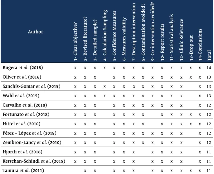
Table I shows the score obtained by the studies selected using the instrument proposed by Law et al. [6]. It aims to classify the quality of studies and has 15 items.
However, item 4 does not score, as it is only to distinguish the type of study, so item 4 was removed from our analysis and item 5 became 4 and so on, totaling 14 items that were scored below in the table. A quality cut-off point of 10 points was defined, that is, the article that did not score at least 10 items would be eliminated from the review. The items that were scored were marked with an “x” while those that were not scored were left blank.
The profile of the 12 selected studies that met the inclusion and exclusion criteria was described in Chart 1. The total number of participants was 224 individu-als, 74.2% (166) of whom were male and only 13.8% (31) women. Two studies did not report the gender of the sample. The age range of study participants ranged between 18 and 65 years.
Regarding study designs, 75% (9) were cross-sectional, while only 25% (3) evaluated biological responses in a longitudinal way. The respon-se of myokines by stimulating resistance exercirespon-ses (RE) was evaluated in 50% (6) studies. In turn, 50% (6) studies analyzed aerobic exercises (AE). Finally, the Elisa Kit was the most used enzymatic method of analysis in research.
424
Carvalho et al.
Myocins and physical exercise
Figure 1 - Flowchart of the steps of the systematic review.
Table I - Score of studies in the critical review form by Law et al. (1998).
Author 1- Clear objectiv e? 2- R evised lit er atur e? 3- Detailed sample ? 4- Calculation Sampling 5- C onf idenc e Measur es 6- Measur es v alidilt y 7- Description int erv ention 8- C ontamination a voided ? 9- C o-int erv ention a voided ? 10- R eport r esult s
11- Statistical analysis 12- Clinic R
ele vanc e 13-Dr op out 14-C onclusions Total Bugera et al. (2018) x x x x x x x x x x x x x x 14 Oliver et al. (2016) x x x x x x x x x x x x x 13 Sanchis-Gomar et al. (2015) x x x x x x x x x x x x x 13 Wahl et al. (2015) x x x x x x x x x x x x x 13 Carvalho et al. (2018) x x x x x x x x x x x x 12 Fortunato et al. (2018) x x x x x x x x x x x x 12 Hittel et al. (2010) x x x x x x x x x x x x 12
Pérez – López et al. (2018) x x x x x x x x x x x x 12
Zembron-Lancy et al. (2010) x x x x x x x x x x x x 12
Hjorth et al. (2016) x x x x x x x x x x x 11
Kerschan-Schindl et al. (2015) x x x x x x x x x x x 11
Rev Bras Fisiol Exerc 2020;19(5):421-435
Chart 1 - Profile of the selected studies.
Author / year Sample (N and sex) Analysis method Myokines analyzed / Type of exercise
Bugera et al.
(2018) N = 9 M; 18-35 years old; physically active, (at least 1 year in resis-tance training).
Human ELISA Kit KHC0061, Thermo Fisher Scientific, Waltham, WA, EUA;
IL-15
Bilateral knee extension Exercise with and without blood flow restriction Oliver et al.
(2016) N = 10 W; 27 ± 4 years old; healthy, trained in resistance training.
R&d custom
Premixed magnetic be-ad-based multiplex kit, fcstm14-02 (r&d systems inc., minneapolis, mn). IL-6 e IL-15 Squat S a n c h i s G o -mar et al. (2015) N = 15 H; 27 ± 5 years ol; Professional Soccer Players.
CSB-EL007669HU, Cusabio, Wuhan, China and EIAAPC, RayBiotech, Norcross, GA, EUA; respectively.
Alepin
A soccer session.
Wahl et al.
(2015) N = 13; 24,8 ± 3,7 years old; healthy, non-s-mokers. Quantikine HS ELISA--HS600B, R & D Systems, EUA. IL-6 e BDNF Cycloergometer Carvalho et al.
(2018) N = 61 (30W; 31M); 20-45 years old; Sedentary with BMI and obese individuals.
Enzyme-linked immunosor-bent assay (ELISA);
Quantikine human immu-noassays (r&d systems, inc. Minneapolis, usa).
Myostatin
Maximum treadmill test; Evaluation of strength and resistance in isokinetic equipment
Fortunato et
al. (2018) N = 20 M; Healthy; 18-35 years old; TG and UTG in resistance training.
Hmyomag-56k milliplex®
map and luminex® Apelin e BDNFLeg press, knee extension and leg flexion
Hittel et al.
(2010) N = 10 M; 40-60 years old; overweight an h y p e r i n s u l i m e m i c (BMI = 27.9).
ELISA Kit Myostatin
Moderate aerobic exercise
Pérez – Lopez
et al. (2018) N = 14; 18-35 years old; trained in resistance training (at least one year).
ELISA kit (r&d systems,
min-neapolis, mn, usa) IL-15Leg press
Zembron-Lac-ny et al. (2010) N = 16 M; 20,7 ± 0,9 years old; Not trained, physically active.
Enzyme immunoassay com-mercial kits (r&d systems, usa)
IL-6
Treadmill + Treamill 10% in-clined
Hjorth et al.
(2016) N = 24 M; 40-65 years old; Sedentary, CG (n = 13) normal blood glu-cose and dysglycemic (n=11).
Taq- man assays applied
biosystems MyostatinCycloergometer; Resistance Training
K e r
-schan-Schindl et al. (2015)
N = 19 (18M; 1W); 41-48 years old; ultrama-rathon athletes.
Colorimetric competitive immunoassay, immundiag-nostik; bensheim, Germany.
Myostatin ultramarathon Tamura et al.
(2011) N = 13 M; Heathy, un-trained, physically ac-tive.
Human IL-15 quantiglo ELI-SA Kit, R&D systems, Minne-apolis, MN, USA.
IL-15 Treadmill
BMI = body mass index; CG = Control Group; DG = Dysglycemic group; M = men; MR = maximum repe-tition; VO2 = maximum oxygen consumption; TG = Trained Group; UTG Untrained Group = Grupo não treinado); W = women.
426 Chart 2 shows the myokines response concentrations from the exercise stimulus. It is observed that IL-6, IL-15 and myostatin were the target of 4 studies each one, standing out, as the myokines of greatest interest in literature when it comes to response through exercise.
Chart 2 - Biological responses to physical training.
Author / year Exercise applied /
Training Weekly frequency Biological responses to exercise / training Conclusion
Bugera et al.
(2018) Bilateral knee extension.BFR-RE:30 reps at 30% 1MR + 3 sets of 15 reps, with 30s of interval
HL-RE: 4 sets of 7 repetitions com 1’ interval betwe-en series at 80% of 1 ME
LL-RE: replication of BRF-RE but without blood flow restriction.
1 session; cross-
sectional study. IL-15 did not show* difference between exercise protocols neither 1h and 23 post exercise There was no difference in the concentration of IL-15 post-exerci-se in any of the groups.
Oliver et al.
(2016) 4 sets of 10 reps at 70% of 1 MR.TRD: 180s intra ser interval.
Cluster: 30s interval intra reps and 150s intra sets.
1 session; cross-
sectional study. There was no ↑ * to IL-15 or IL-6 under any conditions and at any time There was no ↑ *of myokines IL-15 and IL-6
Sanchis-Go-mar et al. (2015)
A competitive soccer session (from January to May) 6 months;
longitu-dinal study; In the competitive period (January-March) there was ↑ * of apelin from 341.8 ng / mL to 433.3 ng / mL. However, on returning from vacation (May-July) no changes * were found.
The follow-up to a season of professional football showed ↑ * of apelin only in the compe-titive period and there was no correlation with performance Wahl et al.
(2015) 3 Conditions:A = Cycling (C);
B = cycling and electrostimulation (C + E); C = electrostimulation (E).
60 Min / 70% of peak power.
1 session;
cross--sectional study C + E and E ↑ * IL-6 levels at times, 0’, 30’ and 60 ’after EF.30´ after C + E the values of IL-6 ↑ * compa-red to C. The levels of IL-6 ↓ * at moments 0´, 30´ and 60´ after E compared to C + E and C. C + E and C ↑ * BDNF at 0´ after exercise compared to pre. BDNF levels ↑ * at times 0´ and 30´ after E compared to C + E and C. 240´ after exercise C + E ↑ * compared to C.
IL-6 showed higher ↑ after C + E followed by C and E. Serum BDNF levels were * higher in C and C + E, while E induced non-changes *.
Carvalho et al.
(2018) 1 min concentric knee extension and 5 sec isome-tric extension in a dynamometer. Isokinetic test set at 70º with a speed of 60º/s, for the isometric test the fixed position was 60º.
1 session;
cross--sectional study Normal weight group; Obese healthy group; Obese unhe-althy group. Myotastin was considered elevated only in unhealthy obese individuals,
Women had higher values than men for myostatin myostatin had a weak positive * correlation with metabo-lic syndrome (r = 0.26); Insulin levels (r = 0.25) and TNF a (r = 0.39), and negative associations with adiponectin (r = 0.40 and insulin sensitivity (r = 0.27)
Myostatin levels can be used to identify unhealthy phenotypes in young adults with obesity
Fortunato et
al. (2018) 4 sets of leg press, knee extension and leg flexion at 65% of 1MR, with 90s of recovery 1 session; cross--sectional study Two groups: (GNT) and the trained group (GT).Strength training ↑ * levels of apelin in the GNT at the times 2 hours after and 24 hours after, the brain-derived neurotrophic factor (BDNF) ↑ * only in the GT at the time immediately after.
An acute session of strength trai-ning increased * levels of apelin and BDNF levels
Hittel et al.
(2010) Moderate aerobic training 40-55% VO2máx (tread-mill, elliptical and bicycle) 9 months total (3 months of adaptation and 6 of training)
9 months;
longitu-dinal study A negative correlation was found in the pre-workout be-tween myostatin and insulin sensitivity (r2 = -0.82). After training, this correlation was (r2 = 0.49)
Myostatin ↓ * from 28.7 ± 3.1 to 22.8 ± 2.0 ng / mL with aerobic exercise
Myostatin levels decreased after 6 months of aerobic training.
Pérez-López et
al. (2018) Bilateral leg resistance exercise: 4 sets of leg press and 4 sets of knee extension in the extension chair at 75% of 1RM until concentric failure.
1 session; cross-
sectional study Serum IL-15 ↑ * at ~ 5.3 times after EF.IL-15 concentration was negatively correlated with mus-cle strength in the leg press (r = -0.80) and the extension chair (r = -0.63)
The signaling of IL-15 and its IL-15 receptor is activated in response to a single session of ER, increasing * the concentration of IL-15 post--exercise.
Zembron-Lac-ny et al. (2010) Ex.1: 90 ’running at 65% VO2máx and then at Ex.2: 90’ running at 65% VO2máx, 15 ’eccentric
phase. The period between Ex.1 and Ex.2 was 3 months.
1 session; cross-
sectional study ↑* IL-6 in the eccentric phase. IL-6 correlated with NO concentration after Ex.2 (r = 0.66). Eccentric contraction is an im-portant factor in increasing the concentration of cytokines consi-dering that in the eccentric phase, IL-6 levels increased *.
Hjorth et al.
(2016) 12 weeks of training in healthy individuals with supervised dysglycemia with 2 sessions / week of interval cycling (60 ’) and 2 sessions / week of full body strength training (60’).
Before and after the 12 weeks of training, a test was performed on the bicycle, lasting 45 minutes at 70% of VO2máx measured before, shortly after and 2h after the two tests
Longitudinal study Aerobic training 2x / week; Resis-tance training 2x / week;12 weeks;
Plasma concentration of myostatin ↓ * in 7.5% after 12 we-eks of training
Myostatin expression correlated positively with fast-ac-ting fibers (r = 0.53) and negatively with slow-acfast-ac-ting fi-bers (r = -0.52)
There was a negative correlation between myostatin ex-pression in the muscle and insulin sensitivity (r = -0.53)
Myostatin expression in muscle correlated with impaired insulin sensitivity
Kerschan-S-chindl et al. (2015)
Ultramarathon (246 km); Venous blood collection 3 times: the day before the test, 15 ’after the end of the test and 3 days after the test.
U l t ra m a ra t h o n ; c ro s s - s e c t i o n a l study
Average levels of myostatin ↑ * post-test compared to pre--test levels. Average prepre--test levels: 23.73 (ng / ml); Average post-test levels: 26.73 (ng / ml)
Increased serum myostatin levels appear to reflect muscle catabo-lism induced by extreme exercise. Tamura et al.
(2011) 30 min on the treadmill at 70% of HRmax. 1 session; cross--sectional study The serum concentration of IL-15 ↑ * in 10 ’after the 30’ run on the treadmill compared to pre-exercise levels. IL-15 concentration returned to baseline after 3 hours.
Extreme high-intensity exercise does not seem to be necessary to increase circulating levels of IL-15. *Significant differences; BFR = Blood flow resistance; BFR-RE = exercise with blood flow restriction; BDNF = Brain derived neurotrofic factor; BMI = body mass index; C = Cycling CLU = Cluster Test; E = Electro-stimulation; GNT = group non-trained; HL-RE = High load resistance exercise; HRmax = maximun heart rate; IL-6 = Interleukin-6; IL-15 = Interleukin-15; LL-RE = low load resistance exercise; pg/Ml = Picogram per milliliter; ng/mL = Nanogram per milliliter; NO = Nitric Oxide; TNF-α = Tumor Necrosis Factor alfa; VO2 = oxygen con-sumption. HRmax = maximum heart rate; YAU = Yukon arctic ultra.
Author / year Exercise applied /
Training Weekly frequency Biological responses to exercise / training Conclusion
428
Carvalho et al.
Myocins and physical exercise
Discussion
The aim of the present study was to systematically review the response of myokines after exercise training. To better understanding the data, we will initially address the actions of the myokines that will be discussed, to later describe the ef-fects of exercise on them.
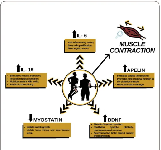
Myokines actions
IL-6, the first myokine described, was showed by Steensberg et al. [8] in 2000. In addition to being produced by SM as a result of muscle contraction, is also produ-ced in other tissues, as liver, for example. There is evidence that IL-6 acts stimulating the proliferation of satellite cells after acute damage in the SM, and, therefore, having a role in muscle hypertrophy [9].
When in physical exercise, the release of IL-6 occurs independently of release of TNF- α [10], possessing, thus, AI capacity. In this conditions, IL-6 acts inhibiting the production and secretion of TNF-α and its soluble receivers, as well, blocking IL-1 and IL-10 receivers [11]. Chronically, the levels of IL-6 are lowed after an exercise pro-gram, however, is also reported a better AI state in individuals that practiced exercise [12]. A possible explanation to this paradox, may be the fact that exercise modulates the release of IL-6 in other tissues, as in the immune system, consequently, lowering the release of IL-6 associated to TNF-α. The scientific literature shows that higher circulating levels of IL-6 are associated with physical inactivity and higher risk of metabolic syndrome [3,11,13].
Another myokine that has been studied in recent years is the IL-15. This myokine, similar with many others molecules produced in the body, promotes seve-ral effects in different organs. These effects include signaling to muscle hypertrophy, in additional to acting on lipidic metabolism [14], reducing the deposition of lipids and reducing the mass of white adipocytes cells. In the immune system, IL-15 acts mobilizing natural killer (NK) cells, that in its turn, act reducing tumor growth [15]. In bone tissue, associated with fibroblast growth factor (FGF) 21, IL-6 helps with bone mineralization, consequently aiding bone formation and repair after fracture [3,16].
We know that not all substances produced by muscular secretome assists in synthesis of other tissues, and myostatin is a good example of a myokine that acts limiting the muscle hypertrophy. Myostatin belongs to the family of transforming growth factor and with higher plasma levels of this myokine being observed in obese and sedentary individuals [17,18]. In addition to this limiting effect on muscle grow-th, myostatin has an important role in bone tissue and adipose tissue. In bone tissue, myostatin has acts opposite to IL-15, making mineralization and post-fracture repair difficult. In adipose tissue, therefore, evidence shows that myostatin triggers the sig-naling to hypertrophy of adipose tissue cells [16].
When performed acutely and chronically, the exercise downregulates myosta-tin levels in the tissues previously mentioned [19]. Evidence points to a large reduc-tion of myostatin levels after one single session of exercise (56%), and in a longitudi-nal way, exercise training can reduce 34% of myostatin levels. In addition, a reduction of 48% was observed in elderly after an exercise program of training [20].
Another important myokine modulated by exercise is the brain-derived neu-rotrophic factor (BDNF), which in central nervous system, acts to maintain or impro-ve cognitiimpro-ve activity by regulating neuronal survival, facilitating synaptic plasticity,
Rev Bras Fisiol Exerc 2020;19(5):421-435 neurogenesis and improving the memory process. BDNF also has a role in neuropro-tection against anxiety and depression [16,21].
Finally, apelin is a myokine that has receptors in various organs, as the kid-neys, lungs, adrenal gland, heart, pancreas and brain. It is important to increase the cardiac inotropism and is associated with insulin metabolism. Apelin also improves the mitochondrial capacity in SM and reduces muscle damage [22].
Myokines and exercise
Interleukin 6 (IL-6)
The study conducted by Oliver et al. [14] evaluated myokines acute late res-ponses after traditional squat and in the cluster squat (with 30s intra-series interval and 150s interval between sets), both at 70% of a maximum repetition (1MR). This study demonstrated a significant increase in the IL-6 concentration after exercise, but there were no significant differences between the kinds of squat. These findings can be partially explained by the fact that IL-6 can both acts as an anti-inflammatory factor and as an energy sensor in the cell [24]. Once having a bigger energy demand and increased gluconeogenesis, IL-6 is released, and both, the intensity and volume of exercise, affect its releasing.
In a study conducted by Wahl et al. [25], when analyzed the responses of three different situations 1) cycling with an effort at 70% of peak power, over a period of 60 minutes, 2) cycling plus electrostimulation and 3) only electrostimulation. The au-thors observed that the concentration of IL-6 increased significantly during cycling both with, and without electrostimulation, but not in isolated electrostimulation. These findings corroborate with the idea that the production and release of IL-6 is related to the mechanotransduction stimulus trigged by muscle contraction.
In a study conducted by Zembron-Lacny et al. [26] related the late acute res-ponse of IL-6 in a normal running and running with eccentric emphasis. They found higher Il-6 concentrations after running with eccentric emphasis. These findings can be explained to complementary factors. First, in eccentric contractions there a bigger tendency to muscle damage. In second, IL-6 also acts in muscle repair, inducing the proliferation of satellite cells [9]. In this way, exercise with eccentric emphasis can increase the expression of IL-6 messenger ribonucleic acid (mRNA) in the muscle.
Bugera et al. [27] evaluated bilateral knee extension with and without blood flux restriction in low intensity and without blood flux restriction in high intensi-ty in strength training experienced individuals. The researchers did not find serum detectible levels of IL-6. Since there is little time of exposure to exercise and that the fact that IL-6 together with AMP-activated protein kinase (AMPK) are the most powerful energy sensors in the cell. Increasing its expression when there is a high metabolic demand compatible with cyclic training of greater volume, in resistance training there is a greater activation of Phosphoinositide 3-kinases-Protein Kinase B- mammalian target of rapamycin (PI3K-AKT-mTOR) pathway, that, which, in addition to signaling for protein synthesis, inhibits AMPK pathway [28].
The studies described above corroborate with the literature about the fact that IL-6 acts as a metabolic sensor and its concentration increases while the glyco-gen concentrations drop both in the muscle as in the liver. In another hand, chro-nically, evidences point, to decrease in plasma concentration levels of IL-6 decrease after physical exercise [12].
430
Carvalho et al.
Myocins and physical exercise
IL-15
We found studies analyzing IL-15 myokine only acutely. In the study conduc-ted by Perez-Lopez et al. [29] was found a significance increase in IL-15 levels after leg press and in bilateral knee extension. IL-15 was more than 5 folds higher after exerci-se. This finding can be explained due to the mediating role of IL-15 in the elevation of myofibrillar protein synthesis observed in SM after a single session of a resistance training. This finding corroborates the study by Oliver et al. [23] who also found a significant post-EF increase for lower limbs.
On the other hand, Bugera et al. [27] when evaluating the resistance training with and without blood flow restriction, found no significant difference in IL-15 concentrations after exercise. Contrary to the two researches previously mentioned, Bugera et al. [27] adopted a submaximal exercise protocol and the total exercise vo-lume was also lower. In our opinion, these results point to the need for high vovo-lume application to stimulate the response of this myokine. In addition, fatigue appears to play an important role in IL-15 secretion, as in the studies by Oliver et al. [23] and Pe-rez-Lopez et al. [29], since this myokine plays a role in the response to muscle fatigue. Tamura et al. [30] evaluated the acute response of IL-15, 30 minutes after an exercise performed on the treadmill at 70% of maximum heart rate, finding a sig-nificance increase in IL-15 levels after 10 minutes of recovery. The aforementioned studies indicate that regardless of the type of exercise, whether aerobic or resistance training, the contractile activity of the SM can triggers the production and release of IL-15, which may influence the mediation of systemic and local benefits from the exercise. From these studies, it seems to us that volume is a more important variable in resistance training than in aerobic training for the release of IL-15.
Miostatin
In an interesting study conducted by Carvalho et al. [18] the myostatin res-ponse was acutely evaluated after a maximal treadmill test and isokinetic exercise for lower limbs in three distinct groups composed of eutrophic individuals (EI), metabo-lically healthy obese individuals (MHOI) and obese individuals metabometabo-lically unhe-althy (OMUH). Being classified as OMUH individuals who had insulin resistance and at least three of the five criteria for Metabolic Syndrome according to Panel III of adult treatment of the National Cholesterol Education Program [31].
The results showed that myostatin was elevated only in OMUH, because unhealthy obesity was associated with events such as insulin resistance, metabolic syndrome, TNF-α and low muscle mass. In addition, and perhaps more importantly, the authors also determined in this study the ideal cutoff point for myostatin con-centration, which is> 505.1 pg / ml. These findings may prove to be useful in future studies and also in the monitoring of cardiometabolic disorders.
Myostatin response was evaluated in a longitudinally way by Hitel et al. [32] in a program of moderate exercise, where after each exercise session was measured myostatin levels. The authors analyzed myostatin levels after a 9 months exercise pro-gram in sedentary hyperinsulinemic individuals, using two different methods: wes-tern blotting and ELISA. In the weswes-tern blotting method was found 37% reduction in myostatin levels, however, through the ELISA method, a 21% reduction in these levels was found. In our view, this discrepancy in values should be observed with caution, because most of the studies reviewed here adopted the ELISA method as a way of quantifying myokine concentrations.
Hjorth et al. [33] evaluated the myostatin concentrations during 12 weeks of exercise, both resistance and aerobic training in healthy individuals and in
individu-Rev Bras Fisiol Exerc 2020;19(5):421-435 als with dysglycemia. The authors found a significant drop of 7.5% in the group with dysglcemia that was in the exercise training. However, acutely, the myostatin levels was found increased, in addition, a moderate positive correlation was found with gly-colytic fiber, indicating that the greater the glucose consumption, more myostatin is produced and released. This find is corroborated by the moderated negative correla-tion for myostatin concentracorrela-tion and slow contraccorrela-tion oxidative fibers.
Kerschan-Schindl et al. [34] evaluated myostatin levels after 246 km mara-thon, the authors found a 12% increase in myostatin levels in the post-race compared to the pre-race. Despite this study finding higher levels of myostatin after exercise, possibly due to the level of effort required in an ultramarathon, the trend shown in the studies cited above is that myostatin appears reduced after physical training per-formed chronically. Still, the scientific literature is not clear when explaining the re-ason for this reduction, but it is known that there is a crosstalk between the skeletal muscle and the liver, where, in this case, the release of follistatin is increased by the liver and this substance acts by inhibiting the production and release of myostatin by the SM, which could chronically lead to these findings [3].
Brain Derived Neurotrofic Factor (BDNF)
In the study by Fortunato et al. [35], there was an increase in BDNF expression only for the group trained in resistance training, compared to the control group. In the study by Wahl et al. [25], the authors found greater increases in BDNF concentra-tion after 60 minutes on the isolated cycle ergometer, followed by the cycle ergome-ter plus electrostimulation condition. These findings contribute to the notion that muscle contraction is a potent stimulator of BDNF release. Recently, the functioning of two pathways of crosstalk between muscle-brain has been discussed.
In the first pathway, moderate to high intensity exercise stimulates the se-cretion of cathepsin B, which manages to cross the blood-brain barrier (BBB) and stimulate the production of BDNF messenger ribonucleic acid (mRNA) [36]. In the second pathway, exercise stimulates the release of irisin into the bloodstream and irisin, in turn, would be able to cross the BBB and stimulate the production of BDNF in the hippocampus region [37].
Apelin
In the study by Fortunato et al. [35] it was shown that resistance training was able to increase plasma levels of apelin in the group with people not trained in resis-tance exercises, 2 hours and 24 hours after the end of session. In the study by Sanchis--Gomar et al. [38] apelin was evaluated longitudinally during a professional football season, with a significant increase in its concentration in the first three months of the season. However, although this myokine is related to the improvement of mi-tochondrial capacity [39], this increase was not correlated with the players’ sports performance. Based on that, the authors consider that this myokine should not be considered as a performance biomarker. In our opinion, more studies need to be car-ried out with this theme, not only in football, but in other sports.
Taking into account the acute results of the studies above. We consider im-portant to highlight the hypotensive effect of apelin already demonstrated in the literature and how its secretion can benefit hypertensive individuals. This is due to phosphorylation of the enzyme nitric oxide synthase endothelial, consequently cau-sing an increase in the production of nitric oxide [40]. In hypertensive subjects, the levels of apelin are decreased, mainly due to hemodynamic changes caused by the pathology [41]. Longitudinally, the study by Izadi et al. [42] demonstrated that
hi-432
Carvalho et al.
Myocins and physical exercise
gh-intensity interval training has the ability to increase the secretion of apelin and nitric oxide in hypertensive individuals.
Figure 2 - Summary of myokine responses and actions.
Limitations and future directions
We highlight as limitations, the fact that of the selected articles, a small number of studies (only three), evaluated the responses of myokines to exercise in a chronic way and that different intervention methodologies resulted in difficulty in comparing the findings. As future directions, we suggest that a pattern in the inter-vention methodology, with respect to volume and intensity, be replicated in different studies, with the aim of verifying whether there is a difference between the results, and that studies investigating the effect of different environmental temperatures are produced and exercise conditions in the responses of myokines to increase the exter-nal validity and application of the exercise prescription considering the concentra-tions of myokines.
Conclusion
Based on the findings of this review, the ability of both aerobic training and resistance training to stimulate changes in the concentrations of different myokines is evidenced. It is also observed that the volume and intensity of exercise play a regu-latory role in the production and secretion of myokines.
In addition, it was possible to observe that, both acutely and chronically, the practice of exercise provided significant changes in the release of myokines and that not all respond in the same way, such as IL-6 and BDNF, which increases after the exercise session, however, on the other hand, myostatin tends to decrease.
Rev Bras Fisiol Exerc 2020;19(5):421-435 It was also possible to verify that most studies analyzed IL-6, IL-15 and myos-tatin, which suggests a specific interest in the literature to investigate the concentra-tions of these myokines. On the other hand, this creates a gap in the study of other myokines that should be further investigated, such as apelin and BDNF.
Potential conflict of interest
No conflicts of interest with potential potential for this article have been reported.
Financing source
Pernambuco State Science and Technology Support Foundation (FACEPE).
Contribuição dos autores
Conception and design of the research: Carvaho LPC. Data collection: Alves HB, Gomes MBC, Oliveira
ICS. Obtaining financing: Not applicable. Writing of the manuscript: Alves HB, Gomes MBC, Carvalho LPC, Oliveira ICS, Santos PHS, Oliveira II AC, Lorena Mariel González Vitavar. Critical review of the
manuscript for intellectually important content: Carvaho LPC.
References
1. Abreu P, Leal-Cardoso JH, Ceccatto VM. Adaptação do músculo esquelético ao exercício físico: con-siderações moleculares e energéticas. Rev Bras Med Esporte [Internet]. 2017;23(1):60-5. http://doi. org/10.1590/1517-869220172301167371
2. Pedersen BK, Steensberg A, Fischer C, Keller C, Keller P, Plomgaard P et al. Searching for the exercise fac-tor: is IL-6 a candidate? J Muscle Res Cell Motil 2003;24(2):113. https://doi.org/10.1023/A:1026070911202 3. Pedersen BK, Febbraio MA. Muscles, exercise and obesity: skeletal muscle as a secretory organ. Nat Rev Endocrinol 2012;3;8(8):457–65. https://doi.org/10.1023/A:1026070911202
4. Giudice J, Taylor JM. Muscle as a paracrine and endocrine organ. Curr Opin Pharmacol 2017;34:49-55. https://doi.org/10.1016/j.coph.2017.05.005
5. Pedersen BK, Febbraio MA. Muscle as an Endocrine Organ: Focus on Muscle-Derived Interleukin-6. Physiol Rev 2008;88(4):1379-406. https://doi.org/10.1152/physrev.90100.2007
6. Law M, Stewart D, Letts L, Pollock N, Bosch J, Westmorland M. Guidelines for critical review of qua-litative studies. McMaster Univ Occup Ther evidence-based Pract Res Gr 1998;
7. Silva RS, Paes ÂT. Por Dentro da Estatística: teste de concordância de Kappa. Educ Contin Saú-de Einstein 2012;10(4):165–6. Disponível em: papers2://publication/uuid/3E5F4C37-E639-43D6-89C-9-96597CA6AB40
8. Steensberg A, van Hall G, Osada T, Sacchetti M, Saltin B, Pedersen BK. Production of interleukin-6 in contracting human skeletal muscles can account for the exercise-induced increase in plasma inter-leukin-6. J Physiol 2000;529(1):237-42. https://doi.org/10.1111/j.1469-7793.2000.00237.x
9. Toth KG, McKay BR, De Lisio M, Little JP, Tarnopolsky MA, Parise G. IL-6 Induced STAT3 signalling is associated with the proliferation of human muscle satellite cells following acute muscle damage. Smith J, editor. PLoS One 2011 9;6(3):e17392. https://doi.org/10.1371/journal.pone.0017392
10. Fischer CP. Interleukin-6 in acute exercise and training: what is the biological relevance? Exerc Immunol Rev 2006;12:6-33. Available from: http://www.ncbi.nlm.nih.gov/pubmed/17201070
11. Petersen AMW, Pedersen BK. The anti-inflammatory effect of exercise. J Appl Physiol 2005;98(4):1154-62. https://doi.org/10.1152/japplphysiol.00164.2004
12. Oberbach A, Lehmann S, Kirsch K, Krist J, Sonnabend M, Linke A, et al. Long-term exercise training decreases interleukin-6 (IL-6) serum levels in subjects with impaired glucose tolerance: effect of the −174G/C variant in IL-6 gene. Eur J Endocrinol 2008;159(2):129-36. https://doi.org/10.1530/EJE-08-0220 13. Moldoveanu AI, Shephard RJ, Shek PN. The cytokine response to physical activity and training. Sports Med 2001;31(2):115-44. https://doi.org/10.2165/00007256-200131020-00004
14. 14. Nielsen AR, Pedersen BK. The biological roles of exercise-induced cytokines: 6, 8, and IL-15. Appl Physiol Nutr Metab 2007;32(5):833–9. https://doi.org/10.1139/H07-054
434
Carvalho et al.
Myocins and physical exercise
15. Idorn M, Hojman P. Exercise-Dependent regulation of NK cells in cancer protection. Trends Mol Med 2016;22(7):565–77. https://doi.org/10.1016/j.molmed.2016.05.007
16. Hoffmann C, Weigert C. Skeletal muscle as an endocrine organ: the role of myokines in exercise adaptations. Cold Spring Harb Perspect Med 2017;7(11):a029793. https://doi.org/10.1101/cshperspect. a029793
17. McPherron AC, Lawler AM, Lee SJ. Regulation of skeletal muscle mass in mice by a new TGF-beta superfamily member. Nature 1997;387(6628):83–90. https://doi.org/10.1038/387083a0
18. Carvalho LP, Basso-Vanelli RP, Di Thommazo-Luporini L, Mendes RG, Oliveira-Junior MC, Vieira RP, et al. Myostatin and adipokines: The role of the metabolically unhealthy obese phenotype in muscle function and aerobic capacity in young adults. Cytokine 2018;107:118–24. https://doi.org/10.1016/j. cyto.2017.12.008
19. Allen DL, Hittel DS, McPherron AC. Expression and function of myostatin in obesity, diabetes, and exercise adaptation. Med Sci Sports Exerc 2011;43(10):1828-35. https://doi.org/10.1249/MSS.0b013e-3182178bb4
20. Kim J, Cross JM, Bamman MM. Impact of resistance loading on myostatin expression and cell cycle regulation in young and older men and women. Am J Physiol Endocrinol Metab 2005;288(6):E1110-9. https://doi.org/10.1152/ajpendo.00464.2004
21. Fortunato AK. Elevação do padrão inflamatório sistêmico após sessão de treino de força em jovens treinados e não treinados [Dissertação]. Ouro Preto: Universidade Federal de Ouro Preto; 2019. 22. Bae JH, Kwak SE, Lee JH, Yangjie Z, Song W. Does exercise-induced apelin affect sarcopenia? A sys-tematic review and meta-analysis. Hormones 2019;18(4):383–93. https://doi.org/10.1007/s42000-019-00157-x
23. Oliver J, Jenke S, Mata J, Kreutzer A, Jones M. Acute effect of cluster and traditional set configura-tions on myokines associated with hypertrophy. Int J Sports Med. 2016 Sep 27;37(13):1019-24. https:// doi.org/10.1055/s-0042-115031
24. Pedersen BK. Muscular interleukin-6 and its role as an energy sensor. Med Sci Sport Exerc 2012;44(3):392-6. https://doi.org/10.1249/MSS.0b013e31822f94ac
25. Wahl P, Hein M, Achtzehn S, Bloch W, Mester J. Acute effects of superimposed electromyostimula-tion during cycling on myokines and markers of muscle damage. J Musculoskelet Neuronal Interact 2015;15(1):53-9. Disponível em: http://www.ncbi.nlm.nih.gov/pubmed/25730652
26. Zembron-Lacny A, Naczk M, Gajewski M, Ostapiuk-Karolczuk J, Dziewiecka H, Kasperska A, et al. Changes of muscle-derived cytokines in relation to thiol redox status and reactive oxygen and nitrogen species. Physiol Res 2010;59(6):945-51. Disponível em: http://www.ncbi.nlm.nih.gov/pub-med/20533854
27. Bugera EM, Duhamel TA, Peeler JD, Cornish SM. The systemic myokine response of decorin, inter-leukin-6 (IL-6) and interleukin-15 (IL-15) to an acute bout of blood flow restricted exercise. Eur J Appl Physiol 2018;118(12):2679-86. https://doi.org/10.1007/s00421-018-3995-8
28. Fernandes T, Soci UPR, Alves CR, do Carmo EC, Barros JG, de Oliveira EM. Determinantes molecula-res da hipertrofia do músculo esquelético mediados pelo treinamento físico: estudo de vias de sinali-zação. Rev Mackenzie Educ Física e Esporte 2008;7(1).
29. Pérez-López A, McKendry J, Martin-Rincon M, Morales-Alamo D, Pérez-Köhler B, Valadés D, et al. Skeletal muscle IL-15/IL-15Rα and myofibrillar protein synthesis after resistance exercise. Scand J Med Sci Sports 2018;28(1):116-25. https://doi.org/10.1111/sms.12901
30. Tamura Y, Watanabe K, Kantani T, Hayashi J, Ishida N, Kaneki M. Upregulation of circulating IL-15 by treadmill running in healthy individuals: is IL-15 an endocrine mediator of the beneficial effects of endurance exercise? Endocr J 2011;58(3):211-5. https://doi.org/10.1507/endocrj.k10e-400
31. Expert Panel on Detection, Evaluation and T of HBC in A. Executive Summary of the Third Re-port of the National Cholesterol Education Program (NCEP) Expert Panel on Detection, Evaluation, and Treatment of High Blood Cholesterol in Adults (Adult Treatment Panel III). J Am Med Assoc 2001;285(19):2486-97. https://doi.org/10.1001/jama.285.19.2486
32. 32. Hittel DS, Axelson M, Sarna N, Shearer J, Huffman KM, Kraus WE. Myostatin decreases with ae-robic exercise and associates with insulin resistance. Med Sci Sports Exerc 2010;42(11):2023-9. https:// doi.org/10.1016/j.cyto.2017.12.008
33. Hjorth M, Pourteymour S, Görgens SW, Langleite TM, Lee S, Holen T et al. Myostatin in relation to physical activity and dysglycaemia and its effect on energy metabolism in human skeletal muscle cells.
Rev Bras Fisiol Exerc 2020;19(5):421-435 Acta Physiol 2016;217(1):45-60. https://doi.org/ 10.1111/apha.12631
34. Kerschan-Schindl K, Thalmann MM, Weiss E, Tsironi M, Föger-Samwald U, Meinhart J et al. Changes in serum levels of myokines and wnt-antagonists after an ultramarathon race. PLoS One 2015;10(7):e0132478. https://doi.org/10.1371/journal.pone.0132478
35. Fortunato AK, Pontes WM, Souza DMS, Prazeres JSF, Marcucci-Barbosa LS, Santos JMM et al. Stren-gth Training session induces important changes on physiological, immunological, and inflammatory biomarkers. J Immunol Res 2018;2018:1-12. https://doi.org/10.1155/2018/9675216
36. Moon HY, Becke A, Berron D, Becker B, Sah N, Benoni G et al. Running-induced systemic cathepsin B secretion is associated with memory function. Cell Metab 2016;24(2):332-40. https://doi.org/10.1016/j. cmet.2016.05.025
37. Boström P, Wu J, Jedrychowski MP, Korde A, Ye L, Lo JC, et al. A PGC1-α-dependent myokine that dri-ves brown-fat-like development of white fat and thermogenesis. Nature 2012;481(7382):463-8. https:// doi.org/10.1038/nature10777
38. Sanchis-Gomar F, Alis R, Rampinini E, Bosio A, Ferioli D, La Torre A et al. Adropin and apelin fluc-tuations throughout a season in professional soccer players: Are they related with performance? Pep-tides 2015;70:32–6. https://doi.org/10.1016/j.pepPep-tides.2015.05.001
39. Fujie S, Sato K, Miyamoto-Mikami E, Hasegawa N, Fujita S, Sanada K et al. Reduction of arterial sti-ffness by exercise training is associated with increasing plasma apelin level in middle-aged and older adults. Raju R, ed. PLoS One 2014;9(4):e93545. https://doi.org/10.1371/journal.pone.0093545
40. Tatemoto K, Takayama K, Zou M-X, Kumaki I, Zhang W, Kumano K et al. The novel peptide apelin lowers blood pressure via a nitric oxide-dependent mechanism. Regul Pept 2001;99(2-3):87-92. https:// doi.org/10.1016/S0167-0115(01)00236-1
41. Przewlocka-Kosmala M, Kotwica T, Mysiak A, Kosmala W. Reduced circulating apelin in essential hypertension and its association with cardiac dysfunction. J Hypertens 2011;29(5):971–9. https://doi. org/10.1097/HJH.0b013e328344da76
42. Izadi MR, Ghardashi Afousi A, Asvadi Fard M, Babaee Bigi MA. High-intensity interval training lowers blood pressure and improves apelin and NOx plasma levels in older treated hypertensive indi-viduals. J Physiol Biochem 2018;74(1):47–55. https://doi.org/10.1007/s13105-017-0602-0