Interference of raw milk autochthonous microbiota
on the performance of conventional methodologies
for
Listeria monocytogenes
and
Salmonella
spp.
detection
Luı´s Augusto Nero
a,b,, Marcos Rodrigues de Mattos
c, Ma
´rcia de Aguiar
Ferreira Barros
d, Vanerli Beloti
c, Bernadette Dora Gombossy de
Melo Franco
b,aUniversidade Federal de Vic
-osa, Departamento de Veterina´ria, Av. P.H. Rolfs s/n, Centro, 36570 000 Vic-osa, MG, Brazil
bUniversidade de Sa˜o Paulo, Faculdade de Cieˆncias Farmaceˆuticas, Departamento de Alimentos e Nutric
-a˜o
Experimental, Av. Prof. Lineu Prestes, 580, 05508 900 Sa˜o Paulo, SP, Brazil
cUniversidade Estadual de Londrina, Departamento de Medicina Veterina´ria Preventiva, Caixa Postal 6001,
86051 990 Londrina, PR, Brazil
dUniversidade de Brası´lia, Faculdade de Agronomia e Medicina Veterina´ria, Caixa Postal 4508,
70910 970 Brası´lia, DF, Brazil
Received 28 December 2006; received in revised form 21 March 2007; accepted 23 April 2007
KEYWORDS
Autochthonous microbiota; Milk;
Pathogen detection; Listeria
monocytogenes; Salmonellaspp.
Summary
Pathogen detection in foods by reliable methodologies is very important to guarantee microbiological safety. However, peculiar characteristics of certain foods, such as autochthonous microbiota, can directly influence pathogen development and detection. With the objective of verifying the performance of the official analytical methodologies for the isolation of Listeria monocytogenesand Salmonellain milk, different concentrations of these pathogens were inoculated in raw milk treatments with different levels of mesophilic aerobes, and then submitted to the traditional isolation procedures for the inoculated pathogens. Listeria monocytogenes was inoculated at the range of 0.2–5.2 log CFU/mL in treatments with 1.8–8.2 log CFU/mL.
Salmonella Enteritidis was inoculated at 0.9–3.9 log CFU/mL in treatments with
3.0–8.2 log CFU/mL. The results indicated that recovery was not possible or was more
difficult in the treatments with high counts of mesophilic aerobes and low levels of the pathogens, indicating interference of raw milk autochthonous microbiota.
www.elsevier.de/micres
0944-5013/$ - see front matter&2007 Elsevier GmbH. All rights reserved.
doi:10.1016/j.micres.2007.04.003
Corresponding author. Universidade Federal de Vic
-osa, Departamento de Veterina´ria, Campus Universita´rio, Av. P.H. Rolfs s/n, Centro, 36570 000 Vic-osa, MG, Brazil. Tel.: +55 21 31 3899 1463; fax: +55 21 31 3899 2317.
Also to be corresponded to.
This interference was more evident for L. monocytogenes, once the pathogen recovery was not possible in treatments with mesophilic aerobes up to 4.0 log CFU/mL and inoculum under 2.0 log CFU/mL. ForS.Enteritidis the interference appeared to be more non-specific.
&2007 Elsevier GmbH. All rights reserved.
Introduction
Listeria monocytogenes andSalmonellaspp. are pathogenic bacteria which frequently contaminate food products of animal origin, in particular those containing poultry, eggs, meat and milk (Boor, 1997; Jay et al., 2005). These pathogens can represent important hazards in the cited products, once several outbreaks and cases of food poisoning are associated with the consumption of contami-nated dairy and meat products (de Buyser et al., 2001;Leclerc et al., 2002; Jay et al., 2005). Since their presence in these foods at low levels can pose a health risk (Bennett et al., 1998;Hof, 2003), it is important to recover even small concentrations of these bacteria through the recommended isolation methodologies (Beumer and Hazeleger, 2003).
The conventional methods for detection of L. monocytogenes and Salmonella spp. in foods are based in sequential steps of enrichment with selective culture media aiming to allow their growth, followed by their isolation using indicator culture media and identification of suspect colonies by biochemical characteristics (ISO, 1996, 2002;
Wher and Frank, 2004;FDA, 2007). Some particula-rities of each pathogen are exploited in some methodologies, such as the ability ofL. monocyto-genes to multiply at low temperatures and its tolerance to specific antibiotics (ISO, 1996; Wher and Frank, 2004; Jay et al., 2005).
Particular characteristics of some foods can cause significant interferences in the recovery of L. monocytogenesand Salmonella spp. (Jay, 1995, 1996;de Boer, 1998). High microbial contamination in milk, usually observed in some regions (Nero et al., 2004; Kivaria et al., 2006; Adesiyun et al., 2007), and the presence of chemical substance residues (van Schaik et al., 2002) can inhibit the proper development of these pathogens during the isolation stages, and even in this food itself. These interferences could be considered as possible reasons for the great variance in pathogens prevalence in milk observed worldwide (Cordano and Rocourt, 2001;de Buyser et al., 2001; Leclerc et al., 2002;Nero et al., 2004;Jay et al., 2005).
Considering the importance ofL. monocytogenes andSalmonelladetection in milk, the objective of this study was to evaluate the performance of the
officially recommended analytical methodologies for isolation of these pathogens in this food (Wher and Frank, 2004), verifying the interference caused by the autochthonous microbiota present in raw milk.
Material and methods
Bacterial strains and inoculations
Cultures of L. monocytogenes ATCC 7644 and Salmonella Enteritidis ATCC 13076, kept at 41C in Tryptone Soy Agar with 0.6% of Yeast Extract (TSA-YE, Oxoid, Basingstoke, Hampshire, UK) were streaked on plates with TSA-YE added to 7% defibrinated lamb blood and incubated for 24 h at 301C, for L. monocytogenes, and 351C, for S. Enteritidis. After incubation, some colonies of each microorganism were inoculated in Tryptone Soy Broth with Yeast Extract (0.6%) (TSB-YE, Oxoid), and incubated at the same temperatures initially used. After the inoculated TSA-YE became turbid (approximately 18 h), the pathogen concen-tration was estimated by absorbency at 660 nm (Cintra 5 UV visible spectrophotometer, GBC Scien-tific Equipment, Dandenong, VIC, Australia) and proper dilutions were carried out using 0.85% NaCl to obtain different pathogen concentrations for inoculation in the prepared treatments. Different dilutions were used for inoculation, to obtain a diversity of pathogen concentrations to be tested.
Raw milk samples and treatments
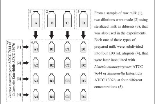
dilution ratios were adopted for each sample, in order to obtain a wide variety of milk autochtho-nous microbiota concentration. Each raw milk sample and its two dilutions were then subdivided into four 100 mL aliquots, and artificially contami-nated with the prepared cultures of L. monocyto-genesandS.Enteritidis, at different concentrations of each pathogen. For each raw milk sample, the sterile milk used as diluent was used as a control and was also subdivided into four 100-mL aliquots that were inoculated with the pathogens. Conse-quently, each raw milk sample generated 12 treatments and further 4 controls, completing 16 tests as shown inFig. 1.
The sterile milk controls inoculated with the pathogens were diluted serially with 0.85% NaCl. Considering the estimated quantity of pathogen inoculated, two dilutions were selected and plated on PetrifilmTM AC (3M Microbiology, St. Paul, MN, USA) and incubated at 351C for 48 h to determine the exact concentration of the pathogen.
Listeria monocytogenes
detection
Immediately after inoculation, the treatments artificially contaminated with L. monocytogenes were submitted to the official detection methodol-ogy for this pathogen in raw milk (Wher and Frank, 2004), using culture medium from Oxoid. A 25 mL aliquot from each treatment or control was added to 225 mL of Listeria enrichment broth (LEB) that
was incubated at 301C for 48 h. After incubation, one aliquot from each culture was streaked on Oxford agar and Palcam agar plates, and incubated at 351C for 48 h. TypicalListeriaspp. colonies were transferred to TSA-YE plates, incubated at 301C for 48 h and identified biochemically (Pagotto et al., 2007). The results were expressed as positive or negative for recovering L. monocytogenes in a 25 mL aliquot of each treatment or control.
Salmonella
Enteritidis detection
Similar to the treatments inoculated with L. monocytogenes, immediately after inoculation, all the treatments artificially contaminated with S. Enteritidis cultures were submitted to the official detection methodology for this pathogen in raw milk (Wher and Frank, 2004), with a modification, using also culture media from Oxoid. A 25-mL aliquot of each treatment was added to 255 mL of lactose broth and after homogenization incubated at 351C for 24 h. After incubation, 0.1 mL was transferred to Rappaport–Vassilidadis broth – RV (incubated at 411C for 24 h) and simultaneously, 1.0 mL was transferred to Tetrationate broth – TT (incubated at 351C for 24-h). Selenite Cystine broth was substituted by RV broth due to its better performance in Salmonella spp. recovering (D’Aoust et al., 1992; Busse, 1995;de Boer, 1998). RV and TT broths were then streaked on Bismuth Sulfite agar, Xylose Lysine Desoxycholate agar and
From a sample of raw milk (1),
two dilutions were made (2) using
sterilized milk as diluents (3), that
was also used in the experiments.
Each one of these types of
prepared milk were subdivided
into four 100 mL aliquots (4), that
were later inoculated with
7644 or Salmonella Enteritidis
concentrations (5).
Listeria monocytogenes ATCC
1 2 3
4 5
A B C D
[1]
[2]
[3]
[4]
Listeria monocytogenes
ATCC 7644 or
Salmonella
Enteritidis ATCC 13076
A1
A2
A3
A4
B1
B2
B3
B4
C1
C2
C3
C4
D1
D2
D3
D4
ATCC 13076, at four different
Hektoen Enteric agar plates and incubated for 24 h at 351C. When suspect Salmonella colonies were present, they were transferred to Lysine Iron agar and Triple Sugar Iron agar slants, and incubated for 24 h at 351C. The results for Salmonella were confirmed through serological tests, using somatic and flagellar polyvalent antiserum (Probac do Brasil, Sa˜o Paulo, SP, Brazil). The results were expressed as positive or negative for recovering S.Enteritidis in a 25 mL aliquot of each treatment or control.
Enumeration of mesophilic aerobes
Before the experimental inoculation, the raw milk samples and their dilutions in sterile milk were submitted to serial decimal dilutions in 0.85% NaCl. From the raw milk samples, the dilutions 1:100,000 and 1:1,000,000 were sown on PetrifilmTMAC plates to enumerate mesophilic aerobes, incubated at 351C for 48 h. Smaller dilutions of the other treatments were selected to enumerate these indicators.
Evaluation of inhibitor presence
All the raw milk samples used in this study were further tested regarding the presence of inhibitory substances of bacterial growth, using the Charm-Farm testTM FVN-100, following the manufacturer instructions (Charm Sciences Inc., Lawrence, MA, USA).
Results
The pathogen was recovered in all the treat-ments carried out with sterile milk artificially contaminated with L. monocytogenes or S. Enter-itidis (the controls), regardless of the inoculum concentration.
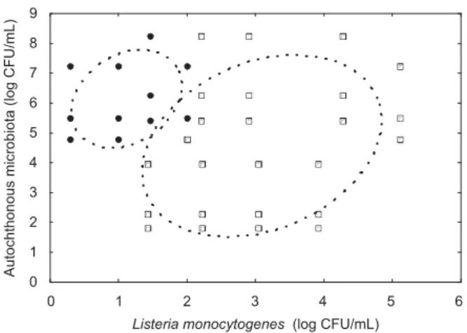
Considering the different levels of contamination by the autochthonous microbiota, considerable variation in the recovery of the inoculated patho-gens was observed, depending on the concentration of the cultures added experimentally (Figs. 2 and 3). Generally, a greater frequency ofL. monocytogenes or S. Enteritidis recovering was observed in the treatments with low contamination levels of the autochthonous microbiota associated with higher inoculum concentrations of the pathogens.
The recovery of L. monocytogenes occurred in all treatments that presented concentrations higher than 2.2 log CFU/mL of the pathogen. In the treatments inoculated with 2.0 log CFU/mL of
L. monocytogenes, pathogen recovery did not occur when the autochthonous microbiota contamination was higher than 5.0 log CFU/mL. At concentrations lower than 2.0 log CFU/mL of L. monocytogenes, pathogen recovery was only possible in the treat-ments that presented mesophilic aerobes at levels below to 4.0 log CFU/mL (Fig. 2).
The recovery of S. Enteritidis was possible in all treatments with concentrations higher than 3.7 log CFU/mL of the pathogen. In the treatments with 2.5–3.0 log CFU/mL ofS.Enteritidis, the patho-gen recovery was possible when the mesophilic aerobes were at levels below 5.5 log CFU/mL. When S. Enteritidis were inoculated at levels below 2.0 log CFU/mL, the pathogen was not recovered in any treatment, except in three cases: one treatment
Listeria monocytogenes (log CFU/mL)
Autochthonous microbiota (log CFU/mL)
0 1 2 3 4 5 6
0 1 2 3 4 5 6 7 8 9
Figure 2. Positive (&) and negative results (K) for
recovering of different levels ofListeria monocytogenes inoculated in raw milk treatments, with different levels of autochthonous microbiota. Ellipses indicate the range of positive and negative results at 0.95.
Salmonella Enteritidis (log CFU/mL)
Autochthonous microbiota (log CFU/mL)
0 1 2 3 4 5
0 1 2 3 4 5 6 7 8 9
Figure 3. Positive (&) and negative results (K) for
that presented low level of pathogen (0.9 log CFU/mL) and autochthonous microbiota (3.1 log CFU/mL), and two treatments with relative low levels of the pathogen (1.2 and 1.9 log CFU/mL), associated with high levels of mesophilic aerobes (8.3 log CFU/mL) (Fig. 3). No sample contained residues of inhibitory substances for bacterial growth.
Discussion
The obtained results indicated that the tested methodologies for isolation of L. monocytogenes and Salmonella in milk were sensitive, without considering possible interfering factors of the samples. In all control treatments the recovery of the inoculated pathogen was possible. Pathogen isolation was possible even in the control treat-ments that were inoculated with the smallest concentrations of the pathogens. Different levels of pathogens and autochthonous microbiota were associated in order to verify possible inter-ferences in the recovery methodologies. These pathogens are not usually quantified in dairy products, but often are present at low levels (0.0–2.0 log CFU/mL) and can represent risks to the consumers (Vlaemynck and Moermans, 1996;
Hof, 2003;Jay et al., 2005).
The treatments with raw milk samples showed that pathogen recovery was not possible on several occasions, probably due to the intrinsic factors of the samples used. The possible interference by antimicrobial substances residues was discarded, since all samples presented negative results for this test. However, the autochthonous microbiota ap-pears as determining factor for pathogen isolation, suggesting poor efficiency of the methodologies in these situations. The current microbiological stan-dard for raw milk in Brazil is 1,000,000 CFU/mL of mesophilic aerobes (Farina et al., 2005), and at this level the recovery of L. monocytogenes was possible when the pathogens were present at high levels of contamination (Figs. 2 and 3).
That Enterococcus and Lactobacillus naturally occurring in milk interfere with the isolation and survival ofL. monocytogeneswas already described by Suh and Knabel (2001), and Jiang et al. (1998)
demonstrated this interference during the enrich-ment phase of detection using LEB. In addition, L. monocytogenes is usually present in milk and dairy products at low levels (Vlaemynck and Moer-mans, 1996), similar to those simulated in the present study in treatments that showed no recovery of the pathogen. RegardingS.Enteritidis, we also observed a reduced recovery of the
pathogen (Fig. 3), but this was not as evident as forL. monocytogenes. In a similar study, only using buffered peptone water instead of lactose broth in the initial phase, no significant interference was observed in the recovery of various concentrations of Salmonella added to raw milk containing mesophilic aerobes at levels of less than 3 log CFU/mL (Jiang et al., 1998). Some atypical and unexpected recovery results were observed (Fig. 3), suggesting differences in the microbial profile of the raw milk samples.
The difficulty of recovering L. monocytogenes andSalmonella in food has been reported, mainly in assessment studies of culture media and meth-odologies to isolate these microorganisms (D’Aoust et al., 1992;Vlaemynck and Moermans, 1996;Jiang et al., 1998; Suh and Knabel, 2001). The auto-chthonous microorganisms are the main interfering factor, since they can generate unfavorable condi-tions for the survival and detection of the patho-gens (Busse, 1995;Vlaemynck and Moermans, 1996;
Jiang et al., 1998; de Boer, 1998)and inhibit their growth by competition (Slade, 1992; Suh and Knabel, 2001). The multiplication of the auto-chthonous microbiota in the initial phases of L. monocytogenes and Salmonella isolation may cause a sharp fall in pH with consequent inhibition in the multiplication and detection of these pathogens. To avoid these interferences, alterna-tive methods suggests the use of different culture media in these phases, such as Half-Fraser broth and Fraser broth forL. monocytogenes(ISO, 1996), and buffered Peptone Water and Selenite Cystine broth for Salmonella spp. (ISO, 2002). These variations aim to benefit the growth of these pathogens during the isolation procedures, espe-cially forL. monocytogenes by using two selective media instead of only one (Wher and Frank, 2004), enhancing their recovery from raw milk samples.
were not submitted to any experimental stress or spoilage, which would certainly also affect recovery.
The results obtained led to the conclusion that the officially recommended analytical methodologies for L. monocytogenesand Salmonella detection in raw milk may suffer direct interference from the auto-chthonous microbiota in the samples to be analyzed. Further studies are necessary to clarify the underlying mechanisms. In addition, the development of more sensitive methods forL. monocytogenesand Salmo-nelladetection in milk is recommended.
Acknowledgments
The authors thank FAPESP (Proc. 01/13076-8) for financial support and for the Ph.D. fellowship to author L.A.Nero, and Patrı´cia H. Nero for useful review of grammar and spelling. The authors are immensely grateful to 3M do Brasil Ltda. for donating the PetrifilmTMplates.
References
Adesiyun AA, Stoute S, David B. Pre-processed bovine milk quality in Trinidad: prevalence and character-istics of bacterial pathogens and occurrence of antimicrobial residues in milk from collection centres. Food Control 2007;18:312–20.
Bennett AR, Greenwood D, Tennant C, Banks JG, Betts RP. Rapid and definitive detection ofSalmonellain foods by PCR. Lett Appl Microbiol 1998;26:437–41.
Besse NG. Influence of various environmental parameters and of detection procedures on the recovery of stressed L. monocytogenes: a review. Food Microbiol 2002;19:221–34.
Beumer RR, Hazeleger WC. Listeria monocytogenes: diagnostic problems. FEMS Immunol Med Microbiol 2003;35:191–7.
Boor KJ. Pathogenic microorganisms of concern to the dairy industry. Dairy Food Environ Sanit 1997;17: 714–7.
Busse M. Media for Salmonella. Int J Food Microbiol 1995;26:117–31.
Carr FJ, Chill D, Maida N. The lactic acid bacteria: a literature survey. Crit Rev Microbiol 2002;28:281–370.
Cordano AM, Rocourt J. Occurrence of Listeria mono-cytogenes in food in Chile. Int J Food Microbiol 2001;70:175–8.
D’Aoust JY, Sewell AM, Warburton DW. A comparison of standard cultural methods for the detection of foodborneSalmonella. Int J Food Microbiol 1992;16: 41–50.
de Boer E. Update on media for isolation of Enterobac-teriaceae from foods. Int J Food Microbiol 1998;45: 43–53.
de Buyser M-L, Dufour B, Maire M, Lafarge V. Implication of milk and milk products in food-borne diseases in France and in different industrialised countries. Int J Food Microbiol 2001;67:1–17.
Farina EMMQ, Gutman GE, Lavarello PJ, Nunes R, Reardon T. Private and public milk standards in Argentina and Brazil. Food Pollut 2005;30:302–15.
FDA, 2007. Bacteriological Analytical Manual Online. Food and Drug Association, Washington. Available at
/http://www.cfsan.fda.gov/ebam/bam-toc.htmlS.
Hof H. History and epidemiology of listeriosis. FEMS Immunol Med Microbiol 2003;35:199–202.
Hudson JA, Billington C, Carey-Smith G, Greening G. Bacteriophages as biocontrol agents in food. J Food Prot 2005;68:426–37.
ISO 11290-1, 1996. Microbiology of food and animal feeding stuffs –horizontal method for the detection
and enumeration of Listeria monocytogenes–Part 1:
detection method. International Organization for Standardization, Geneva, 16pp.
ISO 6579, 2002. Microbiology of food and animal feeding stuffs – horizontal method for the detection of
Salmonella spp. International Organization for Stan-dardization, Geneva, 27pp.
Jay JM. Foods with low numbers of microorganisms may not be the safest foods OR, why did human listeriosis and hemorrhagic colitis become foodborne diseases? Dairy Food Environ Sanit 1995; 15:674–7.
Jay JM. Microorganisms in fresh ground meats: the relative safety of products with low versus high numbers. Meat Sci 1996;43:S59–66.
Jay JM, Loessner MJ, Golden DA. Modern food micro-biology, 7th ed. Gaithersburg: ASPEN Publishers; 2005. 854pp.
Jiang J, Larkin C, Steele M, Poppe C, Odumeru JA. Evaluation of universal preenrichment broth for the recovery of foodborne pathogens from milk and cheese. J Dairy Sci 1998;81:2798–803.
Kivaria FM, Noordhuizen JPTM, Kapaga AM. Evaluation of hygiene quality and associated public health hazards or raw milk marketed by smallholder dairy producers in the Dar es Salaam region, Tanzania. Trop Anim Health Prod 2006;38:185–94.
Leclerc V, Dufour B, Lombard B, Gauchard F, Garin-Bastuji B, Salvat G, et al. Pathogens in meat and milk products: surveillance and impact on human health in France. Livest Prod Sci 2002;76: 195–202.
Lo´pez-Expo´sito I, Minervini F, Amigo L, Recio I. Identifica-tion of antibacterial peptides from bovine k-casein. J Food Prot 2006;69:2992–7.
Nero LA, Mattos MR, Beloti V, Barros MAF, Pontes Netto D, Nogueira Pinto JPA, et al. Hazards in non-pasteurized milk on retail sale in Brazil: prevalence ofSalmonella spp.,Listeria monocytogenes and chemical residues. Braz J Microbiol 2004;35:211–5.
Canada (Available online at: /http://www.hc-sc.gc. ca/food-alimentS).
Riley MA, Wertz JE. Bacteriocins: evolution, ecology, and application. Annu Rev Microbiol 2002;56:117–37.
Slade PJ. Monitoring Listeria in the food production environment. I. Detection of Listeria in processing plants and isolation methodology. Food Res Int 1992;25:45–56.
Suh JH, Knabel SJ. Comparison of different enrichment broths and background flora for detection of heat-injuredListeria monocytogenesin whole milk. J Food Prot 2001;64:30–6.
van Schaik G, Lotem M, Schukken YH. Trends in somatic cell counts, bacterial counts, and antibiotic residue violations in New York State during 1999–2000. J Dairy
Sci 2002;85:782–9.
Vlaemynck GM, Moermans R. Comparison of EB and Fraser enrichment broths for the detection ofListeria spp. andListeria monocytogenesin raw milk dairy products and environmental samples. J Food Prot 1996;59: 1172–5.