UNIVERSIDADE DE LISBOA
Faculdade de Ciências
Departamento de Biologia Vegetal
Analysis of teashirt mutants affecting cell
proliferation in
Drosophila melanogaster
Silvia Alexandra Barbosa Jacome Pimentel
DOUTORAMENTO EM BIOLOGIA
(Genética)
Lisboa
2010
Faculdade de Ciências
Departamento de Biologia Vegetal
Analysis of teashirt mutants affecting cell
proliferation in
Drosophila melanogaster
Silvia Alexandra Barbosa Jacome Pimentel
DOUTORAMENTO EM BIOLOGIA
(Genética)
Supervisors:
Rui Gomes
Laurent Fasano
Lisboa
2010
Por ti,
Alexandre.
This PhD work was support by my PhD grant (SFRH/BD4861/2001 and SFRH/BD/13233/2003) from the POCI 2010 (Fundo Social Europeu_ FSE) approved by Fundação para a Ciência e a Tecnologia (Portugal), and also by the collaboration between the Rui Gomes lab and Laurent Fasano labs (CNRS and the Association pour la Recherche Contre le Cancer and La Ligue, France).
Acknowledgements
I would like to thank to all the people that directly or indirectly contributed for the concretisation of this work:
Ao meu Orientador, Professor Rui Gomes, por me ter acolhido no seu grupo, pela oportunidade que me deu em realizar este trabalho no seu laboratório, pelo seu esforço em arranjar alternativas que permitissem ultrapassar as nosssas limitações financeiras e técnicas, por todas as sugestões e discussões. E pela sua boa disposição e humor.
Au Laurent Fasano, je lui remercie pour m’avoir accueilli dans son labo, pour toute sa disponibilité et patience avec moi, pour me faire confiance et me donner confiance dans mes résultats, pour toutes les suggestions et discussions et pour me montrer une autre forme de faire sience.
À Fundação para a Ciência e Tecnologia (FCT), pelo apoio financeiro, sem o qual teria sido impossível a realização deste trabalho.
To the Centro de Genética e Biologia Molecular (CGBM) and to the Institut de Biologie du Développement du Luminy (IBDML) for giving me conditions for the development of this work.
To all the people that contributed with the necessary reagents needed for the elaboration of this work, mainly to Dr. Alvaro Tavares, Dr. Claudio Sunkel, Dr.Roger Karess, Dr. Steve Kerridge, Dr. Stephen Cohen, Dr. Armel Gallet, Dra. Ruth Lehmann, Dr. E. McCarthy and to Bloomington Drosophila stock Centres.
Aos meus colegas do CGBM, pela entre-ajuda, pela amizade, pelo convívio e pelas “brincadeiras”, que tornaram o nosso dia-a-dia mais agradável e produtivo. Um OBRIGADO especial para os colegas de grupo, Cristina e Paulo, e para a minha “mamã” científica, Isabel, que me revelou o maravilhoso mundo das moscas e me fez dar os primeiros passos no ciclo celular, com muita paciência e enorme boa disposição.
À toutes les personnes qui m’ont aidé dans l’IBDML, sans oublier les personnes des plate-formes techniques qui ont facilité énormément mon séjour à Marseille. Un gros MERCI à tous les membres de l’équipe KF que j’ai connus (Laurent, Natalie, Xavier, Christine, Pierre, Virginia, Hervé, Elise, Steve, Delphine, Mylene, Bernard, Sébastien, Lucile et Fred), pour votre
accueil et patience d’essayer entendre mon luso-français, pour tous les conseils et discussions et pour votre amitié. Je ne peux pas oublier de vous remercier pour tous les bons moments, les blagues, les rigolades et les petites fêtes qui m’ont fait rester presque un année de plus que le prévu! Je vous remercie la magnifique soirée que vous avez organisée avant mon départ et que je ne l’oublierai jamais. Je veux exprimer ma gratification à Laurent et à Christine pour tout le temps dépensé à regarder et à discuter mes résultats et surtout me papier, à Delphine pour tous les protocoles, les anticorps, les solutions et à Steve pour tous les stocks de moches. Un Gros Merci à vous tous !
Ao Dr. Álvaro Tavares e a todos os membros da sua Equipe que tive o prazer de conhecer (à Susana, à Célia, à Mariana, à Cláudia e ao Paulo), gostaria de agradecer o facto de nos terem aberto a porta sempre que precisámos, inclusive para a realização das experiências de RNAi.
A toda a minha família e amigos, inclusive aos que infelizmente já partiram, um obrigado pelos telefonemas e mails que encurtaram a distância e pelo apoio que prestaram. Um agradecimento aos meus pais e irmão por gostarem de mim e pelo carinho que sempre me deram, cada um à sua maneira. Um agradecimento aos quatro avos do Alex por todas as vezes que ficaram com ele para que eu terminasse a tese. E à minha prima Sara pelas enumeras vezes que fiquei em sua casa, nesta minha vida de viagens.
Finalmente, porque os últimos são os primeiros, obrigada César, por me aturares, pelo teu apoio, por estares sempre a meu lado, pelo teu amor e ao Alex, apesar de todo o trabalho e desgaste associado, obrigado por teres nascido e seres meu filho. Os teus sorrisos e beijinhos são a motivação para não desistir e continuar lutando para ultrapassar a pouco e pouco os obstàculos que vão surgindo.
Objectives, Methodological Approach and Structure of the Thesis
The main goal of this thesis was to clarify the role of the transcription factor Teashirt (Tsh) during cell cycle progression and proliferation. Previous work in our lab showed that the late larval lethal mutants tshA3-2-66 and tshNG1 present atrophied brains and strong reduced
imaginal discs with a strong mitotic arrest in a metaphase-like stage (Salazar, 2000). We performed a better characterisation of the mitotic phenotype by immunostaining and by the analysis of the phenotype of tsh in double mutant constructs. The depletion of tsh in S2 cells by the interference RNA technique (RNAi) was performed in order to compare with the phenotype observed in tsh mutant neuroblasts. The analysis of the expression pattern in mutant brains by microarrays was also performed in order to identify an obvious Tsh target among cell cycle genes. Unfortunately, no canonical cell cycle target gene was found to be particularly upregulated or downregulated. Furthermore, our detailed characterisation of the mitotic phenotype revealed only a few structural abnormalities or defects, not enough to cause such a strong mitotic arrest. All these results are presented in chapter II (part IV) of this manuscript.
In order to prove that tsh is the gene responsible for the mitotic arrest observed in
tshA3-2-66 neuroblasts we performed P-element excision mutagenesis to get a revertant. During
this mutagenesis scheme, we obtained news alleles and, among themselves, we identified a potential tsh female sterile mutations causing atrophied ovaries and which do not lay eggs. This interesting phenotype led us to study the tsh role during Drosophila oogenesis. The 5’ region of tsh gene was sequenced in three of the six female sterile lines obtained. It was characterised the ovary phenotype and functional studies have been performed in vivo using the UAS/Gal4 system. The mutagenesis was made in Rui Gomes’ lab and the characterisation of the sterile female mutants was mostly performed in Laurent Fasano lab. All the results are presented in chapter II (part I, II and III) of this manuscript.
Curiously, these independent studies of tsh function had given convergent results. We concluded that tsh is required for the normal progression of the cell cycle in L3 neuroblasts and depleted S2 cells, as in the proliferation of the follicle stem cells during oogenesis (this is detailed discussed in chapter III).
During all this PhD work, the main concern was to prove that the tsh gene was required for cell proliferation since the progression of the cell cycle was affected in the three Drosophila systems (mutant neuroblasts, S2 cells and ovaries). Thus, the introduction of this manuscript will start with a description of the tsh gene and their reported functions during Drosophila development (Chapter I part I). The introduction will be followed by a overview of oogenesis, with special incidence in the principal pathway required for the proliferation and maintenance of the three type of stem cells found in the Drosophila germarium (germline stem cells, follicle
stem cells and escort stem cells; chapter I part II). Finally, a brief review about the cell cycle focalised in the metaphase/ anaphase transition will end the introduction (chapter I, part III).
The results will not be presented by chronological order. The major results were obtained during the study of tsh in the follicle stem cells proliferation. Thus, chapter II (results) will start with P-element excision mutagenesis performed to obtain the new tsh alleles (part I). Next, it will be characterised and described the tsh function in follicle stem cells (part II). A reference about the mutual repression between tsh and their paralog tiptop during oogenesis will be presented afterwards (part III). The results will end with the presentation of all the evidence implicating tsh in mitosis progression (part IV).
A final discussion will be presented in the chapter III. Here, the major conclusion will be discussed, as well as the new questions raised with the results presented in this PhD work.
In this manuscript, I did not dedicated one chapter exclusively to the material and methods used during my PhD work, since they are presented in each part of the results (chapter II).
All the references mentioned in chapters I, II and III are presented in the end of this manuscript.
Objective
The main goal of this thesis was to clarify the role of Tsh in cell cycle progression and to identify putative cell cycle target. For that, it was studied the requirement of Tsh to the progression of cell cycle using three Drosophila model systems: (1) ovaries to study follicle stem cells proliferation during oogenesis and, (2) neuroblasts and (3) S2 cells to analyse the progression of mitosis onset. In addition, during the development of this work, I proposed to characterise, for the first time, the Tsh function in Drosophila oogenesis.
The results presented in this manuscript give evidence that Tsh is required for cell proliferation in Drosophila neuroblasts, S2 cells and ovaries. Furthermore, it was showed that Tsh acts through the Hedgehog pathway to mediate follicle stem cells proliferation in ovaries. Finally, we demonstrated that both in neuroblasts or S2 cells the absence of Tsh arrest cells in metaphase/ anaphase transition.
Table of Contents
Acknowledgements ... VII Objectives, Methodological Approach and Structure of the Thesis ... IX Table of Contents ... XI Liste of Figures... XIII Liste of Tables... XV Liste of Abbreviatures ... XVI Resumo... XVII Palavras Chave: ... XX Abstract ... XVII Key Word: ... XXI
CHAPTER I_ INTRODUCTION
I- The teashirt (tsh) gene... 3
I_1- Characterisation of the tsh gene ...3
I_2- Expression patterning of the tsh mRNA and protein in Drosophila ...4
I_3- Modulators of the tsh expression during embryogenesis ...5
I_4- Tsh functions during Drosophila development...5
I_4.1- Homeotic function of Tsh: the identity of the trunk and the specification of the prothoracic identity...6
I_4.2- Tsh in the segmental polarity: modulation of the Wingless pathway and regulation of the hedgehog pathway target genes ...8
I_4.3- Tsh and the midgut morphogenesis...11
I_4.4- Tsh in the Drosophila Pax6/ So/ Eya network during eye development ...13
I_4.5.2- tsh promotes the identity of the proximal structures of wing and halteres discs ...16
I_5- tiptop as a tsh paralog ...18
I_6- The vertebrate tsh family...20
II- Overview of Drosophila Oogenesis ... 23
II_1- The Drosophila reproductive system ...23
II_2- Drosophila germarium offers the unique opportunity of to study three types of stem cells and their niches ...25
II_3- Origin of GSC, TF and CC...27
II_4- Ovary stem cells and their molecular mechanisms of regulation ...28
II_4.1- Genes and Pathways that control GSC self-renewal, proliferation and differentiation ...28
II_4.1.1- BMP, Piwi and Yb from the niche to regulate the maintenance and division of the GSC...29
II_4.1.2- External factors: Nutrition...32
II_4.1.3- Intrinsic factors in GSC maintenance and differentiation ...33
II_4.1.3.1- Translational factor: Pumilio (Pum) , Nanos (Nos), Vasa (Vas) and Pelota (Pelo) ...33
II_4.1.3.2- MicroRNA (miRNA) and repeat-associated small interfering RNA (rasiRNA) is important for GSC proliferation and maintenance...33
II_4.1.3.3- Junctional proteins: DE-cadherin, Armadillo (Arm) and Zero population growth (Zpg)...34
II_4.1.3.4- Cell cycle regulators: Cyclin B (CycB) and Imitation switch (ISWI) ...35
II_4.1.3.5- Differentiation promoting factors: Bag of marbles (Bam), Benign germ cell neoplasm (Bgcn), Orb, Sex lethal (Sxl) and ovarian tumor (Otu) ...36
II_4.2- Genes and Pathways that control FSC self-renewal, proliferation and differentiation...38
II_4.2.1- Molecular mechanisms shared by GSC and FSC for self-renewal and proliferation ...38
II_4.2.2.1- Wingless (Wg) signalling is essential for FSC maintenance and proliferation ... 40
II_4.2.2.2- Hedgehog (Hh) signalling is essential for FSC maintenance and proliferation ... 40
II_4.3- Genes and Pathways controlling the maintenance of ESC and EC ... 43
II_5- Drosophila ovary as a model system in stem cells studies. ... 43
III- Overview of Drosophila Cell Cycle... 44
III_1- The Cell Cycle... 44
III_2- M-phase... 45
III_2.1- Mitosis Promoting Factor (MPF) and Mitosis entry ... 48
III_2.1.1- CdK1 family ... 49
III_2.1.2- Cyclin A... 50
III_2.1.3- Cyclin B... 51
III_3- The bipolar spindle and the kinetocore-microtubule attachment until the formation of the metaphase plate... 53
III_3.1- The Kinetochore ... 55
III_4- The anaphase promoting complex/ Cyclosome (APC/C)... 56
III_4.1- The APC/C activators and substrate recognition... 57
III_4.2- APC/C regulation ... 57
III_5- The regulation of the cell cycle: the checkpoints ... 58
III_5.1- DNA damage checkpoint ... 59
III_5.2- Metaphase or Spindle assembly checkpoint (SAC) ... 61
III_5.2.1- Models for spindle assembly checkpoint sense the defects ... 63
III_5.2.1.1- Microtubule attachment model... 64
III_5.2.1.2- Tension model ... 65
III_5.2.2- Inhibition of APC/C by the spindle assembly checkpoint... 66
III_5.2.2.1- Recruitment of spindle checkpoint proteins to unattached/ untensed kinetochores ... 68
CHAPTER II_ RESULTS Part I:... 73
New teashirt alleles obtained by P-element excision mutagenesis ... 75
Part II:... 91
Regulation of cell proliferation and patterning in Drosophila oogenesis by Hedgehog signalling depends on teashirt ... 93
Part III:... 117
Teashirt and Tiptop repress each other in Terminal Filament Cells during Drosophila oogenesis... 119
Part IV:... 137
The knock-down of the teashirt gene causes a metaphase-like arrest in Drosophila larval neuroblasts and S2 cells.. 139
CHAPTER III_ DISCUSSION AND PERSPECTIVES 1- Tsh and its paralog Tio repress each other in the tip of the Drosophila ovary, and both are able to regulate En expression... 173
2-Tsh regulates the expression of Hh and En at the Tip of the Drosophila germarium... 174
3-Tsh mediating Hh pathway is required for FSC proliferation ... 175
4-The metaphase/anaphase transition is compromised in the absence of Tsh... 177
5- Tsh/Hh signalling from flies to vertebrates... 179
Concluding Remarks: ... 180
REFERENCE... 183
List of Figures
CHAPTER I_ INTRODUCTION
Figure I_1- Tsh expression patterning during Drosophila embryogenesis ...4
Figure I_2- Tsh as a Hox co-factor in the segment identity of the trunk...7
Figure I_3- Summary of gene interactions in the cells surrounding the middle constriction ...12
Figure I_4- Schematic representation of Drosophila leg imaginal discs and the adult leg...15
Figure I_5- Schematic representation of Drosophila wing imaginal discs and the adult wing ...17
Figure I_6- Embryonic expression of Tio and Tsh ...19
Figure I_7- The Teashirt family in Drosophila and vertebrates ...21
Figure II_1- Diagram and a Drosophila ovary and ovarioles ...24
Figure II_2- Drosophila germarium cross section showing the locations of germ line stem cells (GSC), follicle stem cells (FSC), and their niches...26
Figure II_3- Diagrams of the identified signalling pathways and intrinsic factors that regulate GSC maintenance and differentiation in the Drosophila ovary...31
Figure II_4- Diagrams of the identified signalling pathways and intrinsic factors that regulate follicle stem cells (FSC) and escort stem cells (ESC) maintenance and differentiation in the Drosophila ovary ...39
Figure II_5- The Hedgehog pathway based on what is known in Drosophila ...41
Figure III_1- Cell cycle phases. ...45
Figure III_2- Diagram of mitosis in animal cells ...47
Figure III_3-The regulation of the MPF activity by phosporilation ...48
Figure III_4- Schematic representation of the G2 checkpoint...60
Figure III_5- Relationship of the SAC with the cell-cycle machinery...62
Figure III_6- The attachment process...63
Figure III_7- A Model for the Regulation of APC/CCdc20 by the Spindle Checkpoint...69
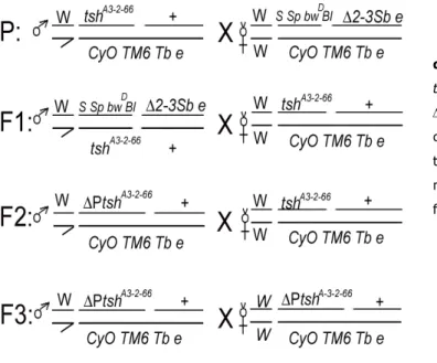
CHAPTER II_ RESULTS Part I Figure 1- Diagram of the flies matting during P-element excision mutagenesis...79
Figure 2- No mitotic anomalies were detected in neuroblasts from 8 P-element excision lines L3 lethal. ...82
Figure 3- No mitotic anomalies were detected in neuroblasts from P-element excision lines lethal during pupae stage. ...82
Figure 4- Adult ∆P48/tshA3-2-66 and ∆P127/tshA3-2-66 ovaries showed a very strong and early arrest in oogenesis...83
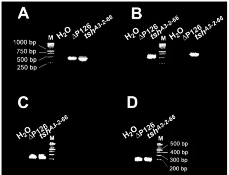
Figure 5- Amplified and sequenced regions of the PlacW in tshA3-2-66 and ∆P126. ...86
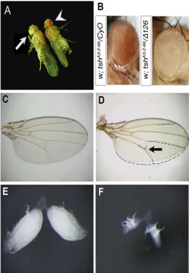
Figure 6- Morphologic aspects of ∆P126/ tshA3-2-66 flies. ...88
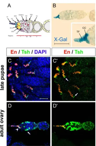
Part II Figure 1- en co-localises with a tsh-subset domain in Drosophila ovaries... 102
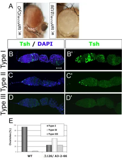
Figure 2- Expression level variation of tsh in ∆126/tshA3-2-66 ovarioles. ... 103
Figure 3- ∆126/ tshA3-2-66 ovaries display abnormal germline cyst encapsulation. ... 105
Figure 4- Cell cycle progression is affected in ∆126/tshA3-2-66 ovarioles. ... 107
Figure 5- Tsh is required for the proper expression of En and Hh in the germarium. ... 109
Figure 6- Ectopic expression of hh and tsh induces similar effects. ... 110
Figure 7- Tsh rescues the imperfect proliferation phenotype of ∆126/tshA3-2-66 mutants and allows the progression of oogenesis until stage S5/S6... 111
Part III
Figure 1- Tsh and Tio co-repress each other. ... 124
Figure 2- Tsh and Tio rescue the imperfect proliferation phenotype of the ∆126/tshA3-2-66 mutant and allow the progression of oogenesis until stage S5/S6... 126
Figure 3- Localisation of En in wild-type, ∆126/tshA3-2-66 and tio473 germaria... 127
Figure 4- Ectopic expression of hh, tsh and tio induces a similar phenotype... 129
Supplementary Figure 1- Tsh and En in wild-type, ∆126/tshA3-2-66, tio473 and ∆126/tshA3-2-66;UAS-tio/hsGAL4 germaria. ... 135
Part IV Figure 1- Molecular organisation in tshNG1 and tshA3-2-66 mutants. ... 150
Figure 2- Expression analysis of Tsh. ... 151
Figure 3- Mitotic phenotypes in tsh mutant larval brains. ... 152
Figure 4- Mitotic figures in preparations of larval CNS cells from wild-type and tsh homozygotes. ... 153
Figure 5- Cyclin A and B levels in wild-type and tsh mutant larval CNS cells. ... 154
Figure 6- Distribution of centromere CID and kinetochore components Bub1 and Rod in wild-type and tshNG1 neuroblast cells. ... 156
Figure 7- Time course analysis of Tsh RNAi in Drosophila S2 Cells. ... 158
Figure 8- Abnormal mitotic figures observed in S2 TSH-RNAi cells... 159
Supplementary Figure 1- Immunolocalisation of the Tsh protein in larval CNS cells... 166
Supplementary Figure 2- In situ cell death detection... 167
Supplementary Figure 3- Cuticules phenotype... 168
Supplementary Figure 4- Immunolocalisation of Polo in larval CNS cells ... 169
Supplementary Figure 5- Analisys of Polo expression by western blot... 169
Supplementary Figure 6- A CNS from L1, L2 and L3 larvae instars expressing tshlacZ stained with X-Gal. ... 170
CHAPTER III_ DISCUSSION AND PERSPECTIVES Figure 1- Tsh signalling to Follicle Stem cells (FSC) proliferation mediating Hh expression... 175
Figure 2- Proposed model to explain Tsh role in cell proliferation, mediating Hh expression signal in FSC and neuroblasts... 183
List of Tables
CHAPTER II_ RESULTS
Part I
Table1- Counting of F2 male that lost P-element based in the colour of the eyes. ...81 Table 2- Classification of the P-element excision line based in their lethality in trans-heterozygous with tshA3-2-66. ....81
Table 3- Fertility of the seven P-element excision lines with viable escapers, in Trans with the tshA3-2-66. ...84
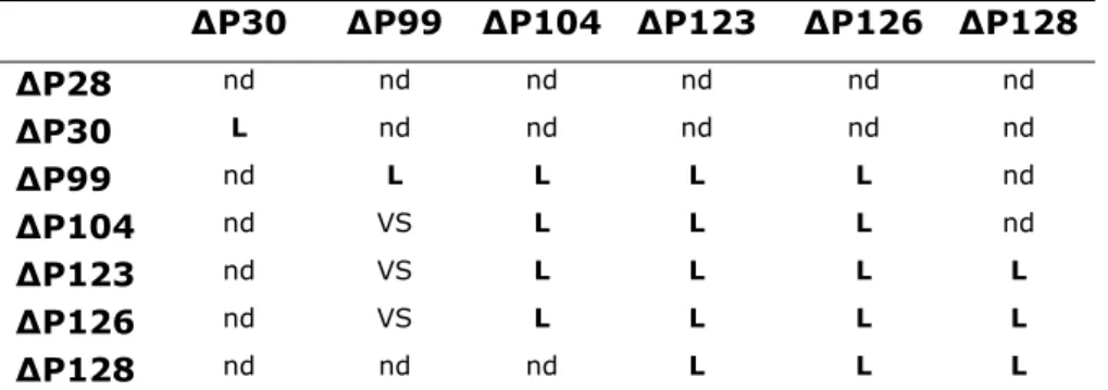
Table 4- Fertility of the combination of the seven P-element excision lines with viable escapers in Trans between
them...85
Table 5- List of the tsh alleles crossed with ∆P126and tshA3-2-66 to test for the viability and fertility of the adult female.
...87
Supplementary table 1- PlacW excision results obtained in two independents mutagenesis from the tshA-3-2-66 allele
by Salazar (2000, refered in chapter IV of the results of this manuscript) and Pimentel (presented in this chapter). ..90
Part II
Table 1- List of the tsh alleles crossed with P{lacW}A3-3-66∆126 and tshA3-2-66 to test for the viability and fertility of the
adult female. ... 104
Table 2- Quantification of the ∆126/ tshA3-2-66and the ∆126/ tshA3-2-66;UAS-tsh13/hs-Gal4 ovaries phenotype. ... 112
Supplementary Table 1- Quantification of the positive P-Histone H3 FC in the first 5 egg chambers of wild-type (n=69) and ∆126/ tshA-3-2-66(n=98) ovaries... 116
Part IV
Table 1- Mitotic index (number of mitotic cells per optic field) and percentage of anaphase figures in the larval CNS of
wild-type, tsh, rod x5, bub1, tsh rodx5 and tsh bub1 mutants... 157
Table 2- List of the most significant gene culters altered in tshNG1 homozygous neuroblats microarrays with the
respectived P value and total number of genes include. ... 160
Table 3- The most up- and down-regulated genes in tshNG1 homozygous neuroblats microarrays, the putative Tsh
target genes are also listed with the respective lower bound of fold change (LBFC), The positive LBFC represent the up regulated genes while the negative LBFC represent the downregulated genes. ... 161
APPENDIX
Table I- Results of the up- or down-regulation genes in tshNG1 homozygous neuroblats which have been ordered by
lower bound of fold change (LBFC), after using a cut-off of 1.3. The positive LBFC represent the up regulated genes while the negative LBFC represent the downregulated genes. Only the genes with the absolute value of LBFC upper than 1.3 are represented. ... 213
Table II- Results of the gene ontology clusters with a P-value <0.001 in tshNG1 homozygous neuroblats microarrays
after using a cut-off of 1.3. The positive lower bound of fold change (LBFC) represents the up regulated genes while the negative LBFC represent the downregulated genes. Only the genes with the absolute value of LBFC upper than 1.3 are represented... 227
Liste of Abbreviatures
µg microgram µl microliter µm micrometer
APC/C Anaphase promoting complex/ cyclosome bp base pair
CC Cap cells
Cdc genes Cell division cycle genes Cdk Cyclin-dependent kinase CNS Central nervous system
D. melanogaster Drosophila melanogaster
DAPI 4’,6’-Diamidino-2-phenilindole DNA Desoxiribonucleic acid
dsRNA double-stranded RNA
en engrailed
ESC Escort stem cells EC Escort cells FSC Follicle stem cells
FC Follicle cells
FITC Flurescein isothiocyanate GSC Germline stem cells
hh hedeghog
hs heat-shock promoter
IGS Inner germarial sheath cells
ihog interference hedgehog
KDa Kilo-Dalton (1KDa=1000 Da) MPF Mitosis promoter factor mRNA messenger Ribonucleic acid
MT Microtubule
NEBD Nuclear envelope breakdown PBS Phosphate buffer saline pH=7.4
PBST PBS with 0,1% Triton X-100 PCR Polymerase chain reaction PPH3 Phospho-histone H3
RNA Ribonucleic acid RNAi RNA interference
SAC Spindle assemby checkpoint SC Stem cells
SDS Sodium dodecyl sulphate Ser Serine
TF Terminal Filament cells Thr Threonine
tio tiptop tsh teashirt
Tyr Tyrosine
UAS Upstream activator sequence
X-Gal X-gal-5-bromo-4-chloro-3-indolyl-beta-D-galactopiranosideo WT Wild-type
Resumo
O gene teashirt (tsh) de Drosophila codifica para uma proteína de 116 KD com três motivos atípicos do tipo Zinc finger (Cx2Cx12HMx4H) espaçados. Este gene foi, inicialmente, referido como sendo necessário para a correcta especificação dos segmentos do tronco, durante a embriogénese de Drosphila, em colaboração com os genes Hox. Adicionalmente, em
Drosophila, o gene tsh foi implicado na morfogénese do intestino médio, durante o
desenvolvimento da parte proximal dos apêndices e durante a especificação dos olhos no adulto.
Este trabalho teve como principal objectivo clarificar a função do gene tsh na proliferação celular em Drosophila. Para tal, estudou-se o efeito da perda de função tsh em três contextos celulares de Drosophila: (1) nos ovários, com o intuito de estudar a proliferação das células estaminais foliculares (FSC) do germarium; (2) nos neuroblastos e (3) nas células S2, com o fim de analisar a progressão da mitose propriamente dita. Caracterizou-se as anomalias apresentadas em neuroblastos e ovários de mutantes hipomorficos tsh e em células S2 defectivas para o mesmo gene através da técnica de interferência de RNA (RNAi). A análise do padrão de expressão de cérebros mutantes para tsh foi obtida, utilizando a técnica de microarrays.
Durante este trabalho, observou-se que o gene tsh é co-expresso com o gene
hedgehog (hh) na extremidade anterior do germário de Drosophila: nas células do filamento
terminal (TF) e nas células cap (CC), ambas são localizadas adjacentemente às células estaminais germinais (GSC) e são descritas como parte integrante do nicho das GSC e FSC. Nos ovários de Drosophila, a via de sinalização Hh é essencial para a auto-renovação e proliferação das FSC. Uma nova mutação tsh, causadora da paragem precoce em oogénese (no estado S2/S3), permitiu mostrar que a proteína Tsh regula a expressão dos genes
engrailed (en) e hh na extremidade do germário, tornando-se, desta forma, crucial para a
normal proliferação das FSC. De acordo com esta observação, as experiências de sobre expressão do gene tsh nos ovários, originaram um fenótipo que se asssemelha ao obtido aquando da sobre expressão do gene hh. As FSC proliferam de forma excessiva, originando um excesso de descendência que se acumula entre as câmaras do ovo, formando as estruturas semelhantes a stalks gigantes. Em adição, nos mutantes defectivos para o tsh, a reposição deste gene, usando o sistema UAS/ hsGal4, permitiu recuperar parcialmente o fenótipo associado à proliferação anormal das FSC, possibilitando a progressão da oogénese até ao estado S5/S6. Consistentemente, a expressão do gene hh através do mesmo sistema UAS/ Gal4, também promoveu uma melhoria do fenótipo nos mutantes tsh, mas de forma menos eficiente.
Assim, conclui-se que o Tsh desempenha um papel crítico na regulação da expressão do
hh, contribuindo, desta forma, para a proliferação e especificação da identidade das FSC nos
ovários de Drosophila. Estes resultados estão de acordo com as interacções entre o gene tsh e a via de sinalização Hh, previamente descritas em embriões de Drosophila. Infelizmente, os resultados obtidos não permitiram concluir se o Tsh actua directamente na regulação da expressão dos genes hh e en, ou se por outro lado, se trata de uma regulação indirecta. Mais, continua por se clarificar, neste caso, se o gene tsh actua como um repressor ou um activador.
Recentemente, o gene tiptop (tio) foi descrito como sendo um novo membro da família
tsh em Drosophila. Tio codifica para uma proteína de 1024 amino ácidos, a qual apresenta um
quarto motivo, tipo zinc finger na região C-terminal. Durante a embriogénese, foi demonstrado que os dois membros da família tsh em Drosophila se reprimem mutuamente. Durante a oogénese, observou-se uma complementaridade na expressão de tsh e tio nas TF: Tio localiza-se nas TF apicais, enquanto o Tsh é preferencialmente detectado nas TF proximais. No entanto, a expressão de tio é ectópica em todas as celulas do TF em ovários ∆126/tshA3-2-66,
assim como a expressão de tsh é generalizasa a todas as TF células em ovários tio473
homozigotas. A expressão de hsGal4>UAS-tioFL permitiu a recuperação do fenótipo dos ovários∆126/tshA3-2-66, tal como foi acima mencionado para o hsGal4>UAS-tsh13. De forma
idêntica, a expressão ectópica de tio induz efeitos similares aos acima descritos para a sobre expressão de tsh, especialmente num contexto mutante para tsh. Desta forma, tal como foi descrito durante a embriogénese, tsh e tio reprimem-se mutuamente na extremidade apical do germário. Sugere-se ainda que Tio desempenha uma função redundante, completamente restituída pela presença de Tsh nos ovários tio473.
Durante este trabalho, reuniu-se várias evidências que indicam que o gene Tsh é indispensável para a progressão em mitose. Duas mutações hipomórficas no gene tsh (tshNG1
and tshA3-2-66) causam, em neuroblastos, uma paragem em metaphase, com a maioria das
figuras mitóticas exibindo cromossomas altamente condensados e associados a fusos mitóticos aparentemente normais e a centrossomas regulares. A análise dos esfregaços destes neuroblastos, com proteínas específicas do ponto de controlo do fuso (como Bub1 e Rod), revelou a normal localização destas proteínas. A imunomarcação com anticorpos contra a proteína CID (homologa da CENP-A) excluiu a ocorrência da separação prematura de cromatídeos irmãos. Mais ainda, nos neuroblastos parados em mitose, a ciclina A aparece normalmente degradada, enquanto que a a ciclina B continua a ser detectada. Duplos mutantes para tsh rod e tsh bub1 apresentam um fenótipo semelhante ao observado nos mutantes tsh simples. Assim, as mutações rod e bub1 não conseguem ultrapassar a forte paragem imposta pela perda de função tsh, continuando inibida a progressão para anáfase. Um papel geral para o Tsh é desconhecido, considerando o seu padrão de expressão localizado no sistema nervoso central (CNS) e as suas bem documentadas funções durante a especificação de estruturas particulares. No entanto, não foi identificado nenhum tipo de
regionalização específica dos defeitos mitóticos no CNS. Além disso, o silenciamento do gene
tsh em células S2 pela técnica de RNAi provoca uma paragem em mitose, imitando o fenótipo
descrito em neuroblastos mutantes para o tsh. Durante as experiências de RNAi, a maioria das células pararam numa configuração semelhante à metafase, o que originou um incrível aumento no índice mitótico. Assim, estes resultados levam a propor que o gene tsh desempenha um papel indispensável na transição metafase/anafase, uma vez que na ausência deste gene, o ponto de controlo do fuso e/ou a actividade do complexo promotor da anafase/ ciclossoma (APC/C) são pertubados. A análise do padrão de expressão de cérebros mutantes para tsh, comparativamente ao selvagem, não permitiu identificar um óbvio gene mitótico alvo. Curiosamente, foi possível estabelecer uma ligação putativa com a via de sinalização Hh através do interference–hedgehog gene (ihog), o terceiro gene com níveis de downregulation mais significativos. IHog é uma proteína transmembranar intermediária na resposta ao sinal activo de Hh, actuando como amplificador deste sinal upstream do receptor negativo desta via de sinalização: Patched (Ptc). O mecanismo, pelo qual o Hh induz a proliferação, depende do tecido em causa, mas inclui a indução e a regulação de componentes do ciclo celular como o das ciclinas: D1, D2, B1 e E, a Cdc25 e o N-Myc. Teoricamente, a Cdc25 (homóloga do String) pode também ser um alvo do Hh nos neuroblastos, uma vez que, de todos os alvos mitóticos conhecidos para a via Hh, a Cdc25 é o unico que se encontra ligeiramente downregulated nos mutantes tsh. No entanto, o sincronismo de paragem em metafase, observado nos neuroblastos tsh, está mais de acordo com alterações nos níveis de expressão do gene polo (um alvo mitótico com níveis de downregulation idênticos aos registados para a Cdc25). De facto, a observação dos neuroblastos tsh faz lembrar o fenótipo mitótico apresentado por mutantes polo (por exemplo homozigotas para o alelo polo9). No entanto, a redução dos níveis
de expressão de polo não é por si só suficiente, para explicar a forte paragem em metafase apresentada pelos mutantes tsh e, por outro lado, o polo nunca foi implicado como sendo um alvo da via Hh. Neste ponto, os resultados aqui expostos falham para identificar um alvo mitótico, no entanto, os significantes níveis de downregulation registados para o iHog sugerem uma interligação entre a forte paragem em mitose e a via de sinalização Hh, fazendo lembrar os resultados observados durante a oogénese. Assim, propõe-se que o gene tsh é necessário para a transcrição do iHog e que este ligando está implicado na modulação da sinalização Hh durante a mitose.
Com o trabalho aqui apresentado, pretende-se demonstrar que o gene tsh afecta a proliferação das FSC através da regulação da expressão do en e hh. Em adição, poder-se-á especular que em neuroblastos e células S2, o Tsh poderá também afectar a progressão da mitose através da sinalização Hh, uma vez que, pelo menos em neuroblastos, pela técnica dos
microarrys, verificou-se que o gene ihog é o terceiro mais donwregulated. Assim, propõe-se
Palavras Chave:
teashirt; tiptop; via de signalização do hedgehog; células estaminais somáticas; neuroblastos; mitose, ponto de controlo do fuso; transição metafase/ anafase, complexo promotor da anafase/ ciclossoma (APC/C).Abstract
Drosophila teashirt (tsh) encodes a zinc finger protein essential during Drosophila
development.
My PhD aimed to clarify the tsh role in cell proliferation studying tsh function in three Drosophila systems: follicle stem cells (FSC), neuroblasts and S2 cells.
I found that Tsh and Hh were co-expressed in the Drosophila terminal filament and cap cells. A new tsh female sterile mutation exhibiting early oogenesis arrest allowed me to show that Tsh acts via Hh-mediated signalling pathway to control FSC proliferation; tsh overexpression mimics the hh overexpression associated phenotype. Additionally, in tsh background, the hsGal4 driven expression of tsh or hh partially rescues the abnormal proliferation phenotype allowing oogenesis progression. These results suggest that Tsh is critical to control hh expression contributing for the regulation of FSC proliferation and specification of somatic cell identity.
In neuroblasts, hypomorphic tsh mutation leads to a metaphase-like arrest with highly condensed chromosomes associated with apparently normal mitotic spindles and centrosomes. Additionally, checkpoint proteins Bub1 and ROD were localised normally and CID imunodetection excluded an eventual premature sister chromatid separation. Furthermore, cyclin-A appears normally degraded, whereas cyclin-B remains detectable. Double tsh rod and
tsh bub1 mutants phenotypes resemble the tsh single mutant. Finally, tsh depletion in S2 cells
mimics the tsh mutant phenotype in neuroblasts. These results suggest that Tsh is critical during mitosis progression and I propose that Tsh loss maintains active the spindle checkpoint and/or unable the APC/C activity.
We proposed a tsh acts through Hh signalling in cell proliferation. During oogenesis, tsh affects FSC proliferation mediating Hh expression. In neuroblasts, tsh could acts through Hh signalling, since expression of interference hedgehog, a positive mediator of Hh signal, was severely downregulated in microarrays-based analysis.
Key Word:
teashirt; tiptop; hedgehog signalling; follicle stem cells; neuroblasts; mitosis, spindle assembly checkpoint; metaphase-anaphase transition, anaphase-promoting complex/ cyclosome (APC/C).CHAPTER I
INTRODUCTION
I- The teashirt (tsh) gene
I_1- Characterisation of the tsh gene
The tsh gene was firstly identified by the enhancer trap technique, in a screen for β-galactosidase activity during embryogenesis, (Fasano et al., 1991). The tsh gene is located at the left arm of chromosome 2 in the cytogenetic region 40A5.
Two size classes of tsh transcripts were detected in Drosophila: a predominant class consisted of transcripts of 5.4 kb, which are expressed throughout development, and a large size class of 8.5 kb transcripts that are detected predominantly, though not exclusively, during embryogenesis (Fasano et al., 1991). The differential splicing is indicated as the responsible, at least in part, for these two different classes of transcript.
Tsh codes for a protein of 993 amino acids, with an estimated molecular mass of 106
KDa (Fasano et al., 1991). The tsh protein (Tsh) is characterised by the presence of three atypical widely-spaced C2-H2 zinc finger motifs, corresponding to the consensus sequence CX2CX3FX5(L,M)X2HMX4H, in which the two histidines are separated by five amino acids and the size of the loop between the second C and the first H is of twelve residues in all Tsh zinc fingers (Fasano et al., 1991). In addition, the protein contains a consensus motif (PLDLS) for the interaction with the C terminal Binding Protein (CtBP) in a complex with Brinker (Brk) and three domains rich in alanine, in the N-terminal (Fasano et al., 1991; Manfroid et al., 2004; Saller et al., 2002). Finally, the acidic rich domain, identified in Tsh, is required for their interaction with Scr (Sex combs reduced) for the identity of the prothorax (Taghli-Lamallem et
al., 2007).
These structural properties are compatible with a role on the modulation of the chromatin. In fact, the gene Su(var)3-7, which encodes a protein with seven zinc of the Tsh zinc finger type, play a role in the modification of the chromatin structure (Cleard and Spierer, 2001). It was shown that Tsh is able to bind to DNA behaving like a transcription factor. In vitro, Tsh binds specifically to a fragment on the promoter of the gene modulo (mod) and recognises two specific binding sites in this mod fragment (Alexandre et al., 1996). The mod expression is repressed by Tsh in the trunk (Alexandre et al., 1996). Moreover, cell culture analyses showed that tsh acts as a transcriptional repressor of mod (Waltzer et al., 2001). Recently, Tsh was identified as a protein that interact with FE65 (Kajiwara et al., 2009). FE65 is an adapter protein that binds to the amyloid protein precursor and this complex can modulated gene expression (Duilio et al., 1991). FE65 could simultaneously recruit SET (a component of the inhibitor of acetyl transferase) and Tsh (that in turn recruit histone deacetylase to produced a powerful gene silencing complex; Kajiwara et al., 2009). The primate specific Caspase 4 (CASP 4) was identified as a target of this FE65/ SET or FE65/ Tsh complex. In addition, chromatin immunoprecepitation showed a direct interaction between FE65 and Teashirt3 with the promoter region of the CASP 4 (Kajiwara et al., 2009).
I_2- Expression patterning of the tsh mRNA and protein in Drosophila
The tsh transcripts are first detected at the beginning of cellularisation (stage 4-5), in a central ring of cells corresponding to the primordium of the thorax (Fig. I_1; Fasano et al., 1991). At the end of cellularisation/ beginning of gastrulation, tsh expression is observed in five bands transient in the central region of the embryo (the three posterior bands are weaker then the two anterior ones). During gastrulation (stage 6), the striped pattern gives a more uniform labelling in a domain of cells that correspond to the presumptive trunk region of the embryo (Fasano et al., 1991). At the beginning of the gastrulation, tsh present a transient expression in 5 bands of the embryo trunk. Than, at stage 9, tsh transcripts present a homogeny distribution in all the presumptive domain of the trunk (to parasegments 3 to 13). This staining is maintained until the end of the embryogenesis, with the tsh transcripts presenting a particular segmental pattern: they are stronger expressed in the posterior region of each trunk segments (Fasano et al., 1991).
Figure I_1- Tsh expression patterning during Drosophila embryogenesis (in <flybase.bio.indiana.edu
/reports/FBgn0003866.html>).
At stage 4-5 tsh transcripts are first dectect in the primordium of the thorax.
At stage 6 (beginning of gastrulation) tsh expression is observed transiently in five band in the central region of the embryo; that correspond to the presumptive trunk region of the embryo.
At stage 9 (the extended germband stage), cells specific to parasegments 3 to 13 are labelled, that correspond to the presumptive domain of the trunk. The trunk staining is maintained until the end of the embryogenesis with the tsh transcripts stronger expressed in the posterior region of each trunk segments.
After stage 15 of embryogenesis, tsh expression is also observed in the ventral nervous cord, in the anterior part of the pharynx, in the visceral mesoderm, in the anal opening and in stellate cells of the Mapighi tubes.
Tsh transcripts are also detected in ventral nervous cord, with a strong intensity in the three thoracic segments, in the anterior part of the pharynx, in the visceral mesoderm and in the anal opening, after the 15 stage of embryogenesis (Fasano et al., 1991).
al., 2003). Tsh is also present in the proximal region of the imaginal discs of the halteres,
wings and legs (Bhojwani et al., 1997; Erkner et al., 1999).
During embryogenesis, the tsh transcript and protein present sensibly the same distribution. The protein is detected within the cytoplasm or the nucleus during the first stages of development. After stage 7 of embryogenesis, the localisation is predominantly in the nucleus (Alexandre et al., 1996).
I_3- Modulators of the tsh expression during embryogenesis
tsh expression is regulated during embryogenesis by Hox genes in ectoderm and
mesoderm derivatives (Roder et al., 1992; Mathies et al., 1994). Moreover, some of these homeodomain transcription factors, including Antennapedia (Antp), Ultrabithorax (Ubx) and abdominal-A (abd A), bind directly to specific enhancers in the tsh regulatory region (McCormik et al., 1995). However, Hox proteins are not required for initiation of the tsh expression, but rather modulate and maintain the expression pattern in a segment-specific manner. Tsh in turn regulates the expression of Hox genes active in the gnathal segment and head: Sex combs reduced (Scr), Deformed (Dfd) and labial (lab; Roder et al., 1992). Thus, tsh works at the same level as Hox.
spalt (sal) and grunge (gug, homologue of vertebrates atrophine genes), are known to
regulate tsh expression (Erkner et al., 2002; Röder et al., 1992). Sal protein acts like a transcriptional repressor of tsh: in the absence of sal, tsh is expressed ectopically in parasegment 2, in the labial and in the tail region (parasegments 14-15, stage 10-11; Röder et
al., 1992). Grunge acts positively to regulate teashirt expression in proximoventral parts of the
leg. Grunge has other regulatory functions in the leg, including the patterning of ventral parts along the entire proximodistal axis and the proper spacing of bristles in all regions (Erkner et
al., 2002). In Gug loss-mutants, the embryo segmentation is affected via a failure in the
repression of at least four segmentation genes known to regulate tsh [hunchback (hb), Krüppel (Kr), knirps (kni) and fushi tarazu (ftz)]; in these embryos, tsh appears in bands pattern in the dorsal region, and the ventral tsh expression is lost (Coré et al., 1997; Erkner et al., 2002; Röder et al., 1992).
I_4- Tsh functions during Drosophila development
Tsh is crucial for the patterning of the trunk identity in collaboration with the Hox genes
(Fasano et al., 1991; Roder et al., 1992). Tsh also acts on the Wingless (Wg) and Hedgehog (Hh) pathways (Angelats et al., 2002; Gallet et al., 1998, 1999) for the specification of the naked cuticule during embryogenesis. In addition, tsh function is required for the midgut morphogenesis (Mathies et al., 1994) and for the development of adult appendages (Bessa et
2000). The molecular mechanisms underlying tsh function are poorly understood. However, in
vitro experiments have shown that, when bound to DNA, Tsh could repress transcription
(Alexandre et al., 1996; Waltzer et al., 2001; Saller et al., 2002). Tsh binds to a specific enhancer of the modulo (mod) gene, a known target of Scr and Ubx (Graba et al., 1994). Tsh inhibits mod expression in the epidermis of the prothorax (or T1, the first thoracic segment) (Alexandre et al., 1996). In the midgut mesoderm, tsh is required for the transcriptional repression of Ubx that is mediated by high levels of Wg in collaboration with the co-repressors Brk and CtBP (Waltzer et al., 2001; Saller et al., 2002). In this case, no sequence similar to the mod enhancer bound by Tsh has been identified. Instead, Brk binds the Ubx enhancer and then recruits Tsh and CtBP into a ternary repressor complex (section I_4.3 of this chapter).
I_4.1- Homeotic function of Tsh: the identity of the trunk and the specification of the prothoracic identity
The absence of any single homeotic gene results in a change in the identity of a specific segment or segments into another type, generating a typical homeotic transformation. Trunk morphology in the Drosophila embryo also depends on the normal function of the tsh gene. However, tsh is different from the classical homeotic genes in at least two aspects: (1) it codes for a zinc finger protein and (2) the analysis of the phenotype of mutations at this locus affect the entire trunk (Roder et al., 1992).
tsh is critically required for the identity of the T1 segment and globally for segmental
identity throughout the entire trunk, whereas the “classical” homeotic genes have more specific roles (Fig. I_2; de Zulueta et al., 1994; Fasano, 1991; Roder et al., 1992).
The specific identity of the anterior prothorax is determined by the simultaneous activities of the tsh and Scr gene products. First, in the absence of the Hox genes from the trunk [Scr, Antp, Ubx, abdA and abdominal B (AbdB)], each thoracic and abdominal segment adopts a T1 identity conferred by the presence of tsh, which is active in all this region (Struhl, 1983). Moreover, in tsh mutants, the T1 segment is transformed in labial, since Scr is unable to promote the T1 identity in the absence of tsh (de Zulueta et al., 1994; Fasano et al., 1991). In the absence of tsh, the Scr expression is affected: Scr is still present in the labial and the dorsal part of T1 segment, but it is ectopic in the ventral region of this T1 segment, showing that tsh controls the posterior limit of the Scr expression in the ventral ectoderm (Fasano et
Figure I_2- Tsh as a Hox co-factor in the segment identity of the trunk (in Laugier, 2005 modified
from Robertson et al., 2004).
In the trunk segments, the Tsh protein repress the expression of disco and target the segments for a trunk identity. Sal (Spalt protein) defines the boundary between head and trunk (broken line) repressing tsh expression in the gnathal segments. In the head segments, disco is expressed and activates the acquisition of gnathal fate. Thus, the distribution of Tsh and Disco regionalize the embryo. The combination of these proteins with Hox genes determines the segment identity. Scr (Sex combs reduced) is expressed in the gnathal domain as well as in the trunk, and the acquisition of different segment identities depend on the co-factor present. Dfd (Deformed) can establish the maxilar ou mandibular identity in function of the presence of the protein cap-n-collar (Cnc; Mohler et al., 1995). Ma: mandibular segment; Mx: maxilar segment; Lb: labial segment; T1: first thoracic segment (or prothorax) and T2: second thoracic segment.
In contrast, the ectopic expression of tsh induces the inverse: the head-to-trunk transformation. The labial segment is transformed in T1 and the maxilar and antennal segments acquire a trunk identity, illustrated by the presence of naked cuticule, without clear segmental identity (de Zulueta et al., 1994). In this genetic context, Scr expression remains unchanged. When tsh and Scr are simultaneously ectopically expressed, the labial, maxilar and antennal segments acquire a T1 phenotype.
Recently, there has been a new insight into the mechanism by which Tsh, in concert with Scr, specifies the prothoracic identity (Taghli-Lamallem et al., 2007). It was shown that Tsh interacts directly with Scr, and this interaction depends, at least in part, on the presence of a short “acidic domain” located on the N-terminal half of Tsh. In vivo, the expression of full length Tsh can rescue the tsh null phenotype throughout the trunk, whereas Tsh lacking the Scr interacting domain rescues all the trunk defects except in the prothorax (Taghli-Lamallem
et al., 2007). Thus, direct interaction between tsh and Scr is indispensable to define the T1
Since tsh activity is required for segment identity of the entire trunk region, it has characteristics in common with the “region specific” homeotic genes spalt and fork head (Jürgens, 1988; Jürgens and Weigel, 1988).
It has been shown that Split ends (Spen), tsh and Antp function in a combinatorial manner to repress the development of head-like sclerites and to promote the development of thoracic identity (Wiellette et al., 1999).
In tsh Antp double mutant embryos, the first two thoracic denticle belts are completely absent and replaced by cuticle typical of the head skeleton (Roder et al., 1992). Conversely, ectopic expression of Antp induces a strong transformation of T1 to T2 (Gibson and Gehring, 1988; Gibson et al., 1990). In vitro, it was recently shown that Tsh interacts directly with the Hox protein Antp (Taghli-Lamallem et al., 2007).
If Tsh is a co-factor for Hox proteins in the trunk, how can different Hox proteins establish different segment identities with the same co-factor (for example, Scr and Antp with Tsh)?
One role for the Tsh–Scr and Tsh–Antp complexes could be to control specific subset of target genes, in order to specify the identity of the T1 and T2 segments. Moreover, the capacity of Tsh∆acid (defective for the acidic domain) to rescue the T2 defects of a tsh8 null
mutant suggests that the collaborative activity of Tsh/Scr and Tsh/Antp does not involve the same domain of Tsh (Taghli-Lamallem et al., 2007).
I_4.2- Tsh in the segmental polarity: modulation of the Wingless pathway and regulation of the hedgehog pathway target genes
The segment-polarity genes, which encode a diversified group of proteins including transcription factors and components of the Hedgehog (Hh; Ingham, 1998) and Wingless (Wg) signal transduction pathways (Klingensmith and Nusse, 1994; Van den Heuvel et al., 1989; Willert and Nusse, 1998) are required for intrasegmental patterning (Baker, 1988; Bejsovec and Martinez-Arias, 1991; DiNardo et al., 1988; Dougan and DiNardo, 1992; Heemskerk et
al., 1991; Martinez-Arias et al., 1988; Noordermeer et al., 1992; Perrimon, 1994).
The wg gene (Wnt in vertebrates), encodes secreted glycoproteins (Klingensmith and Nusse, 1994; Kühl and Wedlich, 1997; Miller and Moon, 1996; Van den Heuvel et al., 1989; Willert and Nusse, 1998). A conserved signal transduction pathway transmits the signal promoted by Wg following its secretion (Klingensmith and Nusse, 1994; Kühl and Wedlich, 1997; Miller and Moon, 1996; Willert and Nusse, 1998). At the end of this pathway, two proteins are responsible for Wg output: Armadillo (Arm; homologue to vertebrate β-catenin; Kühl and Wedlich, 1997; Miller and Moon, 1996; Peifer and Wieschaus , 1990; Peifer et al., 1991; Riggleman et al., 1990; Willert and Nusse, 1998), and the Drosophila T-cell factor (dTcf), which is also known as Pangolin and is analogous to the vertebrate T-cell factor (Tcf) or
1996; Riese et al., 1997; van de Wetering et al., 1997). Tcf is a member of the family of high mobility group (HMG) DNA-binding proteins required for DNA architecture and gene regulation. Both, Arm and dTcf, seem to be crucial for the transmission of all known Wg signalling events. Two pools of Arm exist within cells: one at the cell membrane that is required for cell adhesion (Orsulic and Peifer, 1996; see also section II_4.1.3.3 of this chapter), and which is independent of the Wg signal, and the other, a cytoplasmic pool for transmission of the Wg signal (Pai et al., 1997; Peifer et al., 1991; Riggleman et al., 1990). In the plasmic membrane, Arm binds to E-cadherin and α-catenin in order to maintain the cellular adhesion (Willert and Nusse, 1998). In the cytoplasm, upon reception of Wg, Arm is stabilized and recruited for the
wg transduction signalling (Peifer, 1995).
In the absence of Wg, cytoplasmic Arm is ubiquitinated and degraded by the proteossome, thereby blocking the transmission of the Wg signal (Aberle et al., 1997). In Wg-receiving cells, cytoplasmatic Arm is stabilised and associated with dTcf. The Arm–dTcf complex is transported to the nucleus, where it is thought to regulate the transcription of genes required for cellular patterning (Brunner et al., 1997; Riese et al., 1997; van de Wetering et al., 1997).
Tsh acts like a modulator of the Wg signalling pathway, being required to maintain the expression of the late target (like wg itself), but not the early target (like engrailed, in stages 7–10 of embryogenesis (the cell-stabilisation phase; Gallet et al., 1998). Moreover, ectopic Tsh expression maintains the Wg expression in the gnathal labial and maxillary segments at the 12-13 stage, whereas at this stage, in a wild embryo, wg is no longer detected in these two segments (Gallet et al., 1998; Manfroid et al., 2004). In addition, tsh mutants present a phenotype similar to the one observed in absence of the late components of the Wg pathway and, in fact, wg is not maintained ventrally in the trunk segments of the embryo (Gallet et al., 1998).
Epistasis tests and in vitro interactions showed that Tsh modulates Wg signalling by a direct interaction with the C-terminal domain of Arm (Gallet et al., 1998; 1999). The expression pattern of both proteins is modulated in a similar way depending on Wg. In fact, around stage 10/11, both (Tsh and Arm proteins) accumulate to a high level in the nuclei of posterior cells, receiving Wg signal that will form the naked cuticle. By contrast, in the anterior part of the segment, where Wg does not signal, lower levels of nuclear Tsh are detected, which in part contribute to the patterning of denticles (Gallet et al., 1998). Very high-level of Tsh replaces denticles with naked cuticle. Moreover, the maintenance of wg expression is controlled by tsh in the ventral part of the trunk (Gallet et al., 1998). The intracellular localisation of Tsh depends on phosphorylation: Tsh is hyperphosphorylated in the nucleus and hypophosporylated in the cytoplasm. This phosphorylation and consequent nuclear accumulation seems to depend partially on Wg signalling. Probably, the stabilization of Arm by Wg occurs first and favours interaction between Arm and Tsh in the cytoplasm prior to the
translocation of Tsh to the nucleus. In fact, the accumulation of Arm precedes Tsh accumulation in nuclei (Gallet et al., 1999).
Recently the interaction between Tshz and Wnt signalling has been demonstrated in Xenopus (Koebernick, et al., 2006). XTsh1 is not expressed in the hindbrain, but was found to influence spatially restricted hindbrain genes. Embryos injected with XTsh1 antisense morpholino oligonucleotides (MO) showed reduced Wnt-4 expression in brain and spinal cord. However, ectopic expression of XTsh1 in Xenopus had no effect on Wnt-4 expression (Koebernick, et al., 2006).
The alternate expression of wg, hh, patched (ptc) and rhomboid (rho) is responsible for cellular identity and polarity of the embryonic segments (Alexandre et al., 1999; Gritzan et al., 1999). Hh is required to maintain the expression of wg in a single row of cells (Ingham and Hidalgo, 1993; Hidalgo and Ingham, 1990). The stabilisation of wg expression allows the maintenance of hh transcription in the adjacent cell row and, thus, Hh and Wg reinforce each others expression. In a later step, these secreted molecules will specify the cell fate choices. Wg is directly responsible for the naked cell fate with the requirement of Tsh (as mentioned above), whereas Hh will indirectly govern some of the denticle identities by activating rho transcription in rows of cells posterior to the Hh-secreting cells (Sanson et al., 1999; Gritzan et
al., 1999). rho encodes a transmembrane protein implicated in EGF signal activation and it is
required for denticle diversity during embryogenesis (Szuts et al., 1997; O’Keefe et al., 1997). In addition, to transduce the Hh signal, a third target gene expression, the Hh receptor Ptc, must be maintained by Hh on both sides of its expression domain, thus limiting the range of Hh action by restricting its diffusion (Chen and Struhl, 1996; Hidalgo and Ingham, 1990; Hooper and Scott, 1989; Nakano et al.,1989).
Loss of hh function abolishes wg, rho and ptc expression and gives rise to larvae without any naked cuticle and none denticle diversity. By contrast, hh overexpression promotes the expansion of wg, rho and ptc expression and the larvae displays mirror image duplication of their denticle belts (Alexandre et al., 1999, Gallet et al., 2000; Ingham, 1993; Tabata and Kornberg, 1994).
Cubittus interruptus (Ci; belonging to the vertebrate Gli family) is the most downstream component so far identified in the Hh pathway (reviewed by Aza-Blanc and Kornberg, 1999). It encodes a zinc-finger transcription factor that mediates the majority of the Hh cellular responses in the imaginal discs and embryo. In the presence of Hh, Ci is converted into an active form (155-kDa form, full-length), while the absence of Hh promotes the cleavage of Ci into a repressive form (75-kDa form; Alexandre et al., 1996a; Aza-Blanc et al., 1997; Dominguez et al., 1996; Hepker et al., 1997; Méthot and Basler, 1999; Ohlmeyer and Kalderon, 1998; Von Ohlen et al., 1997; Wang and Holmgren, 1999; see also section II_4.2.2 of this chapter). Thus, Ci is required for both activation and repression of Hh target genes. However, Ci does not mediate all Hh functions during embryogenesis: it is only required for ptc
regulation and for late wg maintenance, but not for early neither for rho expression (Gallet et
al., 2000).
It appears that Tsh is also an actor in Hh signalling, controlling the expression of certain target genes in the embryonic epidermis (Gallet et al., 2000). Moreover, before stage 11, Tsh and Ci are necessary in a redundant way, for the regulation of Wg dependent on Hh (Gallet et
al. 2000). In addition, the expression of the gene rho requires the presence of Tsh.
Different epistasis tests showed that different combinations of the three proteins (Tsh, Ci, and Arm) are required for the specification of the naked cuticle at different positions along the anterior–posterior (A/P) axis (Angelats et al., 2002). Biochemical analyses revealed the presence of complexes including the proteins Tsh, Ci and Arm (Angelats et al., 2002). In vitro, it was previously shown that Ci is able to directly interact with the hyperphosphorylated form of Tsh, while hypophosphorylated Tsh specifically immunoprecipitates with Arm (Gallet et al., 1999). In addition, Arm does not bind directly to Ci, at least in vitro. Therefore, it was suggested that Arm, Tsh, and Ci form protein complexes, with Tsh interacting directly with both Ci and Arm (Angelats et al., 2002).
Tsh takes part in the regulation of target genes of two essential pathways, responsible for the correct polarisation patterning of the ventral part of the segments in the embryo trunk, appearing as an essential regulator for the development of the ventral epidermis. Tsh is a good candidate to link segmental polarity and homeotic segmental identity in the trunk, since Tsh participates in these both aspects of embryogenesis.
I_4.3- Tsh and the midgut morphogenesis
The Drosophila midgut is formed late in embryogenesis and is a tube consisting of two cell layers: an inner endodermal layer and an outer mesodermal layer, encasing the yolk (Skaer, 1993). The gut becomes divided into several compartments through the constriction of the midgut in three places and four tubes.
Drosophila midgut development is regulated by four homeotic genes expressed in the
visceral mesoderm, where two of their identified target genes encode secreted proteins (Bienz and Tremml, 1988; Tremml and Bienz, 1989; Reuter and Scott, 1990). The Ubx gene activates transcription of the decapentaplegic (dpp) gene, while in adjacent mesoderm cells the abd-A gene activates transcription of the wg gene. Dpp is a secreted molecule, homologue to TGFβ (Transforming growth factor β) familly and it is implicated in some processes of proliferation and differentiation (Hoodless et Wrana, 1998). The homeotic genes Antp and Scr act in more anterior midgut regions. Both target genes, dpp and wg, are transcribed in the mesoderm, but their secreted signalling molecules move into the endoderm (van den Heuvel et al., 1989; Panganiban et al., 1990b; Reuter et al., 1990). In the endoderm, both are required for normal expression of the homeotic gene labial (lab; Immerglück et al., 1990; Reuter et al., 1990;
Figure I_3- Summary of gene interactions in the cells surrounding the middle constriction (adapted
from Mathies et al., 1994).
Ubx and abd-A are expressed in adjacent cells in parasegments parasegment 7 and parasegment 8 respectively. abd-A
represses the transcription of Ubx in the cells posterior to parasegment 7, and so no cells express both Ubx and abd-A (Tremml and Bienz, 1989). Ubx activates expression of dpp in parasegment 7, while abd-A activates expression of wg in parasegment 8. abd-A prevents Ubx from activating dpp in the cells posterior to parasegment 7 (Reuter et al., 1990). wg and dpp are both required for activation of tsh in the cells overlying and posterior to the central constriction. dpp influences tsh expression through the activation of wg.
During embryogenesis, perturbations in the tsh expression affect the formation of the midgut. Tsh is required for the formation of the anterior and central structures of the midgut (the 1st and 2sd midgut constrictions) in the visceral mesoderm (Fig. I_3; Mathies et al., 1994).
In this domain, tsh transcription is controled by the Hox genes Antp, Ubx and abdA, and by the Wg and Dpp signalling pathways (Mathies et al., 1994; McCormick et al., 1995). Antp activates
tsh in anterior midgut mesoderm, while in the central midgut mesoderm Ubx, abd-A, dpp, and wg are required for proper tsh expression (Mathies et al., 1994). During embryogenesis,
perturbations in the tsh expression affect the formation of the midgut. Tsh is required for the formation of the anterior and central structures of the midgut (the 1st and 2sd midgut
constrictions) in the visceral mesoderm (Fig. I_3; Mathies et al., 1994). In this domain, tsh transcription is controled by the Hox genes Antp, Ubx and abdA, and by the Wg and Dpp signalling pathways (Mathies et al., 1994; McCormick et al., 1995). Antp activates tsh in anterior midgut mesoderm, while in the central midgut mesoderm Ubx, abd-A, dpp, and wg are required for proper tsh expression (Mathies et al., 1994).
In the embryonic midgut, Ubx and labial are stimulated by low levels of Wg and repressed by high levels of Wg (Bienz 1997). It was shown, that Tsh is also required for this transcriptional repression of Ubx. In response to high levels of Wg, Brinker (Brk) binds to the
two proteins can recruit the corepressor C-terminal Binding Protein (CtBP) and repress the Ubx expression (Waltzer et al., 2001; Saller et al., 2002).
I_4.4- Tsh in the Drosophila Pax6/ So/ Eya network during eye development
The eyes of the fly arise from a larval structure called the eye-antennal imaginal disc.
tsh is also crucial for eye development, which is controlled by the ‘eye selector’ genes: the Pax6 paralogs eyeless (ey) and twin of eyeless (toy) (Czerny et al., 1999; Gehring, 2002).
Both ey and toy have the ability to induce ectopic eye formation when ectopically expressed during larval development (Halder et al., 1995; Czerny et al., 1999).
The eye primordium specification starts with the expression of the early retinal genes:
eyes absent (eya), sine ocullis (so) and Dachshund (Dac). Their coexpression is necessary to
lock-in the eye fate within the eye field, possibly by acting together as a transcriptional complex (Desplan, 1997; Kenyon et al., 2003; Kumar and Moses, 2001; Pichaud et al., 2001). Then, retinogenesis is triggered by hh and the Hh target (Dpp/Bmp4) signals that are produced by the surrounding posterior margin cells, at the so-called ‘firing point’ (Treisman and Heberlein, 1998). The expression of hh and hh-dependent dpp transcription, at the posterior margin of the disc, is a key for the definition of the eye primordium, since they activate the expression of eya and so. The eye-inducing functions of dpp, also include the posterior repression of wg, that by they turn, could repress eye development by promoting the alternative head-capsule fate (Dominguez and Casares, 2005).
Retinal differentiation begins in the posterior region of the eye primordium and proceeds as a wave in a posterior-to-anterior direction (Curtiss and Mlodzik, 2000; Dominguez and Hafen, 1997; Heberlein et al., 1993). During wave progression, undifferentiated cells are anterior to the morphogenetic furrow (MF; an indentation of the main epithelium that mark the border of differentiated/ undifferentiated cells), while differentiating cells are posterior to it (Treisman and Heberlein, 1998). The progression of the MF is driven by the joint action of dpp, expressed within the furrow, and by hh, expressed in cells posterior to the furrow. The induction of the proneural gene atonal (ato) by hh, is the first step towards the definition of the R8 photoreceptor, the founder neuron of the mature eye units or ommatidia (Dominguez, 1999; Dominguez and Hafen, 1997).
tsh expression starts during L2 and is restricted to one of the two epithelial layers that
compose the disc (the ME). tsh expression in L3 eye discs is very similar to that of ey/toy, whose expression is activated anterior to the MF and repressed posterior to it (Bessa et al., 2002; Fasano et al., 1991). The tsh territory can be further subdivided into two domains: a domain far from the MF, in which Homotorax (hth, a transcription factor member of Meis family homeobox gene) expression maintains cells in an undifferentiated state and represses retinal selector gene expression (such as eya); and a domain more close to the MF, in which