Vol.55, n. 6: pp.857-863, November-December 2012
ISSN 1516-8913 Printed in Brazil BRAZILIAN ARCHIVES OF
BIOLOGY AND TECHNOLOGY
A N I N T E R N A T I O N A L J O U R N A L
Hepatoprotective Efficacy of Hypnea muciformis Ethanolic
Extract on CCl
4Induced Toxicity in Rats
Giridharan Bupesh
1*,
Chinnaiah Amutha
3, Sakthivel Vasanth
1, Natesan Manoharan
2,
Ramalingam Senthil Raja
4, Raju Krishnamoorthy
1and Periyasamy Subramanian
11Department of Animal Science and Biotechnology; Bharathidasan University; Tiruchirappalli -24, India. 2Department of Marine Science; Bharathidasan University; Tiruchirappalli -24, India. 3Department of Animal
Behaviour & Physiology; Madurai Kamaraj University; Madurai-21, India. 4Department of Virology; King Institute of Preventive medicine; Guindy,Chennai-32
ABSTRACT
The ethanolic extract of Hypnea muciformis (red algae) was tested for hepatoprotective activity against experimentally induced liver damage by Carbon tetrachloride (CCl4) in male albino rats. The levels of serum
enzymatic and biochemical parameters such as serum glutamate oxaloacetate transaminase (SGOT), Serum glutamate pyruvate transaminase (SGPT), alkaline phosphatase (ALP), lactate dehydrogenase, 5′ nucleotidase,
bilirubin, creatinine, urea, triglycerides, lipid peroxides and albumin were determined. The CCI4 induced lesions in
the liver significant increased the levels of serum marker enzymes SGPT and SGOT, bilirubin, creatinine and decreased urea. The oral treatment with ethanolic extract of H. muciformis exhibited significant hepatoprotective activity by reducing the CCL4 caused changes in the biochemical parameters such as total protein, total bilirubin,
total cholesterol, triglycerides, and urea. These parameters were restored towards the normal levels as shown by the enzymatic tests. In addition, H. muciformis significantly decreased the liver weight of CCl4 intoxicated rats.
Apparently the H. muciformis extract interfered with the free radical formation, which resulted in hepatoprotective activity. Acute toxicity studies revealed that the LD50 value was more than 3 g/kg body weight. These results clearly
indicated that this seaweed contained some active principles in its ethanolic extract which acted as an antidote against the hepatotoxicity induced by CCl4.
Key words:Hypneamuciformis, Hepatoprotective activity, Carbontetrachloride
*Author for correspondence: bupeshgiri@gmail.com
INTRODUCTION
Natural product research is increasingly turning to marine herbs as a source of natural products and is currently in preclinical and clinical evaluation showing promising biological activities in in vitro
and in vivo assays (Konig and Wright 1996;
Blunden 2001). The nutritional value of marine algae has long been recognized in the orient than in the western world with limited use as a dietary part (Indegaard and Ostgaard 1991; Smiddsrod
Seaweeds are low in fats but contain vitamins and bioactive compounds such as terpenoids and sulphated polysaccharides, which are potential natural antioxidants not found in the terrestrial plants (Lahaye and Kaffer 1997). The marine red algae are edible algae as they contain carbohydrates, proteins, vitamin A, C and B12, ash and large amount of sodium and potassium electrolytes (Boyd and Good year 1971). Hypnea muciformis is a marine red algae distributed
throughout the coastal areas of Rameshwaram (Mandapam), western and eastern coasts of India,
Andaman and Nicobar islands. H. muciformis
contains the highest free radical scavenging anti-oxidant activity (Matsukawa, 1997; Yan et al. 1998). It also possesses potent antitumour and antimicrobial activity (Boyd and Bereckzy 1966). CCl4 is one of several chemicals, which cause liver injury. It has long been well documented as a hepatotoxin (Recknagel 1967; Klaassen and Plaa 1969; Harris et al. 1982). It is introduced into the water mainly as industrial wastes from its primary use in the manufacture of chlorofluorocarbons (Borzelleca et al. 1990). CCl4 causes centrilobular necrosis and fat accumulation in the liver. Studies have proved that CCl4-induced hepatotoxicity is catalyzed by the cytochrome P-450 in the endoplasmic reticulum of hepatocytes (Recknagel 1967; Slater 1984). Therefore, the present study was designed to determine the effect of the ethanolic extract of the marine red algae H. muciformis against the experimentally induced
liver damage by CCl4 in albino rat model.
MATERIALS AND METHODS
Preparation of Hypnea muciformis extract
The H. muciformis used in this study was collected
from the Mandampam seashore region, Tamilnadu during the month of February 2003 and the identification was confirmed with the National Facility for marine Cyanobacteria, Bharathidasan University, Tamilnadu. The fresh and healthy part of marine algae was washed in seawater and fresh water thoroughly to remove the epiphytes and other contamination. Then the sample was air-dried in the shade and coarsely powdered. The powder was soaked in alcohol for one week with occasional shaking; then the supernatant was drained. This crude solution was extracted at 45°C under reduced pressure using rotary evaporator (yield: 12–15%) and the resulting extract was then
concentrated and lyophilized to a brownish residue (Anggadiredja et al. 1996).
Animals
Male Wistar albino rats (150–200 g each) were procured from the FIPPT, Chennai and were maintained under standard animal house condition (12 h light and dark cycle at 28 ± 2ºC). They were fed with the balanced rodent pellet diet procured from the Poultry Research Station, Nandam, Chennai, India and dechlorinated tap water was provided ad libitum. The animals were sheltered
for one week prior to the experimentation for acclimatization to laboratory temperature. The rats of uniform size were divided into four groups of six animals each for experimentation.
Experimental Protocol
The CCl4 (SRL Chemicals, India) was dissolved in
gingely oil (Idhayam, India) and was introduced into the stomach of the rats by gavage oral administration through an intragastric tube. The group I (the saline vehicle control group) received only the vehicle (6.25 ml kg-1) orally. The vehicle was made of gingely oil and normal saline solution (10 ml kg-1). The group II (the CCl
4 toxin treatment group) received the suitable dose of CCl4 to induce the chemical hepatitis followed 6 h later by oral saline administration. The group III was treated with the ethanolic extract of H. muciformis (200 mg kg-1, dissolved in 10 ml of
25% of Dimethyl sulfoxide (DMSO) with suitable dose of CCl4).. The DMSO was purchased from SRL Chemicals, India, which was dissolved in 0.9% (v/v) saline. The ethanolic extract was administered with CCl4 to evaluate its effect on CCl4-induced hepatotoxicity. The group IV of rats were treated similarly to group III but instead of seaweed extract, they were administered with the
sylimarin (50 mg kg-1, dissolved in 10 ml of 25%
of DMSO vehicle), a standard drug used to compare the curative effects of H. muciformis.
Assessment of Liver Functions and Biochemical Assays
estimated by the method of Reitman and Frankel (1975). Alkaline phosphatase (ALP) was determined by the method of Kind and King (1971). The lactatedehydrogenase (LDH) was analyzed by the method of King (1965). The nucleotidase (5′NT) activity was analyzed by the method of Luly et al. (1972). The total protein was estimated by the method of Gornall et al. (1949). The total bilirubin was estimated by Malloy and Evelyn method (1937). Triglyceride was estimated by the method of Fossati and Lorenzo (1983). Lipid peroxides was estimated by (Ohkawa et al. 1979) and urea concentration was determined by the method of Bousquet et al. (1971). Immediately after the scarification, the liver was excised from the animals, washed in ice-cold saline and the weight of the liver was measured. All the enzymatic and biochemical assays were taken at particular nm using Shimadzu spectrophotometer, UV-1601 model.
Statistical Analysis
The mean ± SEM was calculated for each parameter. Results were statically analyzed by One
way ANOVA. P < 0.05 indicated significant
differences between the group means.
RESULTS
The ethanolic extract of H. muciformis was
nontoxic when administered orally to the rats. Its LD50 value was higher than 3 g/kg body weight.
Administration of CCl4 to the rats caused
significant liver damage, as evidenced by the altered serum enzymatic and biochemical parameters. The treatment of the rats with the ethanolic extracts of H. muciformis eexhibited
marked protection against CCl4 induced
hepatotoxicity (Table 1, Figs. 1 and 2). The
ethanolic extract of H. mucifomis showed
significant hepatoprotective activity against CCl4 compared to the standard sylimarin.
There was a significant reduction in the liver weight (P < 0.001) in the group IV rats. Table 1
shows the results of H. muciformis treated animals
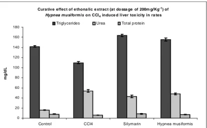
compared to that of the group II CCl4 intoxicated animals. As shown in Figure 2, the biochemical parameters such as serum bilirubin levels were also decreased significantly at a dose level of 200 mg/kg of body weight treated animals (P <0.001),
when compared with the CCl4 intoxicated the group II animals which had the total bilirubin and
urea 14.0 ± 0.26 and 54.0 ± 1.12 mg/dl,
respectively. Figure 1 showed that in the group III, there was a significant increase in the total protein and triglyceride levels in the CCl4 intoxicated and
H. muciformis treated animals (P<0.001),
respectively. Group comparison between H.
muciformis treated (group III) and sylimarin
(group IV) treated animals showed significant
variation in the biochemical parameters, viz.
bilirubin, triglyceride, blood urea, GSH, ALP,
SGOT and SGPT, indicating that H. muciformis
was able to produce 87.5% activity exerted by sylimarin, the positive control in this study.
A significant increase in the serum GOT (325.21±3.41 U/L) and GPT (260.26±3.12 U/L) levels were seen in the group II CCl4 intoxicated animals. These enzymes were reduced to near normal levels such as (178.23±1.32 U/L) and (163.0±1.64 U/L), respectively in the group H. muciformis (50 mg/kg body weight) treated
animals (P < 0.05). Similarly, the elevated ALP
(348.16 ± 3.42 mU/L) and lipid peroxides (2.86 ± 0.24) enzyme levels in the group II CCl4 intoxicated animals were also decreased to 270 ± 1.42 mU/L and 2.06 ± 0.13, respectively in the group III H. muciformis treated rats (Table I).
The LDH (230.0 ± 1.31 U/L) and 5′NT (5.85 ± 0.28 U/L) were also significantly decreased in the group III H. muciformis treated animals when
compared with the group II CCl4 intoxicated animals that showed the elevated levels of LDH (435.38±1.84 U/L) and 5′ NT (9.32±0.20 U/L), respectively (P<0.05). All the parameters were
Table 1 - Hepatoprotective effect of H. muciformis alcoholic extract on biochemical responses of experimental
animals to CCl4.
Parameters Group I control Group II CCl4
Group III
(H. muciformis + CCl4)
Group IV
sylimarin + CCl4
Liver weight (mg/g) SGOT (U/L) SGPT (U/L) ALP (mU/L) LDH (U/L) Lipid peroxides 5′nucleotidase(U/L)
23.14 ± 0.2 134.4 ±1.49
80.2 ± 6.2 262.5 ± 1.40 132.6 ± 0.52 1.42 ± 0.08 6.24 ± 0.26
54.26 ± 0.41 325.21 ± 3.41 260.26 ± 3.12 348.16 ± 3.42
435.38±1.84
2.86 ± 0.24 9.32 ± 0.20
32.64 ± 0.82 178.23 ± 1.32
163.0 ± 1.64 270.0 ± 1.42 230.0 ± 1.31 2.06 ± 0.13 5.85 ± 0.28
28.32 ± 0.1 140.36 ± 1.14
126.2 ± 2.00 226.63 ± 2.10 192.20 ± 1.45 1.83 ± 0.09 6.81 ± 0.13
Values are mean ± SEM; n = 6: a
p< 0.05 compared to control; b
p< 0.05 compared to paracetomol
Each value represents the mean ± SEM. of six treated rats. Values statistically significantly different from those of control group are indicated by
∗(One way Anova, p < 0.05) and ∗∗(p < 0.05). Treatment groups are as follow: No Treatment control (group I); CCl4 (group II); Sylimarin +
CCl4 treatment (group III); H. muciformis + CCl4 (group IV).
Figure 1 - Level of Bilirubin and GSH in ethonalic extract of H.muciformis on CCl4 induced liver
toxicity.
Cura tive effect of e thona lic e xtract (at do ssa ge of 200m g/Kg-1) of Hypnea musiformis o n CCl4 induce d liver tox icity in ra tes
0 20 40 60 80 100 120 140 160 180
Control CCl4 Silymarin Hypnea mus iformis
m
g
/d
L
Triglycerides Urea Total protein
Each value represents the mean ± SEM. of six treated rats. Values statistically significantly different from those of control group are indicated by
∗(One way Anova, p < 0.05) and ∗∗(p < 0.05). Treatment groups are as follow: No Treatment control (group I); CCl4 (group II); Sylimarin +
CCl4 treatment (group III); H. muciformis + CCl4 (group IV).
Figure 2 - Level of Triglyceride, Urea and Total Protein in ethonalic extract of H.muciformis on
DISCUSSION
Carbon tetrachloride is commonly used as a model to study the hepatotoxicity (Chun-Kwan Wong et al. 2004). The CCl4 is bio-transformed by the cytochrome P-450 system in the endoplasmic reticulum to produce trichloromethyl free radical
(•CCl3). Trichloromethyl free radical when
combined with the cellular lipids and proteins in the presence of oxygen form trichloromethyl peroxyl radical, which may attack the lipids on the membrane of endoplasmic reticulum faster than trichloromethyl free radical.Thus, trichloromethyl peroxyl free radical leads to elicit lipid peroxidation, the destruction of Ca2+ homeostasis, and finally, results in cell death (Opoku et al. 2007).
In this present study, the administration of CCl4 decreased the levels of total protein and triglycerides. These parameters were brought back to the normal levels in the group III H. muciformis
treated animals. H. muciformis treatment showed a
protection against the injurious effects of CCL4 that might be due to the interference with cytochrome P-450, resulting in the hindrance of the formation of hepatotoxic free radicals. The site-specific oxidative damage in some susceptible amino acids of proteins is now regarded as the major cause of metabolic dysfunction during the pathogenesis (Uday et al. 1999). The attainment of near normalcy in protein, cholesterol, and triglycerides levels in CCl4 intoxicated and H.
muciformis treated rats confirmed the
hepatoprotective effect of the seaweed extract. The marked elevation of bilirubin and urea level in the serum of group II CCl4 intoxicated rats were
significantly decreased in the group IV H.
muciformis treated animals. Bilirubin is the
conventional indicator of liver diseases (Girish 2004). These biochemical restorations may be due to the inhibitory effects on cytochrome P-450 or/and promotion of its glucuronidation (Cavin et al. 2001).
The health of the liver can assessed by estimating
the activities of serum GOT, GPT, ALP, LDH, 5′
nucleotidase, which are enzymes originally present higher concentration in the cytoplasm. SGPT and SGOT (especially the former) are highly localized in hepatocyte cytosols (Ooi 1996). The crude ethanolic extracts of the seaweed probably acted to preserve the structural integrity of the plasma cellular membrane of the hepatocytes to protect it
against the breakage by the reactive metabolites produced from exposure to CCl4. This prevented further damage to more hepatocytes, and hence reduced further leakage of SGPT and SGOT due to cell destruction. This could explain the lower levels of these transaminases observed in the rats treated with the seaweed extract after exposure to the toxin. The tendency of other marker enzymes
to return towards a near-normalcy in the H.
muciformis treated rats was a clear manifestation
of anti-hepatotoxic effect due to the presence of sulphated polysaccharides and vitamin C in the seaweed extract (Naidoo et al. 2006). The results were comparable to sylimarin. Silymarin is the composite name of three flavonoids isolated from the milk thistle, Sylibum marinum, and is used as
hepatoprotectives against the experimental
hepatotoxicity of various chemicals, including CCl4 (Chhaya and Mishra 1999).
In conclusion, the ethanolic extract of seaweed provided protection from the CCl4 induced liver damage. The protections against the liver damage by the seaweed H. muciformis were comparable to
sylimarin. Possible mechanism that might be responsible for the protection of CCl4 induced liver damage by H. muciformis could involve its
action as a free radical scavenger intercepting those radicals involved in CCl4 metabolism by the microsomal enzymes. By trapping the oxygen related free radicals, the H. muciformis extract
could hinder their interaction with the polyunsaturated fatty acids and would abolish the enhancement of lipid peroxidative processes (Upadhyay et al. 2001). It is well documented that flavonoids and glycosides are strong antioxidants (Natarajan et al. 2006). The antioxidant principles from the natural marine resources are multifaceted in their effects and provide enormous scope in correcting the imbalance through regular intake of a proper diet. Thus, from the results, it could be concluded that H. muciformis could a promising
hepatoprotective agent and this hepatoprotective activity of H. muciformis might be due to its
antioxidant chemicals present in it.
ACKNOWLEDGMENTS
Tamilnadu, India for providing the adequate laboratory facilities in the successful completion of this research work.
REFERENCES
Anggadiredja J, Hasanudin Sidiq AS, Pratomo S, Rudyansyah A. Screening of marine algae from Warambadi seashore Sumba island of Indonesia for antibacterial activity. Phytomedicine.1996; 3: 1-37.
Blunden G. Biologically active compounds from marine organisms. Phytother Res. 2009; 15: 89–94.
Bousquet BF, Julien R, Bon R, Dreux C. Determination of Blood urea. Ann Biol Clin., 1971; 29: 415.
Borzelleca JF, O'Hara TM, Gennings, C, Granger RH, Sheppard MA, Condie LW. Interaction of water contaminants. I.Plasma enzyme activity and response surface methodology following gavage administration of CCL and CHC13 or TCE singly and in combination in the rat. Fundam Appl Toxicol., 1990;
14: 477–490.
Boyd CE, Good year CP. Nutritive quality of food in ecological systems. Arch Hydrobial., 1971; 69: 246–
260.
Boyd EM, Bereckzy GM. Liver necrosis from paracetamol. J Pharmacol, 1996; 26: 606–611.
Cavin C, Mace K, Offord EA, Schilter B Protective effects of coffee diterpenes against aflatoxin B1-induced genotoxicity: mechanisms in rat and human cells. Food Chem Toxicol 2001; 39:549–556
Chhaya Gadgoli, Mishra SH, Antihepatotoxic activity of p-methoxy benzoic acid from Capparis spinosa . J.
Ethnopharmacol. 1999; 66: 187–192
Chun-Kwan W, Vincent EC, Put O, Ang Jr. Hepatoprotective effect of seaweeds’ methanol extract against carbon tetrachloride-induced poisoning in rats. Hydrobiologia., 2004; 512: 267–
270.
Fossati P, Lorenzo P. Estimation of Triglycerides. Clin Chem, 1983; 28: 2077–2080.
Gornall A, Bardawil J, David MM. Determination of Protein by Biuret modified method. Biol Chem.,
1949; 177: 751.
Girish S, achliya, Sudhir, wadodkar G, Avinash, Dorle K, Evaluation of hepatoprotective effect of Amalkadi Ghrita against carbon tetra chloride induced hepatic
damage in rats. J. Ethnopharmacol.2004; 90: 229-232.
Harris RN, Ratnayake V, Garry F, Anders MW, Interactive hepatotoxicity of CHCl3 and CCl4.
Toxicol. Appl. Pharmacol.1982; 63: 281–291.
Indegaard M. Ostgaard K. Polysaccharide for food and pharmaceutical uses in: Seaweed resources in Europe: Uses and potential Wiley, 1991; 169–183.
Klaassen CD, Plaa GL. Comparison of the biochemical alterations elicited in livers from rats treated with CCl4, CHCl3, 1,1,2-trichloroethane and 1,1,1-trichloroethane. Biochem. Pharmacol. 1969; 18: 2019–2027.
Kind PN, King EJ, Invitro determination of serum alkaline phosphatase. J Clin Path., 1971; 7: 322–336.
King J. Practical clinical enzymology. Van Nostrand Co., London. 1969.
Konig GM, Wright AD. Marine natural products research: Current Directions and future potential.
Planta Medica., 1996; 62: 193–211.
Lahaye M, Kaffer B. Seaweed dietary fibers structure physiochemical and Biological properties relevant to intestinal physiology. Sci Aliments, 1997; 17; 563–
564.
Luly P, Branahel O, Tria E. Determination of 5’ Nucleotidase kinectic method. Biochem Biophys Acta, 1972; 283, 447.
Malloy HT, Evelyn KA. The determination of bilirubin with the photoelectric colorimeter. J. Biol. Chem.
1937; 119: 481.
Matsukawa R, A comparison of screening methods for antioxidant activity in seaweeds. J Appl Phycol.,
1997;9: 29–35.
Naidoo K, Naidoo G, Maneveldt K, Ruck K, Bolton JJ. A comparison of various seaweed-based diets and formulated feed on growth rate of abalone in a land-based aquaculture system. Journal of Applied Phycology, 2006; 18: 437–443.
Natarajan, Kavithalakshmi, Madhusudhanan, Narasimhan, Radha Shanmugasundaram, Shanmugasundaram ERB, Antioxidant activity of a salt–spice–herbal mixture against free radical induction. J. Ethnopharmacol.2006; 105: 76–83. Ohkawa Η, Ohishi Ν, Yagi, K, Anal Biochem., 1979;
95: 351.
Ooi VEC. Hepatoprotective effect of some edible mushrooms. Phytotherapy Res. 1996; 10: 536–538. Opoku AR, Ndlovu IM, Terblanche SE, Hutchings AH.
In vivo hepatoprotective effects of Rhoicissus tridentata sub sp. cuneifolia, a traditional Zulu medicinal plant, against CCl4-induced acute liver injury in rats. Sou Afr J Bot., 2007;73: 372–377.
Recknagel R, Pharmacol. Rev. 1967; 19: 145.
Reitman S, Frankel S. Invitro determination of transaminase activity in serum. Am J Clin Path.,
1975; 28: 56–58.
Smiddsrod R, Christensen BE. Molecular structure and physical behaviour of seaweed colloids as compared with microbial polysaccharide in sea weeds resources in Europe uses and potential Wiley, 1991; 185–215. Snedecor GW, Cochran WG. Statistical methods. Vol.
33, IBH Publishing Co., New Delhi 1967.
Siva Kumar K. Seaweed utilization a review. In-
Phycology, principles, processes and applications,
ed. A. S. Ahluwalia. Daya publishing house, New Delhi, 2000; 385–400.
Upadhyay RK, Pandey MB, Jha RN, Pandey VB. Eclalbatin, triterpine saponins from Eclipta alba. J.
Asian Nat. Prod. Res.2001; 3: 213– 217.
Uday B, Dipak D, Banerji ranjit K. Reactive oxygen species: Oxidative damage and pathogenesis. Cur Sci., 1997; 5: 658.
Yan X, Nagata T, Fan X. Antioxidant activities in some common sea weeds. Plant Foods Human Nutr, 1998;
52: 253–262.