Article
Printed in Brazil - ©2017 Sociedade Brasileira de Química0103 - 5053 $6.00+0.00
*e-mail: [email protected]
Warmingiins A and B, Two New Dimeric Naphthoquinone Derivatives from
Sinningia warmingii
(Gesneriaceae)
Vanessa Winiewski,a Maria Helena Verdan,a Marcos A. Ribeiro,b Alvaro J. Hernandez-Tasco,c Marcos J. Salvadorc and Maria Elida A. Stefanello*,a
aDepartamento de Química, Universidade Federal do Paraná (UFPR),
81530-900 Curitiba-PR, Brazil
bLaboratório de Química de Coordenação, Instituto de Química and cDepartamento de Biologia
Vegetal, Instituto de Biologia, Universidade Estadual de Campinas (UNICAMP), 13083-970 Campinas-SP, Brazil
Chemical investigation of Sinningia warmingii (Gesneriaceae) tubers lead to the isolation of
two new dimeric naphthoquinone derivatives, named warmingiins A and B, besides six known compounds, aggregatin E, aggregatin F, tectoquinone, halleridone, cleroindicin B, and cornoside. All compounds were identified by spectroscopic analysis, mainly nuclear magnetic resonance (NMR) and mass spectrometry (MS), and comparison with the literature. The structure of the warmingiin A, which is an artifact of warmingiin B, was confirmed by X-ray diffraction analysis. Antimicrobial activity of the ethanolic extract and fractions of S. warmingii was evaluated
against Staphylococcus aureus, S. epidermidis, Escherichia coli, Pseudomonas aeruginosa, Candida albicans, C. dubliniensis, C. glabrata and C. parapsilosis. All samples were
inactive.
Keywords: Gesneriaceae, Sinningia warmingii, naphthoquinone derivative, antimicrobial
evaluation
Introduction
Sinningiawarmingii (Hiern.) Chautems (Gesneriaceae)
is a rupicolous or terrestrial herb, with erect stems reaching until 120 cm and perennial tubers. It is native to South America where is distributed over several countries. In Brazil this plant occurs in Midwest, Southeast and South regions.1,2 In Peruvian Amazon S. warmingii is known as
“mother potato”, and an infusion of its tubers is used to treat gynecological problems, such as puerperal inflammations and microbial vaginosis.3 A preliminary phytochemical
screening of the tubers detected phenolic glycosides, terpenes, and steroids,3 classes of compounds that are
widespread in Gesneriaceae family.4
Recently we reported the isolation of eight known compounds from S. warmingii, which were identified
as lapachenole, tectoquinone, 7-methoxytectoquinone, 1-hydroxytectoquinone, 7-hydroxytectoquinone, aggregatin C, aggregatin D and halleridone.5 In continuing
the phytochemical investigation of S. warmingii tubers, we
report now the isolation of two new dimeric naphthoquinone derivatives, warmingiin A (1) and B (2), together with six
known compounds identified by comparison with literature as aggregatin E (3),6 aggregatin F (4),6 tectoquinone (5),7
halleridone (6),8 cleroindicin B (7),9 and cornoside (8).10
Compounds 3, 4, 7 and 8 arebeing reported for the first
time in this species (Figure 1). In addition, antimicrobial activity of the ethanolic extract and fractions obtained by partition with solvents was evaluated.
Experimental
General experimental procedures
Optical rotations were measured in CHCl3 on a Rudolph
(AC 200, Avance 400 and/or Avance 600), observing 1H at
200, 400 or 600 MHz and 13C at 50, 100 or 150 MHz. The
solvent used was CDCl3 and the chemical shifts are given
in ppm (d), using TMS as internal reference, with coupling constants (J) in Hz. High-resolution electrospray ionization
mass spectrometry (HRESIMS) data were obtained on a Micromass ESI-Qq Tof mass spectrometer. High-performance liquid chromatography (HPLC) separations were performed in a Waters apparatus equipped with PDA detector and a semi-preparative Nucleosil 100-5 C18
column (250 × 4.6 mm). Silica gel (Merck, 230-400 mesh) was used for column chromatographic separations, while precoated silica gel plates (Macherey-Nagel) were used for thin-layer chromatography (TLC) and preparative TLC (PTLC). Compounds were visualized by exposure under UV254/366 light and spraying with 5% (v/v) H2SO4 in ethanol
solution, followed by heating on a hot plate. X-ray data were collected on a Bruker D8 Venture Photon 100 by using graphite-monochromated Cu Kα radiation (λ = 1.5418 Å) at 100(2) K. Accurate unit cell dimensions and orientation matrices were determined by least squares refinement of the reflections obtained by θ-χ scans. The data were indexed and scaled with the ApexII Suite.11 Bruker Saint
and Bruker Sadabs were used to integrate and scaling of data, respectively. The structure was solved with Olex2,12
using the ShelXT13 structure solution program using Direct
Methods and refined with the ShelXL14 by a full-matrix
least-squares technique on F2. All non-hydrogen atoms
were refined anisotropically. The hydrogen atoms in the compound were added to the structure in idealized positions and further refined according to the riding model; Olex2 was also used for the drawing of molecular graphics and the preparation of figures for publication.
Plant Material
Tubers of Sinningia warmingii (Hiern.) Chautems were
collected from plants growing wild in Londrina City, Paraná
State, Brazil (23°25’32”S, 50°24’50”W) in May 2012. The plant was identified by Clarice B. Poliquesi, who deposited a voucher specimen in the herbarium of Museu Botânico Municipal (MBM 12804).
Extraction and isolation
Dried and powdered tubers (61.3 g) were extracted with ethanol 95% (300 mL for 24 h, three times) at room temperature. The solvent was removed at reduced pressure using a rotaevaporator. The resulting extract (2.82 g) was dissolved in EtOH:H2O 1:1 (150 mL) and subjected to
partition with hexanes (mixture of isomers), EtOAc and 1-butanol (3 × 50 mL, each solvent), successively, giving the respective fractions (A-C) after solvent removal.
Fraction A (0.59 g) was submitted to CC (hexanes:acetone,
9:1 → 0:1, and MeOH) yielding 43 fractions (15 mL
each). After TLC analysis were obtained eleven fractions (A1-A11). Fraction A4 (15.6 mg, eluted with hexanes:acetone
8:2) was purified by HPLC (H2O:MeCN 20:80) to give
3 (Rt (retention time) = 7.83 min, 1.6 mg), 4 (Rt = 8.54 min,
1.5 mg), and 5 (Rt = 10.76 min, 0.4 mg). A8 (48.4 mg,
eluted with hexanes:acetone 3:2) yielded 1 (6.3 mg) and 1 + 2 (4.6 mg) after PTLC in CH2Cl2:EtOAc 95:5. The
mixture 1 + 2 was purified by HPLC (MeCN 100%) to
give 1 (Rt = 3.31 min, 2.7 mg). A10 (13.0 mg, eluted with
hexanes:acetone 3:7) was purified by HPLC (H2O:MeOH
62:38) to give 7 (Rt = 7.59 min, 7.1 mg).
Fraction B (0.298 g) was analyzed by 1H NMR revealing
almost pure 6.
Fraction C (0.198 g) was submitted to CC
(EtOAc:MeOH:H2O 8:1:0.25 → 3:6:0.25 and MeOH)
yielding 36 fractions (15 mL each). After TLC analysis were obtained nine fractions (C1-C9). Fraction C4 (14.2 mg,
eluted in EtOAc:MeOH:H2O 8:1:0.25) was purified by
PTLC in EtOAc:MeOH:H2O 8:1:0.25 to give 7 (1.4 mg).
Fraction C8 (23.5 mg, eluted in EtOAc:MeOH:H2O
6:3:0.25) yielded cornoside14 (8, 3.8 mg) after PTLC in
EtOAc:MeOH:H2O 6:3:0.25.
Antimicrobial assays
The samples were evaluated for antimicrobial activity applying the microdilution method (96-well plates), as previously described,15 to give concentrations between 10 to
500 µg mL-1. The minimal inhibitory concentration (MIC)
v/v) served as the negative control. Each sensitivity test was performed in duplicate for each microorganism evaluated and repeated three times. The strains of bacteria and fungi tested were Escherichia coli ATCC 10799, Pseudomonas aeruginosa ATCC 27853,Staphylococcus aureus ATCC
14458, Staphylococcus epidermidis ATCC 12228,
Candida albicans ATCC 10231, Candida glabrata
ATCC 30070, Candida dubliniensis ATCC 778157 and
Candida parapsilosis ATCC 22019.
Warmingiin A (1)
Colorless solid; [α]D20 + 50.0 (c 0.003, MeOH); UV-Vis
(MeOH) λmax / nm (log ε) 206 (4.0), 218 (3.9), 269 (3.5);
CD (c 0.01, MeOH) λmax (θ) 251 (−15.3), 300 (+12.6),
366 (+4.5); 1H and 13C NMR data, see Table 1; HRESIMS m/z, observed:501.1308; C30H22O6Na [M + Na]+ requires:
501.1314. Crystallographic data: compound 1 was
crystallized from acetonitrile by slow solvent evaporation. A colorless block crystal was selected for crystallographic measures. For the supplementary crystallographic data, see Supplementary Information section.
Warmingiin B (2)
Colorless solid; 1H and 13C NMR data, see Table 1;
HRESIMS m/z, observed:513.15641; C30H25O8 [M − H]−
requires: 513.15549.
Results and Discussion
Compound 1, named warmingiin A (Figure 1), was
isolated as a colorless solid. Its HRESIMS in positive mode displays an ion at m/z 501.1308 [M + Na]+, indicating
the molecular formula C30H22O6, which is consistent with
twenty indices of hydrogen deficiency. In the 1H NMR data,
signals of four aromatic protons, in a spin system typical of an aromatic ring 1,2-dissubstituted (dH 7.54-8.10), two
doublets of olefinic protons (dH 6.35 and 6.45, J 9.7 Hz),
two doublets of oxymethylene protons (dH 3.86 and 3.99, J 6.5 Hz), and one singlet of a methyl group (dH 1.71) were
observed (Table 1). These data were very similar to those of aggregatin E (3), previously reported for S. aggregata6
and also isolated here, but differing mainly for the absence of the signal of H-6. Analysis of heteronuclear single quantum correlation (HSQC) and heteronuclear multiple bond correlation (HMBC) showed 15 different carbons, including a carbonyl group (dC 182.7), two oxygenated
carbons (dC 74.4 and 80.6) and a dioxygenated carbon
(dC 99.5). Considering the molecular formula, these data
indicate a symmetrical dimeric compound. The correlations observed in the HSQC and HMBC spectra (Table 1) are in accordance with structure 1. In particular, the position of
the linkage between the two moieties was determined by cross-peaks among H-5 (dH 6.35) and carbons at dC 80.6
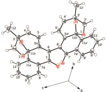
(C-3), 99.5 (C-11b), 126.4 (C-6) and 147.4 (C-5a). Analysis by X-ray diffraction of a single crystal of 1 confirmed the
structure and led to assign its absolute configuration as 3R,
3’R, 11bS, 11’bS (Figure 2). The CD of 1 was very similar
to that reported for aggregatins E (3) and F (4),6 indicating
the same absolute configuration (Figure S5).
Compound 2, named warmingiin B (Figure 1), was
isolated as a colorless solid in admixture with 1. Its
1H NMR spectra showed signals in duplicate, with different
intensities. The less intense signals were identical to those of 1, while the more intense ones were attributed to
compound 2. These signals indicate that 2 has the same
groups as in 1 (Table 1). The main difference was observed
for the chemical shifts of H-4/H-4’ and H-5/H5’, which had higher chemical shifts in 1 than in 2. From integrals
of H-4 and H-5, the relative proportion of 2 and 1 was
determined as 1:0.7. The HRESIMS spectra in negative mode showed peaks at m/z 477.13517 ([M − H]−, relative to compound 1) and 513.15641 ([M − H]−, relative to compound 2, molecular formula C30H25O8). All efforts to
separate this mixture failed, resulting only in the isolation of 1. After six weeks, a new record of 1H NMR spectra
showed that the proportion of 2 in the mixture changed
from around 60 to 30%. These observations led to the conclusion that 1 was an artifact, formed from 2 by loss of
two unities of water during the isolation process. Despite the impossibility of getting compound 2 pure, its chemical
shifts could be assigned by careful analysis of HSQC and HMBC data (Table 1). The absolute configuration of 2 was
Figure 2. View of assymetric part of unit cell of compound 1. Atomic
assigned as 3R, 3’R, 11bR, 11’bR by comparison with 1,
considering a change in the priority order of the groups around C-11b.
Considering the use of S. warmingii in folk
medicine,3 ethanolic extract and the fractions obtained
by partition were tested for antimicrobial activity against
Staphylococcus aureus, S. epidermidis, Escherichia coli, P s e u d o m o n a s a e r u g i n o s a, C a n d i d a a l b i c a n s, C. dubliniensis, C. glabrata and C. parapsilosis, using
the methodology previously reported.15 All samples were
inactive (MIC > 500 µg mL-1).
Conclusions
The new compound 1 seems derivate from 2 by
acid-catalyzed dehydration during isolation process. This reaction is easy, considering the proximity among the hydroxy groups in 2. Compound 2 is structurally
formed by coupling of two unities of aggregatin E (3),
a hemiketal with a rare carbon framework. To date three compounds of this type have been reported in the literature, namely aggregatins D-F. All have been isolated for the first time from S. aggregata,6,16 and now were found in S. warmingii. Aggregatin E also was recently reported in S. allagophylla,17 suggesting a close relationship among
these three species. Our results of antimicrobial activity did not support the traditional use of S. warmingii in infectious
diseases. However, this assessment can not be considered
final, because chemical composition of this plant may vary along its dispersion area.
Supplementary Information
Supplementary information (1D and 2D NMR, HRESIMS and CD data) is available free of charge at http://jbcs.sbq.org.br.
Supplementary crystallographic data are deposited at CCDC 1439549. These data can be obtained free of charge from the Cambridge Crystallographic Data Centre (www.ccdc.cam.ac.uk/data_request/cif).
Acknowledgments
We are grateful to CAPES, FAEPEX-UNICAMP, FAPESP and CNPq for scholarships and financial support, CNPq by authorization for access of samples from the Brazilian genetic heritage (process 010087/2012-5), Clarisse B. Poliquesi, at Museu Botânico Municipal de Curitiba, for collection and identification of the plant, and A. A. Stefanello for English revision.
References
1. Ferreira, G. E.; Chautems, A.; Waechter, J. L.; Acta Bot. Bras.
2015, 29, 310.
2. Araujo, A. O.; Chautems, A.; Lista de Espécies da Flora do
Table 1. NMR data (600 MHz, CDCl3) for the compounds 1 and 2
Position 1 2 HMBC
dH (J in Hz) dC dH (J in Hz) dC
2, 2’ 3.86 (1H, d, 6.5)
3.99 (1H, d, 6.5)
74.4 3.86 (1H, d, 6.6)
4.01 (1H, d, 6.6)
74.2 4, 12
3, 3’ 80.6 80.6
4, 4’ 6.45 (1H, d, 9.7) 141.4 6.39 (1H, d, 9,7) 140.8 2, 5a, 12
5, 5’ 6.35 (1H, d, 9.7) 124.2 6.20 (1H, d, 9.7) 124.6 3, 6, 12
5a, 5a’ 147.4 147.2
6, 6’ 126.4 125.8
7, 7’ 182.7 182.2
7a, 7a’ 131.3 131.2
8, 8’ 8.10 (1H, ddd, 7.8, 1.3, 0.8) 126.7 8.13 (1H, ddd, 7.8, 1.2, 0.5) 126.6 7, 10, 11a
9, 9’ 7.54 (1H, ddd, 7.8, 7.6, 1.3) 130.0 7.55 (1H, ddd, 7.8, 7.6, 1.3) 129.8 7a,11
10, 10’ 7.67 (1H, ddd, 7.8, 7.6, 1.3) 133.1 7.68 (1H, ddd, 7.8, 7.6, 1.2) 132.9 8, 11a
11, 11’ 7.81 (1H, ddd, 7.8, 1.3, 0.8) 126.0 7.82 (1H, ddd, 7.8, 1.3, 0.5) 125.8 7a, 9, 12
11a, 11a’ 137.4 137.2
11b, 11b’ 99.5 99.2
Brasil; Instituto de Pesquisas Jardim Botânico do Rio de Janeiro: Rio de Janeiro. Available at http://floradobrasil.jbrj. gov.br/jabot/floradobrasil/FB7935, accessed in April, 2016. 3. Casana, C. F. D.; Cruz, P. L. B.; Cruz, K. L. B.; Teixeira, B. J.;
Medina, M. D. P.; Effio, P. J. C.; Pueblo Cont. 2012, 23, 345. 4. Verdan, M. H.; Stefanello, M. E. A.; Chem. Biodiversity 2012,
9, 2701.
5. Verdan, M. H.; Ehrenfried, C. A.; Scharf, D. R.; Cervi, A. C.; Salvador, M. J.; Barison, A.; Stefanello, M. E. A.; Nat. Prod. Commun. 2014, 9, 1535.
6. Verdan, M. H.; Souza, L. M.; Carvalho, J. E.; Costa, D. B. V.; Salvador, M. J.; Barison, A.; Stefanello, M. E. A.; Chem. Biodiversity 2015, 12, 148.
7. Moreira, R. Y.; Arruda, M. S.; Arruda, A. C.; Santos, L. S.; Müller, A. H.; Guilhon, G. M. S. P.; Santos, A. S.; Terezo, E.; Rev. Bras. Farmacogn. 2006, 16, 392.
8. Messana, I.; Sperandei, M.; Multari, G.; Galeffi, C.; Marini-Bettolo, G. B.; Phytochemistry 1984, 23, 2617.
9. Tian, J.; Zhao, Q.; Zhang, H.; Lin, Z.; Sun, H.; J. Nat. Prod.
1997, 60, 766.
10. Yamamoto, H.; Hori, M.; Kuwajima, H.; Inoue, K.; Planta
2003, 216, 432.
11. APEX2 Version 2014/1-1, Bruker AXS Inc.: Madison, WI, USA, 2014; SAINT Version 8.34A, Bruker AXS Inc.: Madison, WI, USA, 2013.
12. Dolomanov, O. V.; Bourhis, L. J.; Gildea, R. J.; Howard, J. A. K.; Puschmann, H.; J. Appl. Cryst. 2009, 42, 339.
13. Sheldrick, G. M.; Acta Cryst. 2015, A71, 3.
14. Sheldrick, G. M.; Acta Cryst. 2008, A64, 112.
15. Salvador, M. J.; Ferreira, E. O.; Pral, E. M. F.; Alfieri, S. C.; Albuquerque, S.; Ito, I. Y.; Dias, D. A.; Phytomedicine 2002, 9, 566.
16. Verdan, M. H.; Barison, A.; Sá, E. L.; Salvador, M. J.; Poliquesi, C. B.; Eberlin, M. N.; Stefanello, M. E. A.; J. Nat. Prod. 2010,
73, 1434.
17. Scharf, D. R.; Verdan, M. H.; Ribeiro, M. A.; Simionatto, E. L.; Sá, E. L.; Salvador, M. J.; Barison, A.; Stefanello, M. E. A.; J. Nat. Prod. 2016, 79, 792.
Submitted: April 12, 2016
Published online: July 8, 2016