Vol.56, n.6: pp. 980-984, November-December 2013
ISSN 1516-8913 Printed in Brazil BRAZILIAN ARCHIVES OF
BIOLOGY AND TECHNOLOGY
A N I N T E R N A T I O N A L J O U R N A L
Polymerase Chain Reaction (PCR) Identification of
Terverticillate
Penicillium
Species
Isolated
from
Agricultural Soils in Eski
ş
ehir Province
Rasime Demirel
1*, Nalan Yilmaz Sariozlu
1and Semra
İ
lhan
21
Department of Biology; Faculty of Science; Anadolu University; Eskişehir-Turkey. 2Department of Biology; Faculty of Science and Art;Eskişehir Osmangazi University; Eskişehir-Turkey
ABSTRACT
In the present study, nine terverticillate Penicillium isolates (P. griseofulfum, P. puberulum, P. crustosum, P. aurantiogriseum, P. chrysogenum, P. primulinum, P. expansum, P. viridicatum, Eupenicillium egyptiacum) from 56 soil samples were characterized genetically by a PCR method. The DNAs of the strains were isolated using the glass beads and vortexing extraction method and then used for PCR amplification with the internal transcribed spacer 1 (ITS1) and ITS4 universal fungal specific primers. The ITS regions of fungal ribosomal DNA (rDNA) were sequenced through the CEQ 8000 Genetic Analysis System. ITS-5.8S sequences obtained were compared with those deposited in the GenBank Database. The results indicated that the identification of Penicillium species with PCR based methods provided significant information about the solution to taxonomy and improve food safety and to protect the users from harmful contaminants such as mycotoxins, which must be controlled during the production of agricultural materials as well as during the processing of food and feed.
Key words:Penicillium, identification methods, PCR, ITS
*Author for correspondence: rasime.demirel@gmail.com
INTRODUCTION
Penicillium is a genus of Hyphomycetes,
characterized by the production of conidia in chains from the verticils of the phialides. The most complex penicillus characteristically has three branch points. These penicillus types are defined as terverticillate (Pitt 2000). This group includes important food and feed spoilage moulds, pathogens of mature fruits and cereal grains. In addition to causing deterioration and quality loss, these moulds may contaminate agricultural products with potential mycotoxins (Logrieco et al. 1990).
The taxonomy of terverticillate penicillia has been considerably studied (Pitt 1979). However, there
moulds is mainly performed by phenotypic criteria, including micro- and macro-morphology and requires considerable expertise. This procedure may take several days and is dependent on the growth and sporulation, as well as on individual interpretation. Even then, related species may be confused and rare species may remain unidentified (Ciardo et al. 2007). Though many biochemical and physiological techniques have provided information useful for the classification, the nucleotide sequence and the organization of DNA are the most likely to give a clear and sensitive discrimination between the organisms and also to indicate clearly their evolutionary and phylogenetic relationship (Croft et al. 1990).
Comparative studies of the nucleotide sequences of ribosomal RNA (rRNA) genes provide a means for analyzing the phylogenetic relationships over a wide range of taxonomic levels (White et al. 1990). The small subunit rRNA gene has been used extensively for phylogenetic analysis, and for the identification purposes at genus, or species level in bacteria and eukaryotes. The ITS region of the rRNA operon is located between the 18S and 28S rRNA genes and includes the two intervening regions, ITS1 and ITS2, and the highly conserved 5.8S rRNA gene (White et al. 1990). This work aimed to study the use of ITS sequences for the identification of the closely related terverticillate
Penicillium species isolated from agricultural soils
in Eskişehir province. In addition to molecular characterization of Penicillium species, the study also aimed to study the potential of Penicillium
species as a risk factor because of their mycotoxin, spoilage and pathogenic properties in agricultural soils in Eskişehir province.
MATERIALS AND METHODS
Fungal strains
All terverticillate Penicillium strains (Table 1) used in this study were obtained from the agricultural soils in the Eskişehir province and identified using the traditional methods according to Pitt (1979) and Demirel et al. (2005). All the strains were stored in suitable conditions at the Culture Collections of KUKENS (WDCM101), the Centre for Research and Application of Culture Collections of Microorganisms. Cultures were maintained at 4ºC on potato dextrose agar (PDA).
Table 1 - List of the terverticillate Penicillium species in this study.
Species Isolate number
Penicillium griseofulvum Dierckx T2
Penicillium puberulum Bainier T3
Penicillium crustosum Thom T4
Penicillium aurantiogriseum Dierckx T5
Penicillium chrysogenum Thom T7
Penicillium primulinum Pitt T8
Eupenicillium egyptiacum (J.F.H. Beyma) Stolk & D.B. Scott
T9
Penicillium expansum Link T10
Penicillium viridicatum Westling T11
DNA extraction
Genomic DNA was isolated from the cultures grown on PDA for seven days. About 50-100 mg of mycelia were scraped from the agar surface and placed in a 1.5 mL micro-centrifuge tube containing 400 µL lysis buffer (2% Tripton X-100, 1% SDS, 100 mM NaCl, 10 mM Tris pH 8.0, 1 mM EDTA). Lysis of the was achieved by the addition of 500 mg acid-washed 0.4-0.6 mm diameter glass beads, 400 µL
phenol/chloroform/iso amyl alcohol
(Phe/Chl/IAA) (25:24:1), and continuous vortexing for 30 min at the highest intensity setting (Van Burik et al. 1998) using a vortex (IKA Vortex GENIUS 3). The sample was then centrifuged at 13,000 rpm for 5 min. The aqueous (top) layer was transferred into a new tube and re-extracted with an equal volume of Phe/Chl/IAA (25:24:1) two times. At the re-extraction stage, following the addition of Phe/Chl/IAA (25:24:1), the sample was gently mixed for 5 min at mixer (Multi KS-60 BIOSAN Programmable Rotator-mixer 90º), and then centrifuged at 13,000 rpm for 5 min. The top aqueous layer was transferred to a clean 1.5 mL micro-centrifuge tube and the DNA was precipitated with the addition of 0.1 volume 3 M sodium acetate, followed by two volumes cold 98% ethanol. After incubation at – 20ºC for 20-30 min, the DNA was pelleted by centrifugation at 13,000 rpm for 5 min. The ethanol was decanted and the pellet was re-suspended in 200 µL distillated water. DNA was precipitated by the addition of 0.1 volume 0.3 M sodium acetate, followed by two volume cold 98% ethanol. After incubation at – 20ºC for 20-30 min, the DNA was pelleted by centrifugation at 13,000 rpm for 10 min, the ethanol was decanted, and the pellet was air dried in a laminar flow hood. DNA pellet was re-suspended in 50 µL T10E1 (10 mM Tris-HCl, 1
estimated visually in 1% agarose gels containing 5 µg/mL ethidium bromides by comparing band intensity with known quantities of DNA high range markers.
Polymerase chain reaction (PCR)
The nuclear ribosomal ITS1-5.8S-ITS2 region was amplified with the primers ITS1 (5´-TCCGTAGGTGAACCTGCGG-3´) and ITS4 (5´-
TCCTCCGCTTATTGATATGC-3´). Reactions
were performed in 25 µL volumes containing 1 µL genomic DNA, 2.5 µL 2.5 µM ITS1, 2.5 µL 2.5 µM ITS4, 2.5 µL 10X Taq buffer +KCl – MgCl2
(Fermentas), 2.5 µL 25 mM MgCl (Fermentas), 2 µl 2.5 mM dNTPmix, 0.25 µL 5 U/ µL Taq DNA polymerase (Fermentas), and 11.75 µL sterile deionized water. DNA amplification was performed in a thermocycler with an initial denaturation step for 2 min at 95ºC, followed by 35 cycles of denaturation for 1 min at 95ºC, anneling for 45 sec at 55ºC, and extension for 1 min 30 sec at 72ºC. A final extension at 72ºC was performed for 5 min. To confirm the amplification of solely the ITS, 5 µL of PCR product together with marker (GeneRuler DNA Lader 50 bp Fermentas) was resolved by gel electrophoresis on 1% agarose gel containing 5 µg/mL ethidium bromide in 1X TAE buffer. The gel samples were photographed in the Gel Documentation System (Uvitec M02 4611).
Sequencing
Template DNA for sequencing was prepared as follows: agarose gel blocks containing DNA fragment were cut out and purified with Promega Wizard® SV Gel and the PCR Clean-Up System. PCR products for sequencing were obtained with the GenomeLab DTCS Quick Start Kit and sequences were determined with the primers ITS1 and ITS4. Cycle sequencing products were purified with Dynabeads® Sequencing Clean-Up to remove unincorporated dye-labeled nucleotides. Direct sequencing was performed with Beckman Coulter CEQ 8000 Genetic Analysis System.
Phylogenetic analysis
The sequences were blasted with the Genbank sequences to ensure the identifications; the closest Blast results are reported below each taxon (Table 2). New sequences from these groups were aligned along with the similar GenBank sequences in software ClustalW in the MEGA5 programme (Tamura et al. 2011). Ambiguously aligned regions and sequence ends were manually excluded. The Tajima-Nei model in Neighbor-joining method in MEGA5 programme was used for the analysis of phylogenetic relationship with 1000 bootstrap and Penicillium aurantiogriseum
(DQ249210) was used as an outgroup in the ITS analysis.
Table 2 - Newly generated ITS sequences with their closest GenBank sequences (according to Blast searches).
Species Collection accession number GenBank Closest Blast hit (% identity/% coverage)
Penicillium griseofulvum Dierckx T2 KF293650 Penicillium griseofulvum JX091409 (99/100)
Penicillium puberulum Bainier T3 KF293652 Penicillium tricolor JN942703 (99/97)
Penicillium crustosum Thom T4 KF293653 Penicillium chrysogenum KC492574 (99/96)
Penicillium aurantiogriseum
Dierckx
T5 KF293654 Penicillium chrysogenum KC492574 (99/96)
Penicillium chrysogenum Thom T7 KF293651 Penicillium sp. JX982473 (98/96)
Penicillium primulinum Pitt T9 KF293655 Penicillium sp. JX997060.1 (99/96)
Eupenicillium egyptiacum (J.F.H. Beyma) Stolk & D.B. Scott
T8 KF293657 Penicillium chrysogenum JQ724529 (94/96)
Penicillium expansum Link T10 KF293658 Penicillium brevicompactum JQ781814 (82/76)
Penicillium viridicatum Westling T11 KF293656 Penicillium polonicum KC692223 (99/96)
RESULTS AND DISCUSSION
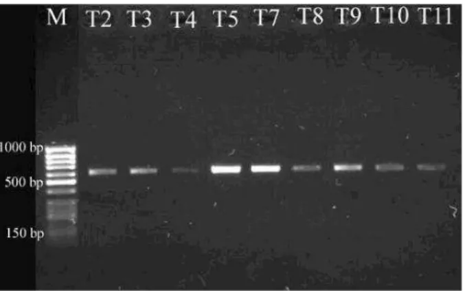
Using the universal fungal primers (ITS1/ITS4), PCR products (570 bp) were obtained from all of the species. Figure 1 showed that the sizes obtained for the full ITS region amplified different terverticillate Penicillium species.
Figure 1 - Full ITS PCR products amplified from different terverticillate Penicillium species with ITS1/ITS4 primers. M, molecular-weight markers (50 bp GeneRuler DNA Lader, Fermentas).
The rDNA genes, generally used in the taxonomic and identification studies, were confirmed in the present study. ITS primers 1 and 4 were used in this study to amplify the entire 5.8S rDNA gene, both ITS1 and ITS2. The most similar ITS sequences up to 82% similarity values were
determined by BLAST N comparison of ITS to other ITS sequences from penicillia in the GenBank database. In this comparison, the T2 strain that was identified as P. griseofulvum by the traditional identification methods showed a similarity to P. griseofulvum strains with 99% maximum identification. The T3 strain identified
as P. puberulum showed a similarity to P. tricolor
strains with a 99% maximum identification with P. tricolor. Thus, useful results were obtained about this strain with sequences of the ITS region. The T4, T5 and T8 strains identified as P. crustosum,
P. aurantiogriseum and Eupenicillium egyptiacum,
respectively had a 94-99% similarity to P.
chrysogenum. The T9 strain identified as P.
primulinum had 99% similarity to Penicillium sp. strains. The T7, T10 and T11 strains identified as
P. chrysogenum, P. expansum and P. viridicatum
had 98, 82 and 99% similarities to Penicillium sp.,
P. brevicompactum and P. polonicum,
respectively.
T8 Penicillium chrysogenum Penicillium chrysogenum JQ724529 T5 Penicillium chrysogenum
Penicillium sp. 2 JX982473 T9 Penicillium sp.
T4 Penicillium chrysogenum T7 Penicillium sp.
Penicillium chrysogenum KC492574 Penicillium sp. JX997060
T2 Penicillium griseofulvum Penicillium griseofulvum JX091409
T11 Penicillium polonicum T3 Penicillium tricolor Penicillium tricolor JN942703 Penicillium polonicum KC692223
Penicillium brevicompactum JQ781814
T10 Penicillium brevicompactum Penicillium aurantiogriseum DQ249210 (Outgroup)
64
36
33 46 75
53 20
55
64
0.01
Figure 2 - Best-scoring Neighbour joining tree calculated using MEGA5.0 based on ITS sequences showing the relationships of the newly generated sequences in this study with previously known taxa in NCBI GenBank. The tree is rooted with Penicillium aurantiogriseum
Phylogenetic tree analyses showed that the studied terverticillate penicillia were prominently divided to four clades. One of them was included as P.
chrysogenum, Penicillium species and closely
related isolates as T4, 5, 7, 8 and 9. Furthermore, T5 and T8 were distinctly differentiated from other clade members in the phylogenetic tree. The isolate T2 and P. griseofulvum occurred as another clade in phylogenetic tree. This species has been distinguished from P. chrysogenum with short phialides and strongly divergent branching. P. tricolor and P. polonicum were clustered into the same clade; besides, these species were member of
Penicillium section Viridicata series Viridicata. The last clade belonged to P. brevicompactum. This species was distinguished from others with appressed all elements.
The studied terverticillate penicillia species had some distinguishable characters but the discrimination of these characters was very difficult by microscopic investigation. On the other hand, PCR identification techniques provided highly useful information about the molecular identification and distinguishing of fungi, especially closely related fungi.
CONCLUSIONS
This study resulted reliable information using universal fungal primers. In addition, information regarding the closely related terverticillate
Penicillium species were clarified. Through the
use of molecular techniques correct descriptions and individual selections were achieved.
ACKNOWLEDGEMENTS
We would like to thank Prof. Dr. Kiymet Guven, Assoc. Prof. Dr. M. Burcin Mutlu, Asst. Prof. Dr. Muhittin Arslanyolu for providing chemical materials and Asst. Prof. Dr. Cem Ozic for his help in DNA sequencing.
REFERENCES
Bridge PD, Hawsksworth DL, Kozakiewicz Z, Onions AHS, Paterson RRM, Sackin MJ, et al. A reappraisal of the terverticillate Penicillia using biochemical, physiological and morphological features. In: Samson RA, Pitt JI, editors. Modern Concepts in Penicillium
and Aspergillus Classification. New York and
London: Plenum Press; 1990. p. 139-147.
Ciardo DE, Schär G, Altwegg M, Böttger EC, Bosshard PP. Identification of moulds in the diagnostic laboratory—an algorithm implementing molecular and phenotypic methods. Diagn Micr Infec Dis. 2007; 59: 49–60.
Croft JH, Bhattacherjee V, Chapman KE. RFLP analysis of nuclear and mitochondrial DNA and ITS use in Aspergillus systematics. In: Samson RA, Pitt JI, editors. Modern Concepts in Penicillium and
Aspergillus Classification. New York and London: Plenum Press; 1990. p. 309-320.
Demirel R, Ilhan S, Asan A, Kınacı E, Oner S. Microfungi in cultivated fields in Eskişehir provience (Turkey). J Basic Microbiol. 2005; 45: 279-293. Logrieco A, Peterson SV, Wicklow DT. Ribosomal
RNA comparisons among taxa of the terverticillate Penicillia. In: Samson RA, Pitt JI, editors. Modern Concepts in Penicillium and Aspergillus
Classification, Plenum Press, New York and London, USA. (1990) p. 343-355.
Mullaney EJ, Klick MA. A review of molecular biological techniques for systematic studies of
Aspergillus and Penicillium. In: Samson RA, Pitt JI editors. Modern Concepts in Penicillium and
Aspergillus Classification, New York and London: Plenum Press; 1990. p. 301-307.
Pitt JI. The Genus Penicillium and its Telemorphic States Eupenicillium and Talaromyces. London: Academic Press; 1979.
Pitt JI. A Laboratory Guide to Common Penicillium
Species. North Ryde: Food Science Australia NSW; 2000.
Tamura K, Peterson D, Peterson N, Stecher G, Nei M, Kumar S. MEGA5: molecular evolutionary genetics analysis using maximum likelihood, evolutionary distance, and maximum parsimony methods. Mol Biol Evol. 2011; 28: 2731-2739.
Van Burik JA, Schreckhise RW, White TC, Bowden RA, Myerson D. Comparison of six extraction techniques for isolation of DNA from filamentous fungi. Med Mycol. 1998; 36: 299-303.
White TJ, Bruns T, Lee S, Taylor J. Amplification and direct sequencing of fungal ribosomal RNA genes for phylogenetics. In: Innis MA, Gelfand DH, Sninsky JJ, White TJ, editors. PCR Protocols: A Guide to Methods and Applications. New York: Academic Press; 1990. p. 315-322.