www.rsbmt.org.br ̶ www.scielo.br/rsbmt
277
Case Report
Major Article
Revista da Sociedade Brasileira de Medicina Tropical 46(3):277-280, May-Jun, 2013http://dx.doi.org/10.1590/0037-8682-0024-2013
INTRODUCTION
Address to: Dr. Marlon Cezar Cominetti. Lab. Biologia Molecular/Sanidade Animal/EMBRAPA Gado de Corte. Avenida Rádio Maia, Vila Popular, 79106-550 Campo Grande, MS, Brasil.
Phone: 55 67 3368-2173
e-mail: mccominetti@gmail.com
Received 5 February 2013 Accepted 12 June 2013
Monitoring
Trypanosoma cruzi
infection in triatomines
using PCR in Mato Grosso do Sul, Brazil
Marlon Cezar Cominetti
[1],
Robson Ferreira Cavalcante de Almeida
[1],
Guilmara Maria do
Amaral Gonçalves
[2]and Renato Andreotti
[3][1]. Programa de Pós Graduação em Doenças Infecciosas e Parasitárias, Faculdade de Medicina, Universidade Federal de Mato Grosso do Sul, Campo Grande, MS. [2]. Laboratório Regional de Entomologia, Núcleo Regional de Saúde, Secretaria de Estado de Saúde, Campo Grande, MS. [3]. Sanidade Animal, Empresa Brasileira de Pesquisa Agropecuária Gado de Corte, Campo Grande, MS.
ABSTRACT
Introduction: The aim of the present study was to assess the polymerase chain reaction (PCR) as a method for detecting
Trypanosoma cruzi infection in triatomines that had been previously determined by microscopic examination in the State of Mato Grosso do Sul, Brazil. Methods: In total,515 specimens were collected. Material from the digestive tract of each triatomine was analyzed for the presence of T. cruzi by microscopic examination and PCR using the 121/122 primer set. Results: Among the 515 specimens tested, 58 (11.3%) were positive by microscopy and 101 (19.61%) were positive by PCR and there was an association between the results of the techniques (χ2 = 53.354, p = 0.001). The main species of triatomine identifi ed was T. sordida (95.5%)
Conclusions: The use of PCR in entomological surveillance may contribute to a better assessment of the occurrence of T. cruzi
in triatomine populations.
Keywords: Epidemiology. Triatomine infection monitoring. Polymerase chain reaction. Microscopy examination. Trypanosoma cruzi.
Trypanosoma cruzi is a protozoan belonging to the order Kinetoplastida and the Trypanosomatidae family. T. cruzi is the causative agent of Chagas disease and is transmitted by triatomines, which are Hemiptera insects of the Reduviidae subfamily that are characterized by hematophagy (both males and females) from juvenile to adult stages1.
Among the 138 described species of triatomines, only four play a direct role in the epidemiology of the parasite2:
Triatoma brasiliensis (Neiva, 1911), Panstrongylus megistus
(Burmeister, 1835), Triatoma pseudomaculata (Corrêa and Espínola, 1964) and Triatoma sordida (Stal, 1859). In a recent study of Triatominae in the State of Mato Grosso Sul (MS),
T. sordida was frequently found to be parasitized by fl agellate
protozoa.3 In the same study, the presence of three major species
of triatomines was confi rmed in MS: T. brasiliensis (Neiva, 1911), P. megistus (Burmeister, 1835) and T. sordida (Stal, 1859). Infestation rates for domiciliary and peridomestic areas
were only signifi cant for Triatoma sordida (9.3% and 86.6%, respectively), whereas T. brasiliensis and P. megistus exhibited less than 0.2% infestation3.
T. cruzi in triatomines is identifi ed using optical microscopy
and material extracted from the digestive tract of the insect. Although this method is inexpensive and widely used, various drawbacks have been reported, particularly in relation to
sensitivity and specifi city4,5.
The aim of the present study was to evaluate the frequency of infection with T. cruzi in triatomines in the State of Mato Grosso do Sul, Brazil using polymerase chain reaction (PCR) and microscopic examination (ME).
METHODS
Study area
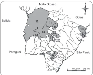
Insects were collected in the municipalities of Jaraguari (May to August 2009 and September 2011), Rochedo, Caarapó, Douradina, Antônio João, Dourados, Terenos, São Gabriel do Oeste, Aparecida do Taboado, Paranaíba, Rio Verde de Mato Grosso, Corumbá, Miranda and Aquidauana (August 2011 to November 2012) in the State of Mato Grosso do Sul
(Figure 1) using the method described by Cominetti et al.
Triatomine identifi cation and microscopic examination
Triatomines were identifi ed using the dichotomous keys
www.rsbmt.org.br ̶ www.scielo.br/rsbmt 278
Cominetti MC et al - PCR to monitor Trypanosoma cruzi in triatomines
RESULTS
DISCUSSION
TABLE 1 - Comparison of the results obtained by PCR and microscopy in triatomines from the municipalities of Mato Grosso do Sul, Brazil.
PCR
ME positive negative total
Positive 58 0 58
Negative 43 414 457
Total 101 414 515
ME: microscopic examination; PCR: polymerase chain reaction.
Bolívia
Paraguai São Paulo Mato Grosso
Goiás
1 2
3 5
6 7
8
4
10 9
0 117,5 km 215 km
11 12
13
4 5
9 8
6 7 14
Campo Grande
FIGURE 1 - Map of the state of Mato Grosso do Sul, Brazil, highlighting municipalities where insects were captured. 1: Jaraguari; 2: Rochedo; 3: Caarapó; 4: Douradina; 5: Antônio João; 6: Dourados; 7: Terenos; 8: São Gabriel do Oeste; 9: Aparecida do Taboado; 10: Paranaíba; 11: Rio Verde de Mato Grosso; 12: Corumbá; 13: Miranda; 14: Aquidauana.
DNA extraction and PCR
Genomic deoxyribonucleic acid (DNA) was extracted from the insects as described by Westenberger et al. The integrity of the DNA sample was determined using 0.8% agarose gel electrophoresis, staining with ethidium bromide (0.5µg/ml) and visualization under ultraviolet light. Additionally, one glass slide containing material from the digestive tract of the insects was
analyzed. After the fi xed material was scraped from the slide,
DNA extraction was performed as described above.
The following primers described by Wincker et al10
were used for molecular identification of T. cruzi: 121 (5'-AAATAATGTACGGG (T/G) GAGATGCATGA-3') and 122 (5'-GGTTCGATTGGGGTTGGTGTAATATA-3'). This set
of primers allows the amplifi cation of 330 bp of kDNA from T. cruzi10. The amplifi cation scheme utilized was previously
described by Schijman et al. Under natural conditions, T. cruzi
and Trypanosoma rangeli frequently co-infect triatomines. Therefore, the samples were also subjected to PCR for T. rangeli
using the primers TrF3 (5’-CCCCATACAAAACACCCTT-3’) and TrR8 (5’-TGGAATGACGGTGCGGCGAC-3’), which target a conserved subtelomeric region in T. rangeli (SubTr,
GenBank accession number: AF426020). The amplifi cation profi le utilized was previously described by Chiurillo et al. All amplifi cation reactions were performed on an Eppendorf AG
22331 thermocycler. Control DNA (T. cruzi as positive and
T. rangeli as negative) was kindly donated by Dr. Marta M.G. Teixeira (Universidade de São Paulo, Brazil). Furthermore, ultrapure water was used as an additional negative control.
The reactions were performed in a fi nal volume of 25μl
containing 1X PCR buffer [10mM Tris-HCl (pH 8.3), 50mM KCl], 1.5mM MgCl2, 0.2mM dNTP mix, 5pmol of each primer, 1U of Taq DNA polymerase (Platinum®, Invitrogen) and 20ng of genomic DNA.
The amplifi cation products were visualized under ultraviolet
light after electrophoresis on an agarose gel (2%) and staining with ethidium bromide.
Statistical analysis
The results were compared using the chi-squared test (χ2) with a signifi cance level of 5%. Statistical analysis was
performed using MedCalc 12.4.0.0.
In total, 515 samples were analyzed. Among the samples, 58
(11.3%) were positive for fl agellated protozoa as determined by
optical microscopy, and 101 (19.6%) were positive for T. cruzi
as determined by PCR.
The main species of triatomine identifi ed was T. sordida, which represented approximately 95.5% of the specimens
collected, thus confi rming the fi ndings of Almeida et al. The
frequency of T. cruzi infection in this species was 10.7% when assessed by microscopy and 18.1% when assessed by PCR.
Association was found between the results obtained by the techniques (χ2 = 53.354, p = 0.001) (Table 1) and the number
of positive samples by PCR was higher than by ME.
PCR was the only technique that has found T. cruzi in triatomines in the municipalities of Rochedo, Corumbá, Aquidauana and Terenos (Table 2) , since ME only confi rms the
cases in which the parasite is visible in the test, and the PCR
also identifi es the parasite in samples where this was not seen.
Reactions using primers for T. rangeli (data not shown)
produced no overlapping data, thereby confi rming that the amplicons were specifi c to T. cruzi.
www.rsbmt.org.br ̶ www.scielo.br/rsbmt
279
Rev Soc Bras Med Trop 46(3):277-280, May-Jun, 2013
The authors declare that there is no confl ict of interest.
CONFLICT OF INTEREST TABLE 2 - Triatomines positive for T. cruzi by PCR and microscopy
in municipalities of the State of Mato Grosso do Sul, Brazil.
Positive
City Species n* ME PCR
Jaraguari T. sordida 260 45 76
Rochedo T. sordida 64 - 3
Caarapó P. megistus 1 1 1
P. geniculatus 4 - 1
Douradina T. sordida 1
-Miranda T. sordida 7
-Terenos T. sordida 48 - 1
Rio Verde de Mato Grosso T. sordida 17 -Aparecida do Taboado T. sordida 54 10 10 São Gabriel do Oeste T. matogrossensis 8
-Paranaíba T. sordida 22
-Dourados P. megistus 1 ** 1
Antônio João T. sordida 1
-Aquidauana T. matogrossensis 9 2 5
T. sordida 12 - 2
Rhodnius sp. 1
-Carumbá T. sordida 5 - 1
Total 515 58 101
*number of triatomines captured; ** inconclusive; ME: microscopic examination; PCR: polymerase chain reaction; P: Panstrongylus; T: Triatoma.
the accurate identifi cation of T. cruzi because other species of trypanosomes are morphologically indistinguishable from
T. cruzi, thereby generating false-positive results. Furthermore, microscopy does not detect the presence of the fl agellates in triatomine as PCR, evidenced in the present study.
Advantages of PCR over ME with respect to sensitivity have been reported for T. cruzi4, Acanthamoeba spp.15, Plasmodium
spp.16, Babesia spp.17, Trypanosoma evansi18 and Leishmania
spp.19. Advantages related to specificity have also been
reported20. Furthermore, PCR is more effi cient than ME for the
detection of T. cruzi in Triatoma infestans feeding on patients with Chagas diseases, with a difference of 46% between PCR and ME20. Previous studies have reported differences of 34.9%
(Rhodniusprolixus), 14.5% (Triatoma dimidiata)21 and 24.6%
(T. infestans)5 between PCR and ME. In the present study, the
difference between the techniques (with respect to the frequency of positive results) was 8.4%. Although this value is lower than that of previous studies, PCR still exhibited highest number of triatomines infected by T. cruzi than ME.
A combination of PCR and optical microscopy to monitor
T. cruzi in triatomines may ensure better results in terms of the presence of the parasite, as shown by the results in the municipalities of Rochedo, Dourados, Corumbá, Aquidauana and Terenos. However, considering the true sensitivity and
specifi city of microscopic examination, questions may arise
regarding the current distribution of fl agellated protozoa among
triatomines, e.g., what is the true distribution of the parasite among triatomines?
In the present study, the proportion of false-negative results in microscopic examination was approximately 8.2%. Considering this issue and taking the data published by Almeida et al. regarding the frequency of triatomines positive for
T. cruzi by ME in the State of Mato Grosso do Sul as an example, the number of insects positive for the parasite should be 724 (8.2%) rather than 15 (0.2%) found by Almeida et al. Similar or higher values have been reported in other studies comparing these techniques4,5,20,21, which demonstrates that the PCR results
may be different from the results reported by the current offi cial surveys. Additionally, results obtained from fi rst- and
second-stage nymphs using ME are underestimated because of the
diffi culty in obtaining feces during these stages. This diffi culty
does not exist when PCR is performed5.
In the municipality of Dourados, ME was performed using fi xed material on a microscope slide that was previously classifi ed as
inconclusive for T. cruzi. Inconclusive results in ME may be caused by parasite deformation during preparation on the microscope slide,
which hinders the identifi cation of the protozoan. In the present study, the identity of the fl agellated protozoa found by microscopy was confi rmed by PCR, which demonstrates the effectiveness of
this tool, particularly with respect to controversial points that may
arise during the identifi cation process. Other studies have shown
that PCR may detect the DNA of parasites on microscope slides even years after preparation22,23.
False-positive results are a potential problem with PCR when
used for identifi cation of T. cruzi24. To reduce the possibility of
false-positive results, a negative control was used in the present study. In addition, the different steps of the technique were performed in different rooms.
Although the epidemiological importance of ME is clear in terms of disease control, the use of sensitive techniques, such as PCR, can increase the accuracy of the epidemiological
investigation and enable the optimal allocation of fi nancial
resources. Furthermore, PCR may be used in epidemiological surveys of other agents of public health importance without
signifi cant additional implementation costs.
The frequency of capture of T. sordida was higher than that
of the other triatomines, which confi rms the results of previous
entomological surveys in which this species was frequently found3,14,25,26. Although previous studies have reported a low
rate of T. cruzi infection2,3,25, the present study suggests that
these values may be underestimated and that PCR is essential
as a tool for detection in offi cial surveys.
Clearly, one detection technique does not exclude the other. The utilization of ME and PCR in combination contributes
greater effi cacy in identifying outbreaks of the parasite and also
www.rsbmt.org.br ̶ www.scielo.br/rsbmt 280
ACKNOWLEDGMENTS
The authors wish to thank the Laboratório Regional de Entomologia, Núcleo Regional de Saúde, Secretaria de Estado de Saúde for their assistance and Dr. Marta M.G. Teixeira (Universidade de São Paulo, Brazil) for the generous gift of
T. cruzi and T. rangeli genomic DNA.
FINANCIAL SUPPORT
Embrapa Gado de Corte, Conselho Nacional de
Desen-volvimento Científi co e Tecnológico (CNPQ) and Fundação de
Apoio ao Desenvolvimento do Ensino, Ciência e Tecnologia do Estado de Mato Grosso do Sul (Fundect-MS). PhD scholarship provided by Coordenação de Aperfeiçoamento de Pessoal de Nível Superior (CAPES).
REFERENCES
1. Rey L. Parasitologia: parasitos e doenças parasitárias do homem nos trópicos, 4th ed. Rio de Janeiro: Guanabara Koogan; 2008.
2. Ministério da Saúde, Secretaria de Vigilância em Saúde, Departamento de Vigilância Epidemiológica. Guia de vigilância epidemiológica, 7th ed. Brasília: Ministério da Saúde; 2009.
3. Almeida PS, Ceretti Júnior W, Obara MT, Santos HR, Barata JM. Survey of Triatominae (Hemiptera: Reduviidae) fauna in domestic environments and natural infection by Trypanosomatidae in the State of Mato Grosso do Sul. Rev Soc Bras Med Trop 2008; 41:374-380.
4. Braz LMA, Raiz R, Amato Neto V, Alárcon RS, Gakyia E, Okay TS. The detection of Trypanosoma cruzi in Triatoma infestans: comparison of a PCR-based assay with microscopical examination. Ann Trop Med Parasitol 2007; 101:461-465.
5. Pizarro JC, Lucero DE, Stevens L. PCR reveals signifi cantly higher rates of Trypanosoma cruzi infection than microscopy in the Chagas vector,
Triatoma infestans: high rates found in Chuquisaca, Bolivia.BMC Infect Dis 2007; 27:66.
6. Cominetti MC, Andreotti R, Oshiro ET, Dorval ME. Epidemiological factors related to the transmission risk of Trypanosoma cruzi in a Quilombola community, State of Mato Grosso do Sul, Brazil. Rev Soc Bras Med Trop 2011; 44:576-581.
7. Carcavallo RU, Rodrigues MEF, Galvão C, Rocha DS, Girón IG, Arocha MAO, et al. Habitats e fauna relacionada. In: Carcavallo RU, Girón IG, Jurberg J, Lent H, editors. Atlas dos vetores da doença de Chagas nas Américas. Rio de Janeiro: Fundação Oswaldo Cruz; 1997. p. 107-244.
8. Souza MA. Morphobiological characterization of Trypanosoma cruzi
Chagas, 1909 and its distinction from other trypanosomes. Mem Inst Oswaldo Cruz 1999; 94:205-210.
9. Westenberger SJ, Sturm NR, Yanega D, Podlipaev SA, Zeledón R, Campbell DA, et al. Trypanosomatid biodiversity in Costa Rica: genotyping of parasites from Heteroptera using the spliced leader RNA gene. Parasitology 2004; 129:537-547.
10. Wincker P, Britto C, Pereira JB, Cardoso MA, Oelemann W, Morel CM. Use of a simplifi ed polymerase chain reaction procedure to detect
Trypanosoma cruzi in blood samples patients in a rural endemic area. Am J Trop Med Hyg 1994; 51:771-777.
11. Schijman AG, Bisio M, Orellana L, Sued M, Duffy T, Jaramillo AMM, et al. International study to evaluate PCR methods for detection of
Trypanosoma cruzi DNA in blood samples from Chagas disease patients. PLoS Negl Trop Dis 2011; 5:e931.
12. Chiurillo MA, Crisante G, Rojas A, Peralta A, Dias M, Guevara P, et al. Detection of Trypanosoma cruzi and Trypanosoma rangeli infection by duplex PCR assay based on telomeric sequences. Clin Diagn Lab Immunol 2003; 10:775-779.
13. Silveira AC. Current situation with Chagas disease vector control in the Americas. Cad Saude Publica 2000; 16:35-42 .
14. Peñaranda-Carrillo R, Moreira EF, Silveira AC, Leite J, Vinhaes MC, Castro C, et al. Evaluation of the impact of vector control programs through serological testing in Mambaí/Buritinópolis, Goiás State. Rev Soc Bras Med Trop 2002; 35:331-338.
15. Yera H, Zamfi r O, Bourcier T, Ancelle T, Batellier L, Dupouy-Camet J, et al. Comparison of PCR, microscopic examination and culture for the early diagnosis and characterization of Acanthamoeba isolates from ocular infections. Eur J Clin Microbiol Infect Dis 2006; 26:221-224 16. Johnston SP, Pieniazek NJ, Xayavong MV, Slemenda SB, Wilkins PP,
Silva AJ. PCR as a confi rmatory technique for laboratory diagnosis of malaria. J Clin Microbiol 2006; 44:1087-1089.
17. Jojima FS, Garcia JL, Vidotto MC, Balarin MRS, Fabretti AK, Gasparini MR, et al. Occurrence and molecular characterization of Babesia species in a canine hospital population in the Londrina Region, Parana State, Brazil J Vet Parasitol 2008; 17:277-283
18. Muieed MA, Chaudhary ZI, Shakoori AR. Comparative studies on the sensitivity of polymerase chain reaction (PCR) and microscopic examination for the detection of Trypanosoma evansi in horses. Turk J Vet Anim Sci 2010; 24:507-512.
19. Azizi K, Soltani A, Alipour H. Molecular detection of Leishmania isolated from cutaneous leishmaniasis patients in Jask County, Hormozgan Province, Southern Iran, 2008. Asian Pac J Trop Med 2012; 5:514-517.
20. Shikanai-Yasuda MA, Ochs DE, Tolezano JE, Kirchhoff LV. Use of the polymerase chain reaction for detecting Trypanosoma cruzi in triatomine vectors. Trans R Soc Trop Med Hyg 1996; 90:649-651.
21. Dorn PL, Engelke D, Rodas A, Rosales R, Melgar S, Brahney B, et al. Utility of the polymerase chain reaction in detection of Trypanosoma cruzi in Guatemalan Chagas vectors. Am J Trop Med Hyg 1999; 60:740-745.
22. Edoh D, Steiger S, Genton B, Beck HP. PCR amplifi cation of DNA from malaria parasites on fi xed and stained thick and thin blood fi lms. Trans R Soc Trop Med Hyg 1997; 91:361-363.
23. Brustoloni YM, Lima RB, Cunha RV, Dorval ME, Oshiro ET, Oliveira ALL, et al. Sensitivity and specifi city of polymerase chain reaction in Giemsa stained slides for diagnosis of visceral leishmaniasis in children. Mem Inst Oswaldo Cruz 2007; 102:497-500.
24. Kirchhoff LV, Votava JR, Ochs DE, Moser DR. Comparison of PCR and microscopic methods for detecting Trypanosoma cruzi. J Clin Microbiol 1996; 34:1171-1175.
25. Paula MBC, Costa IN, Freitas PA, Limongi JE, Pajuaba Neto AA, Pinto RMC, et al. Occurrence of positivity for Trypanosoma cruzi in triatomine from municipalities in Southeastern Brazil, from 2002 to 2004. Rev Soc Bras Med Trop 2010; 43:9-14.
26. Toledo MJO, Kühl JB, Silva SV, Gasperi MV, Araújo SM. Estudo sobre triatomíneos e reservatórios silvestres de Trypanosoma cruzi no estado do Paraná, sul do Brasil. resultados preliminares. Rev Soc Bras Med Trop 1997; 30:197-203.