Ana Catarina da Cruz Rodrigues da Silva
Solid lipid nanoparticles (SLN) for oral delivery of Risperidone
Thesis submitted to the Faculty of Pharmacy of the University of Porto PhD in Pharmaceutical Sciences
Pharmaceutical Technology
This work was developed under the supervision of Professor Domingos Ferreira (Faculty of Pharmacy, University of Porto), Professor Delfim Santos (Faculty of
Pharmacy, University of Porto) and Professor Eliana Souto (Faculty of Health Sciences, Fernando Pessoa University)
Acknowledgments
No decorrer da realização deste trabalho, várias foram as pessoas que contribuíram para que esta experiência se tornasse enriquecedora, tanto ao nível científico como pessoal. Deste modo, não posso deixar de expressar os meus sinceros agradecimentos a todos aqueles que de alguma forma fizeram com que este trabalho fosse possível.
Aos Professores Doutores Domingos Ferreira e Delfim Santos por me terem convidado a fazer este Doutoramento, por todo o empenho, incentivo, conselhos, paciência, amizade e confiança que sempre depositaram em mim.
Ao Professor Doutor José Sousa Lobo por me conceder a oportunidade de desenvolver este projeto no Laboratório de Tecnologia Farmacêutica da Faculdade de Farmácia da Universidade do Porto, por toda a simpatia, amizade, apoio e valiosos conselhos, que tornaram este percurso bem mais fácil.
To Professor Benjamin Forbes from King’s College London, I would like to thank for the opportunity that he gave me to collaborate with its research group and for receiving me so well during my visits to London.
A las Profesoras Marisa García y Maria Antonia Egea de la Facultad de Farmacia de la Universidad de Barcelona, por la oportunidad concedida de hacer parte de los experimentos en su laboratorio, y también por sus consejos, simpatía y amistad manifestadas durante mis visitas a Barcelona.
À Professora Doutora Helena Amaral agradeço pela amizade, apoio e conhecimentos transmitidos, que se revelaram fundamentais na execução deste trabalho.
Ao Professor Doutor Paulo Costa pela disponibilidade demonstrada e importantes conhecimentos científicos que me facultou.
A la Dra. Elisabet González-Mira por su amistad incondicional, sus palabras de ánimos siempre en los momentos más difíciles y, sobretodo, por los conocimientos que me ha transmitido relativos a las nanopartículas lipídicas. Muchísimas gracias Eli, esta tesis es un poco tuya también!
Aos Professores do Laboratório de Tecnologia Farmacêutica, Prof. Doutora Maria Fernanda Bahia, Prof. Doutor Maurício Barbosa, Prof. Doutor Paulo Lobão, Prof. Doutor José Silva, Prof. Doutora Isabel Almeida e Dr. José Silveira, por toda a amabilidade e simpatia com que sempre me trataram.
À Dra. Rosa Pena pela sua amizade, simpatia e especial bom humor, que tanto me ajudaram, sobretudo nos momentos mais difíceis.
Ao Joel Fonseca pela sua amizade e apoio durante estes longos anos de trabalho. Obrigada pelas horas extra de trabalho no laboratório, pelas palavras de incentivo nos momentos mais difíceis e por saberes sempre onde estava tudo o que era necessário. À D. Conceição pela sua ajuda na manutenção das boas condições de funcionamento do Laboratório de Tecnologia Farmacêutica.
Aos meus colegas estudantes de Doutoramento e a todos os investigadores do Laboratório de Tecnologia Farmacêutica, entre os quais destaco a Dra. Susana Martins, com quem partilhei o mundo das nanopartículas lipídicas além-fronteiras. Deixo também uma palavra de gratidão ao Professor Doutor Bruno Sarmento pelo apoio prestado.
À Doutora Joana Araújo, pela sua amizade e companheirismo, que foram fundamentais durante as minhas estadias em Barcelona. Obrigada por partilhares, por me ajudares e por me ouvires Juanita!
A lo Dr. Sasha Nikolic por su amistad, simpatía y por los conocimientos y consejos muy inteligentes acerca del tema de las nanopartículas lipídicas.
A la Dra. Veva y a Fran de la Facultad de Farmacia de la Universidad de Barcelona, por su simpatía, buen humor y por las palabras de ánimos, siempre que estuve en Barcelona.
À Professora Doutora Rita Oliveira por toda amizade e excelentes conselhos.
À Sra. Engenheira Elisa Soares pela ajuda fundamental nos ensaios de HPLC e pelas palavras de apoio e incentivo.
À Dra. Renata Silva pelo auxílio nos ensaios de toxicidade.
À Joana Macedo pela preciosa ajuda com as imagens e formatação desta tese.
À Professora Doutora Eliana Souto pelo apoio prestado.
À Doutora Joana Gomes e à Doutora Sandra Rocha, do Departamento de Engenharia Química da Faculdade de Engenharia da Universidade do Porto pela utilização do DLS.
À Professora Doutora Salete Reis do Departamento de Química-física da Faculdade de Farmácia da Universidade do Porto por facultar o aparelho de DLS.
À minha mãe e ao meu irmão por me terem apoiado sempre e me terem incentivado a não desistir. Ao meu pai por me ter dado condições para iniciar este projeto. À minha tia Mimi pelas palavras de incentivo, que sempre me deram força.
Às minhas amigas Andreia, Guida e Guidinha por todas as palavras de incentivo e por terem aguentado tantas recusas por causa deste trabalho. À Guida agradeço também por ter lido o resumo desta tese. A todos os outros meus amigos aqui fica também um thank you very very much!
Para quem esteve sempre disponível para me ajudar nos momentos mais difíceis, aqui ficam os meus sinceros agradecimentos. Sem esse apoio e incentivo, a conclusão deste trabalho não seria possível.
À Faculdade de Ciências da Saúde da Universidade Fernando Pessoa do Porto por me ter autorizado a realizar parte do trabalho experimental nas suas instalações.
À Fundação para a Ciência e Tecnologia (FCT) pelo suporte financeiro referente à bolsa de Doutoramento com a referência SFRH/BD/30576/2006.
Abstract
Apart from some particular situations, the oral route is the first choice for drug administration. This preference is related with its easy access and non-invasive nature, which improves patient compliance and, therefore, facilitates treatments. However, the poor water solubility of several drug molecules and/or the risk of degradation throughout the gastrointestinal tract (GIT), turn into impossible their oral administration. In particular, drugs like risperidone (RISP) that belongs to the Biopharmaceutical Classification System (BCS) class II (i.e. low water solubility and high intestinal permeability) exhibit poor oral biopharmaceutical properties. Moreover, the administration of RISP for a long period of time can lead to the incidence of unpleasant side effects, which decreases patient adherence and, therefore, increases costs of therapy. To overcome these drawbacks, new oral RISP formulations are required and the exploration of alternative ways of administration should also be considered (e.g. in the oral mucosa). In this perspective, efforts have been done in order to improve the oral bioavailability of poorly water-soluble drugs, by means of developing new colloidal delivery carriers. Among these systems are the so-called solid lipid nanoparticles (SLN), which have been showing promising results. The main reason for this is related with the well-known conception that lipids promote oral drug absorption, because they undergo the same physiological mechanisms of food lipid digestion.
The objective of the present work was to study the use of SLN systems as an alternative to improve the oral delivery of RISP, by means of peroral and mucosal routes.
A review regarding the most important issues that should be addressed during the development of an effective oral drug delivery system is initially presented. The state of the art of RISP delivery systems and toxicological concerns related with the use of colloidal carriers are also highlighted.
(4,5-dimethylthiazol-2-yl)2,5-dyphenyl-tetrazolium bromide (MTT) assay. The results revealed that both techniques originate stable SLN formulations with expected good long-term stability. Moreover, DSC and WAXS confirm the RISP encapsulation within the SLN systems and MTT demonstrate good biocompatibility.
The second experimental work was about the development and validation of a simple reverse-phase high performance liquid chromatography (HPLC), in order to determine the encapsulation parameters (encapsulation efficiency and drug loading) and to study the RISP release profile from the SLN. The in vivo performance of RISP-loaded SLN
was predicted by kinetic modeling (zero order, Higuchi, Korsmeyer-Peppas and Baker-Lonsdale). A robust, specific, accurate, and intra-day and inter-day precise method was obtained. A linear response range, with small detection and quantification limits were also reached. High encapsulation parameters values and an expected in vivo
anomalous non-Fickian transport (i.e. diffusion and erosion) for the RISP-loaded SLN were achieved.
The third experimental work had the objective of prepare two different types of SLN-based hydrogels (HGs) formulations for oral transmucosal delivery of RISP. The suitability of the HGs for the purpose stated was evaluated by means of rheological and textural analysis. The potential increase in SLN particle size after HGs preparation was evaluated by means of laser diffractometry (LD) and cryo-scanning electron microscopy (cryoSEM). The RISP release profile from the HGs was fitted by kinetic models in order to predict the in vivo performance. Both HGs revealed a plastic behavior with thixotropy and increased adhesiveness, which are desired features for oral transmucosal application. LD and cryo-SEM analysis revealed that SLN preserved their colloidal size after HGs preparation. The expected in vivo RISP release
mechanisms from the HGs were pH dependent Fickian diffusion alone or combined with erosion.
The fourth part of the experiments deals with the preparation and long-term stability studies of RISP-loaded SLN formulations to study its potential as oral delivery system. Particle size, PI, ZP, TEM and encapsulation efficiency (EE) analysis were performed. Stable SLN systems with high EE and analogous shape were obtained after two years of storage. An in vivo classical Fickian diffusion is expected for RISP release.
Biocompatibility and increased RISP uptake across Caco-2 cells were observed. The results demonstrated the feasibility of RISP-loaded SLN systems as a stable manufactured dosage form.
Resumo
De um modo geral, a via oral é a primeira escolha para a administração de fármacos. Esta preferência está relacionada com o seu fácil acesso e natureza não invasiva, o que aumenta a adesão à terapêutica por parte dos doentes, facilitando os tratamentos. Contudo, a baixa solubilidade aquosa de algumas moléculas de fármacos e/ou o risco de sofrerem degradações no trato gastrointestinal, faz com que seja impossível a sua administração oral. Em particular, fármacos como a risperidona (RISP), que pertencem à classe II do sistema de classificação biofarmacêutica (baixa solubilidade aquosa e elevada permeabilidade intestinal), apresentam limitações nas suas propriedades biofarmacêuticas orais. Por outro lado, a administração de RISP durante longos períodos de tempo pode levar à incidência de efeitos secundários indesejáveis, o que diminui a adesão à terapêutica, aumentando os custos dos tratamentos. Para ultrapassar estas desvantagens, é necessário o desenvolvimento de novas formulações orais de RISP, bem como o estudo de vias alternativas de administração, como por exemplo, a administração na mucosa oral. Neste sentido, têm sido desenvolvidos novos sistemas coloidais de transporte de fármacos. Entre estes, as nanopartículas de lípidos sólidos (SLN) têm vindo a demonstrar resultados promissores. Para este sucesso contribui o fato dos lípidos promoverem a absorção oral de fármacos, dado que sofrem os mesmos mecanismos fisiológicos da digestão dos lípidos provenientes dos alimentos.
O objetivo do presente trabalho foi o estudo da aplicação de sistemas de SLN como alternativa para promover a libertação oral de RISP, através das vias de administração peroral e na mucosa oral. Inicialmente foi efetuada uma revisão bibliográfica, enfatizando os assuntos mais importantes, que devem ser considerados aquando do desenvolvimento de um sistema de libertação oral de fármacos. O estado da arte dos sistemas de libertação de RISP, bem como as inquietações toxicológicas relacionadas com o uso de transportadores coloidais de fármacos foram também referidos.
biocompatibilidade em culturas celulares. Ambas as técnicas permitiram obter sistemas de SLN estáveis e biocompatíveis, com previsão de elevada estabilidade ao longo do tempo. A encapsulação da RISP nas SLN foi também confirmada.
O segundo trabalho experimental teve como objetivo o desenvolvimento e validação de um método simples de cromatografia líquida de elevada resolução (HPLC), com o intuito de determinar os parâmetros de encapsulação (eficácia de encapsulação e capacidade de carga) e de estudar o perfil de libertação da RISP a partir das SLN. O comportamento in vivo dos sistemas foi previsto pela aplicação dos modelos cinéticos
de ordem zero, Higuchi, Korsmeyer-Peppas e Baker-Londsdale. O método desenvolvido revelou-se robusto, específico, exato e preciso. Uma gama linear de resposta com baixos limites de deteção e de quantificação foi obtida. Os parâmetros de encapsulação foram elevados e é esperado in vivo um transporte não-Fickiano
anómalo (difusão e erosão), para a RISP a partir das SLN.
O terceiro trabalho experimental focalizou-se na preparação de dois tipos de hidrogeles (HGs) contendo SLN para a administração de RISP na mucosa oral. A aptidão dos HGs para o uso proposto foi estudada através de ensaios reológicos e de textura. O tamanho das SLN após a inclusão nos HGs foi avaliado através de difratometria de laser (LD) e de crio-microscopia de varrimento eletrónico (cryoSEM). O perfil in vivo de libertação da RISP a partir dos HGs foi estudado por modelos cinéticos. Ambos os HGs revelaram um comportamento plástico com tixotropia e elevada adesividade, que são características desejáveis para a aplicação na mucosa oral. Os ensaios de LD e de cryoSEM demonstraram que as SLN mantiveram os seus tamanhos coloidais após a preparação dos HGs. O mecanismo in vivo esperado para
a libertação da RISP a partir dos HGs foi dependente do pH e corresponde a uma difusão de Fick isolada ou combinada com um mecanismo de erosão.
O quarto trabalho experimental relaciona-se com a preparação e estudo da estabilidade ao longo do tempo de sistemas de SLN contendo RISP para a administração oral. Análises de tamanhos de partículas, PI, ZP, TEM e eficácia de encapsulação (EE) foram efetuadas. Sistemas de SLN estáveis com elevada EE e forma análoga foram obtidos após dois anos de armazenamento. Uma difusão clássica de Fick é esperada in vivo para a RISP a partir das SLN. Biocompatibilidade e elevada
absorção através de células Caco-2 foram também observadas para as SLN. Os resultados revelaram a adequação dos sistemas de SLN como formas de dosagem estáveis para a RISP.
Table of contents
Acknowledgments ... v
Abstract ... ix
Resumo... xi
Table of contents ... xiii
Index of Figures ... xix
Index of Tables ... xxiii
List of Abbreviations and Symbols ... xxvii
Aims and Organization of the Thesis ... xxix
CHAPTER 1 ... 31
Introduction ... 31
1. Drug delivery via the oral route ... 33
1.1 The gastrointestinal tract (GIT) ... 33
1.2 Peroral administration ... 35
1.3 Drug administration in the oral cavity ... 37
2. Strategies to improve oral drug delivery ... 39
3. Oral delivery of drugs by means of lipid nanoparticles ... 40
3.1 Outcome of lipids in oral delivery ... 40
3.2 Lipid nanoparticles... 42
3.3 Toxicological concerns ... 50
4. Drug delivery systems for risperidone: state of the art ... 51
5. References ... 53
CHAPTER 2 ... 65
Preparation, characterization and biocompatibility studies on risperidone-loaded solid lipid nanoparticles (SLN): high pressure homogenization versus ultrasound ... 65
Abstract ... 68
1. Introduction ... 69
2. Experimental ... 71
2.1. Materials ... 71
2.2. Methods ... 71
2.2.2. Preparation of SLN ... 72
2.2.3. Differential scanning calorimetry (DSC) ... 73
2.2.4. Particle size analysis and zeta potential (ZP) measurements ... 74
2.2.5. Optical assays ... 74
2.2.6. Shape ... 75
2.2.7. Wide angle X-ray scattering (WAXS) ... 75
2.2.8. Biocompatibility studies: MTT assay ... 75
2.2.9. Statistical analysis ... 76
3. Results and discussion ... 77
3.1. Lipid solubility studies ... 77
3.2. Production of SLN ... 78
3.3. Optical analysis ... 80
3.4. Shape ... 82
3.5. Crystallinity and polymorphism of SLN ... 83
3.6. Cell viability: MTT assay... 86
4. Conclusions ... 88
References ... 89
CHAPTER 3 ... 93
Risperidone release from solid lipid nanoparticles (SLN): validated HPLC method and modelling kinetic profile ... 93
Abstract ... 96
1. Introduction ... 97
2. Materials and methods ... 99
2.1. Materials ... 99
2.2. Methods ... 99
2.2.1. Instrumentation ... 99
2.2.2. Chromatographic conditions ... 99
2.2.3. Preparation of standard and sample solutions ... 100
2.2.4. Method validation ... 100
2.2.5. Preparation of SLN ... 102
2.2.6. Method applicability ... 103
2.2.6.2. In vitro drug release studies ... 104
2.2.6.3. Drug release data modelling ... 105
3. Results ... 107
3.1. Method validation ... 107
3.1.1. System suitability ... 107
3.1.2. Linearity ... 107
3.1.3. Precision ... 108
3.1.4. Specificity... 110
3.1.5. Accuracy ... 112
3.1.6. Detection limit (DL) and quantification limit (QL) ... 112
3.1.7. Robustness ... 112
3.2. Method applicability ... 113
3.2.1. Assessment of encapsulation parameters of SLN formulation ... 113
3.2.2. In vitro drug release studies ... 114
3.2.3. Drug release data modelling ... 115
References ... 118
CHAPTER 4 ... 123
Solid Lipid Nanoparticles (SLN) - based hydrogels as potential carriers for transmucosal delivery of Risperidone: preparation and characterization studies ... 123
Abstract ... 126
1. Introduction ... 127
2. Materials and methods ... 129
2.1. Materials ... 129
2.2. Methods ... 129
2.2.1. Preparation of SLN ... 129
2.2.2. Preparation of HGs ... 129
2.2.3. Particle size analysis ... 130
2.2.4. Cryo-scanning electron microscopy (cryoSEM) ... 131
2.2.5. Rheological measurements ... 131
2.2.6. Texture analysis ... 132
2.2.7. Chromatographic conditions ... 132
2.2.8. In vitro drug release studies ... 133
2.2.9. Drug release data modelling ... 133
3. Results and discussion ... 134
3.2. Cryo-scanning electron microscopy (cryoSEM) ... 135
3.3. Rheological measurements ... 136
3.4. Texture analysis ... 138
3.5. In vitro drug release studies and data modelling ... 139
4. Conclusions ... 142
References ... 143
CHAPTER 5 ... 147
Long-term stability, biocompatibility and oral delivery potential of Risperidone-loaded solid lipid nanoparticles ... 147
Abstract ... 150
1. Introduction ... 151
2. Materials and methods ... 153
2.1. Materials ... 153
2.2. Methods ... 153
2.2.1. Lipid solubility studies... 153
2.2.3. Particle size analysis and zeta potential (ZP) measurements ... 155
2.2.4. Transmission electron microscopy (TEM) studies ... 156
2.2.5. Assessment of encapsulation efficiency (EE) ... 156
2.2.6. In vitro drug release studies ... 157
2.2.7. Drug release data modelling ... 157
2.2.8. Long-term stability studies ... 158
2.2.9. Caco-2 cell cultures ... 158
2.2.10. MTT assay ... 158
2.2.11. Oxidative potential (OP) ... 159
2.2.12. Drug uptake studies ... 160
2.2.13. Statistical analysis ... 160
3. Results ... 161
3.1. Lipid solubility studies ... 161
3.2. Long-term stability studies: particle size, polydispersity index, zeta potential and encapsulation efficiency ... 162
3.3. Particle size in cell culture medium ... 164
3.5. In vitro drug release studies and data modelling ... 166
3.6. MTT assay ... 167
3.7. Oxidative potential (OP) ... 169
3.8. Drug uptake ... 170
4. Conclusions ... 172
References ... 173
CHAPTER 6 ... 179
Conclusions ... 179
APPENDIX 1 ... 183
Related review articles and book chapters ... 183
1. Advances in Nanoparticulate Carriers for Oral Peptides and Proteins: Polymeric vs. Lipid Nanoparticles ... 185
2. Nanopartículas Lipídicas ... 200
APPENDIX 2 ... 238
Other published articles ... 238
1. Oral delivery of drugs by means of solid lipid nanoparticles (SLN) ... 239
Index of Figures
Chapter 1
Figure 1: Anatomy of digestive system (gastrointestinal tract and exocrine glands) .. 33
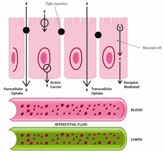
Figure 2: Possible mechanisms of intestinal epithelial drug transport. Adapted from Stenberg et al. [9], with permission ……….. 35
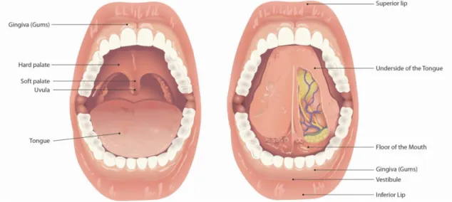
Figure 3: Schematic representation of the anatomy of the oral cavity ……….. 36
Figure 4: Schematic representation of the mechanisms of gastrointestinal lipid processing and successive systemic absorption by means of portal or lymphatic circulation. Adapted from Porter and Charman [35], with permission ……… 40
Figure 5: Lipid nanoparticles: Solid lipid nanoparticles, SLN (left); Nanostructured lipid carriers, NLC (right). Adapted from Müller et al. [43], with permission ……….. 42
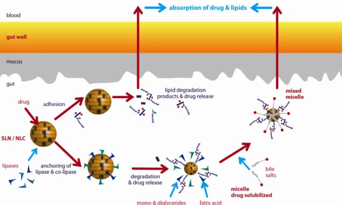
Figure 6: Schematic representation of the mechanisms of intestinal absorption of drugs from lipid nanoparticles. Adapted from Müller and Keck [45], with permission . 45
Chapter 2
Figure 1: DSC patterns of bulk drug (RISP), bulk lipid and physical mixtures of drug and lipid (Imwitor®900K) ……… 76
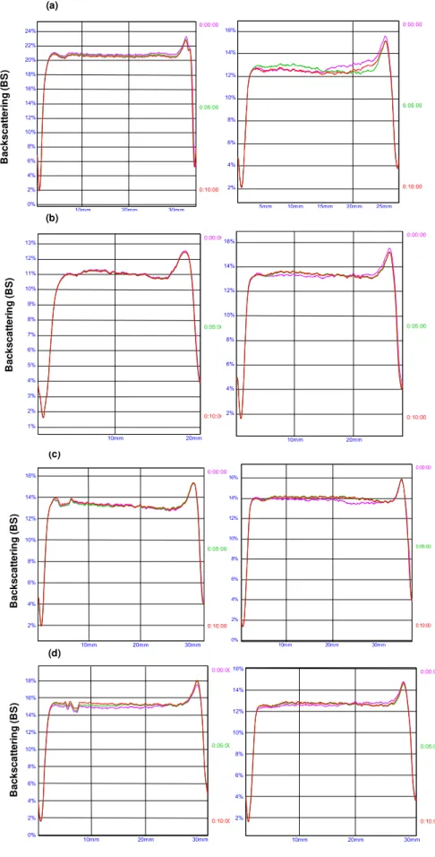
Figure 2: BS profiles of SLN dispersions, measured across the height of the sample
cell during 10 min, on day 0 (n = 3). Dispersions prepared by US (left) and HPH (right): (a) DF; (b) DL1; (c) DL2; (d) DL3……… 80
Figure 4: WAXS diagrams obtained on day 1 after production: (a) bulk Imwitor® 900K; (b) bulk RISP; (c) drug-free (DF) and drug-loaded (DL3) formulations prepared by US; (d) drug-free (DF) and drug-loaded (DL3) formulations prepared by HPH technique .. 84
Figure 5: Cell viability determined by MTT assay, after exposition to drug-free (DF) and drug-loaded (DL3) SLN formulations (concentration range 0–10 µg/ml), prepared by US and HPH technique. The results are average values (n = 3), compared to control cells (unexposed to SLN) ……… 85
Chapter 3
Figure 1: Chemical structure of Risperidone [1] ………... 97
Figure 2: Standard calibration curve of peak areas (mean) against RISP concentration (0.25 - 10.00 µg/ml) ……… 107
Figure 3: (A) Chromatogram of the supernatant of placebo-SLN formulation; (B) Chromatogram of standard 5.00 µg/ml RISP solution ……… 110
Figure 4: Cumulative RISP release profiles from SLN and commercial oral suspension (Risperdal®) (mean values ± SD, n=3) ……….. 114
Chapter 4
Figure 1: LD analysis of HGi and HGd SLN-based hydrogel formulations, measured on days 1 and 30 (mean values, n = 3): (a) HGi (up DF, down DL) and (b) HGd (up DF,
down DL) ……… 133
Figure 2: CryoSEM images of the prepared hydrogel formulations, performed on day 1: (a) blank-HG and HGi (HGi-DF, HGi-DL) and (b) HGd (HGd-DF, HGd-DL) ……….. 134
Figure 4: Shear stress as a function of the shear rate of the blank-HG and HGi SLN-based formulations, evaluated on days 1 and after 30 of storage at 25 ± 1 ºC (mean values, n = 3): (a) Day 1 and (b) Day 30. Upper and lower curves present ascending and descending measurements, respectively, in each formulation ……….. 136
Figure 5: Texture analysis of the HGi and HGd SLN-based formulations, evaluated on days 1 and 30 (mean values ± SD, n = 3). Blank-HG was used for comparison: (a)
firmness and (b) adhesiveness ……….. 137
Figure 6: Comparative results of the cumulative RISP release profiles from the SLN -based HGi-DL and HGd-DL with the SLN dispersion (SLN-DL), in two different media (mean values ± SD, n = 3): (a) phosphate buffer (pH 6.8) and (b) HCl (0.1 N) …….. 139
Chapter 5
Figure 1: TEM images of SLN formulations, acquired on day 8 (left) and after 2 years of storage at 4ºC (right): (a) placebo (RF); (b) RISP-loaded (RL). Black bars are equal to 200 nm ………... 164
Figure 2: Cumulative RISP release profiles from SLN and commercial oral suspension (Risperdal®) (mean values ± SD, n = 3) ……… 165
Figure 3: The effect of placebo (RF) and RISP-loaded (RL) SLN formulations on the viability of Caco-2 cells, after 24h exposure. Cell viability was calculated as a percentage of the control (non-exposed) over a SLN concentration range of 40.6 - 550.0 µg/cm2. The data represent the mean values ± SD (n = 3) ……… 167
Figure 4: Oxidative potential (OP) of placebo (RF) and RISP-loaded (RL) SLN formulations in water and in the presence (+) or absence (-) of DTPA. Copper oxide particles were used as positive control. The data represent mean values ± SD (n = 3)
Figure 5: Release of RISP from Caco-2 cells between 4 - 24 h following exposure to RISP-loaded SLN formulations (RL). The concentrations of SLN formulations tested were 40.6 and 550.0 µg/cm2 (1 and 8 mg of RISP, respectively). The data represent the mean values ± SD (n = 3) ………. 169
Index of Tables
Chapter 1
Table 1: Examples of drugs encapsulated in lipid nanoparticles systems (SLN and NLC) and their potential benefits for application in oral delivery ………. 47
Chapter 2
Table 1: Composition of SLN formulations (DF, Drug-free SLN dispersions; DL1, 1% Drug-loaded SLN dispersions; DL2, 2% Drug-loaded SLN dispersions DL3, 3% Drug-loaded SLN dispersions) ……… 71
Table 2: DSC parameters of bulk drug (RISP), bulk lipid (Imwitor® 900K) and physical mixtures of drug and lipid ……….. 77
Table 3: Z-ave and PI values of DF, DL1, DL2 and DL3 formulations, measured on the production day, for US and HPH techniques (mean ± SD, n = 3) ………... 78
Table 4: LD diameters of DF, DL1, DL2 and DL3 formulations, measured on the production day, for US and HPH techniques (mean ± SD, n = 3) ………... 78
Table 6: DSC parameters of bulk lipid (Imwitor® 900K), drug-free (DF) and drug-loaded (DL3) formulations, prepared by ultrasound (US) and high pressure homogenization (HPH) techniques, measured on day 1 ………... 83
Chapter 3
Table 1: Composition of SLN Formulations (DF, Drug-Free; DL Drug-Loaded) ….. 101
Table 2: Kinetic Models and Mathematical Equations Applied to Study the Drug Release Profile of RISP from SLN [7] ………. 105
Table 3: System Suitability Parameters Obtained for the HPLC System ………….. 106
Table 4: Results Achieved for the Intra-Day and Inter-Day Precision of the Method. 108
Table 5: Results Obtained for the Instrumental Precision ………...……….. 108
Table 6: Results Found for Method Accuracy ………. 111
Table 7: Results Achieved for the Method Robustness ………. 112
Table 8: SLN Encapsulation Efficiency (EE) and Loading Capacity (LC) …………... 113
Chapter 4
Table 1: Composition of the prepared hydrogel formulations (blank hydrogel, blank-HG; HGi-DF, placebo SLN; HGi-DL, RISP-loaded SLN; HGd-DF, placebo SLN; HGd-DL, RISP-loaded SLN) ……… 129
Table 2:Comparison of the kinetic data obtained after fitting the release profile of RISP
in two different media (phosphate buffer, pH 6.8 and HCl, 0.1 N), from the HGi-DL and HGd-DL, with the RISP-loaded (SLN-DL) dispersion. The presented kinetic parameters mean: K, release kinetic constant; R2, determination coefficient; n, diffusion release
exponent ………. 140
Chapter 5
Table 1: Composition of SLN formulations (RF, RISP-free SLN; RL, RISP-loaded SLN) ……….. 153
Table 2: Results of lipid-RISP solubility ……… 160
Table 3: DSC results of bulk RISP, bulk lipid (Compritol® 888ATO) and mixtures of RISP and lipid (Compritol® 888ATO) ………. 161
Table 4: Mean values for groups of placebo SLN (RF) formulations in homogeneous subsets after application of Tukey post-tests ………... 162
Table 6: Z-ave, PI and ZP results of placebo (RF) and RISP-loaded (RL) SLN formulations, before and after admix with cell culture medium (mean ± SD, n = 3) .. 163
List of Abbreviations and Symbols
ANOVA Analysis of Variance
ATCC American Type Culture Collection
BCS Biopharmaceutical Classification System BS Backscattering
CMC Critical Micellar Concentration cryoSEM Cryo-scanning Electron Microscopy DL Detection Limit
DLS Dynamic Light Scattering
DMEM Doubelco’s Modified Eagle’s Medium DMF Dimethylformamide
DSC Differential Scanning Calorimetry DTPA Diethylene Triamine Pentaacetic Acid EE Encapsulation Efficiency
FBS Fetal Bovine Serum GIT Gastrointestinal Tract
GRAS Generally Recognized as Safe HBSS Hank's Buffered Salt Solution
HGs Hydrogels
HPH High Pressure Homogenization
HPLC High Performance Liquid Chromatography ICH International Conference on the Harmonization
K Release Rate Constant LC Loading Capacity
LC-MS-MS Tandem Mass Spectrometry LD Laser Diffractometry
LDC Lipid-Drug Conjugate
MEME Minimum Essential Medium Eagle’s
MTT (4,5-imethylthiazol-2-yl)2,5-dyphenyl-tetrazolium bromide
n Diffusion Release Exponent
NCS Nanotoxicological Classification System NEAA Non-essential Amino Acids
NLC Nanostructured Lipid Carriers OP Oxidative Potential
PBS Phosphate Buffered Saline
PI Polydispersity Index
PLN Polymer-Hybrid Nanoparticles QL Quantification Limit
SEDDS Self-Emulsifying Drug Delivery Systems SDS Sodium Dodecylsulphate
R2 Determination Coefficient RF Risperidone Free
RI Recrystallization Index RL Risperidone Loaded
RSD Relative Standard Deviation RI Recrystallization Indices RISP Risperidone
SEM Scanning Electron Microscopy SD Standard Deviation
SLN Solid Lipid Nanoparticles
TEM Transmission Electron Microscopy TEER Trans-epithelial Electrical Resistance US Ultrasound
UV Ultraviolet
VLDL Very Low Density Lipoproteins ZP Zeta Potential
WAXS Wide Angle X-ray Scattering
Aims and Organization of the Thesis
The conception of a delivery system to improve drug biopharmaceutical properties should start with the selection of the most appropriate dosage form, followed by the evaluation of the physico-chemical compatibility between system and drug. Afterwards, an efficient production method should be selected, according to the laboratory availabilities and, if possible, with its suitability for lab-scale, to conquer the interest of pharmaceutical industries. Once prepared, the system ought to be extensively characterized and its long-term stability must be assured, in order to guarantee its feasibility for the purposed application. When overcome these processes, the studies must pursue to the in vitro (e.g. cell cultures), ex vivo (e.g. tissues and organs), in vivo
(e.g. rats) and finally to the humans clinical trials. As can be easily understood, this is a long-way to go through. For this, a multidisciplinary share of knowledge between several scientific areas (e.g. physical, medical, pharmaceutical, biological and engineering) is crucial. Therefore, regarding the pharmaceutical technologists competences, the aim of this work was to perform the preparation, characterization and study of the long-term stability of solid lipid nanoparticles (SLN) formulations, intended to improve the oral delivery of risperidone (RISP), a poorly water-soluble drug.
The thesis is organized in four main parts, namely abstract, five chapters, conclusions and appendix.
More in detail:
- Chapter 1 includes the theoretical bases of anatomy and physiology of the gastrointestinal tract (GIT), the relevant topics required for the development of an efficient oral lipid-based nanocarrier system, the current status of the RISP-loaded delivery systems and some toxicological concerns regarding the use of colloidal carriers.
- Chapter 3 encompasses the results obtained from the development and validation of a simple high performance liquid chromatography (HPLC) method, according to the International Harmonization Guidelines (ICH). The method was further employed for the quantification of RISP from SLN systems, by means of drug release studies and encapsulation parameters (encapsulation efficiency and drug loading). The former was used to suggest the model of drug incorporation in SLN, and fitted under kinetic models, to predict the in vivo
performances of the referred systems. The last data was used to evaluate the efficiency of the SLN system for RISP encapsulation.
- Chapter 4 involves the use of semi-solid systems as an alternative to the peroral administration of RISP. Two different SLN-based hydrogels (HGs) were prepared and characterized with the aim of study their feasibility for RISP oral transmucosal delivery. For this, rheological, texture, cryo-scanning electron microscopy (cryoSEM) and particle size analysis were performed on the HGs.
CHAPTER 1
___________________________
1. Drug delivery via the oral route
Despite the scientific and technological progresses achieved in the new millennium to create new drug delivery systems, the continuous discovery and subsequent therapeutic needs of active molecules requires a constant update and development of strategies. Therefore, pharmaceutical technologists should always keep in mind two main subjects: the best formulation approach to deliver the active in the local of the therapeutic action (i.e. targeted drug delivery), and the choice of the most pleasant and efficient administration route for patients. In this context, the oral route remains the most preferred, essentially because of its easy administration, non-painful, and low cost of production processes [1-4].
This chapter deals with the most important issues that should be addressed during the development of an effective oral drug delivery system. Moreover, the use of lipid-based nanocarriers is described as a promising alternative to improve oral delivery of poorly water-soluble therapeutics, by means of peroral and mucosal routes.
1.1 The gastrointestinal tract (GIT)
For the conception of a successful oral drug delivery system, knowledge about the anatomy and physiology of the gastrointestinal tract (GIT) is essential [5].
digestive enzymes (e.g. proteases, lipases, amylases) to digest food components (carbohydrates, fats, proteins and nucleic acids) and bicarbonate ions to neutralize the acidic chyme from the stomach; liver produces the bile, which contains the bile salts and phospholipids that act as surfactants to emulsify lipids from food, increasing their absorption.
Figure 1: Anatomy of digestive system (gastrointestinal tract and exocrine glands).
Together the GIT components have the role of process ingested foods before occurring systemic absorption of their molecular components, by direct passage to the blood stream, via portal circulation, or indirectly, passing first trough the lymphatic circulation, according to their physicochemical nature (e.g. dietary fats and lipid soluble vitamins) [6, 7].
According to the GIT anatomy and physiology, and the objective of reach systemic circulation, the drug delivery via the oral route could be performed by two main sub-routes [1, 4, 7, 8]: (i) peroral administration, which means taken the substances through the mouth with absorption elsewhere in the other regions of the GIT; (ii) administration in the oral cavity, that includes drug absorption by means of the oral mucosa for a local (mucosal) or systemic effect (transmucosal).
1.2 Peroral administration
According to the previous description of food absorption processes (Section 1.1), and considering the delivery of drugs administered throughout the mouth, we expect the occurrence of similar sequential events.
mostly by lymphatic circulation, when comparing to portal circulation, which avoids the risk of occurring liver first-pass metabolism [4, 7].
Figure 2: Possible mechanisms of intestinal epithelial drug transport. Adapted from
Stenberg et al. [9], with permission.
should be evaluated (e.g. fed and fasted state, gender, age and health/disease circumstances) [11].
1.3 Drug administration in the oral cavity
The drug administration in the oral cavity offers some advantages over the peroral route, like the avoidance of occurring chemical degradation and rapid first-pass metabolism, since the high vascularisation of some parts of the lining mucosa allow direct passage of drugs to the systemic circulation. Moreover, the enzymatic barrier present in mouth is less effective than the ones present in the GIT [1, 8, 12].
Local and systemic effects could be achieved by means of drug delivery in the oral mucosa, namely by mucosal and transmucosal administration, respectively [8]. According to the aim of this work, we will focus only on transmucosal delivery. For a better understanding of the transmucosal drug delivery process, a brief description of the oral cavity anatomy and physiology is required (Figure 3).
Figure 3: Schematic representation of the anatomy of the oral cavity.
responsible for most mechanical properties of the oral mucosa. At the surface of these layers stays a mucus barrier, which has protective functions and contains mostly water (95-99%) and small amounts of insoluble glycoproteins (1-5%) and other proteins, enzymes, nucleic acids and salts [12].
The transmucosal route comprises drug absorption through sublingual and buccal mucosa, which encompasses about 60% of the overall oral mucosa area. The sublingual mucosa is extremely vascularised and, therefore, is ideal to achieve an immediate systemic effect for high permeable drugs. In contrast, the lower permeability of the buccal mucosa makes it attractive to attain a prolonged drug release from the applied formulations [8, 12].
Similar to the mechanisms of intestinal drug absorption, the oral mucosal epithelial membranes can be across using one or a combination of different mechanisms (Figure 2), such as [1, 8, 12]: (i) passive diffusion by means of a combination of transcellular (intracellular) or paracellular (intercellular) pathways, according to the drug molecular characteristics; (ii) active carrier mediated; (iii) receptor mediated. In between these mechanisms, the passive diffusion has been reported has the most common route of absorption.
Nonetheless, a few drawbacks may occur, such as low drug permeability in some sections of the oral mucosa, related with differences on thickness, degree of keratinisation and the small surface area available for the administration. Additionally, under normal physiological conditions, a low level of saliva is produced continuously, which could originate drug dissolution and increase the risk of swallow the formulation, leading both to a loss in the absorbed amount of the administered drug [1, 12].
At physiological pH, the mucus barrier present within the oral cavity has negative charge. This feature could be used to facilitate the retention of drug delivery systems at the surface of the oral mucosa. For this, mucoadhesive agents that preserve an intimate contact between the formulation and the mucosa, allowing drug absorption through the epithelia, should be applied. In addition, penetration enhancers and enzyme inhibitors could be used concomitantly. Typically, mucoadhesive agents are
polymers, classified as natural/synthetic, water-soluble/water-insoluble and
2. Strategies to improve oral drug delivery
According to the previous description of the GIT parts and respective environments (Section 1.1), is obvious that the oral delivery of poor water-soluble, and enzymatic and metabolic sensitive drugs is a challenge [2]. Examples of these compounds are peptides and proteins, which oral therapeutic feasibility remains open to debate, despite of considerable research efforts and impressive progress made in recent years [14]. Nevertheless, most of the new drug molecules belong to the class II and IV of the Biopharmaceutical Classification System (BCS), which means that they have poor water-solubility. Alternatively, novel molecules could be included in BCS class III, which show good water solubility but low intestinal membrane permeability. Therefore, all these compounds exhibit low bioavailability, since water solubility and membrane permeability are crucial factors for the therapeutic success of peroral administered drugs [2, 15, 16].
3. Oral delivery of drugs by means of lipid nanoparticles
3.1 Outcome of lipids in oral delivery
As previous mentioned (Section 1.2), intestinal lymphatic transport has been presented as an alternative for high lipophilic drugs reach systemic circulation, bypassing hepatic first-pass metabolism. However, considering moderate lipophilic drugs, the variable bioavailability problems remains unsolved, since they mainly undergo portal transport. Therefore, an excellent approach is the administration of these drugs by means of lipid-based systems, since they have been showed very good results in the improvement of lymphatic transport [7, 33]. Moreover, lipids present unique properties (e.g. biocompatibility, GIT absorption enhancement of lipophilic molecules, wide diversity of chemical structures), which make them first-class excipients in drug formulations. For the successful development of lipid-based formulations, knowledge about the role of lipids in oral delivery is fundamental [33].
density lipoproteins, VLDL). Following they pass to lymphatic circulation and finally enter into systemic circulation [7, 33-35].
Figure 4: Schematic representation of the mechanisms of gastrointestinal lipid
processing and successive systemic absorption by means of portal or lymphatic circulation. Adapted from Porter and Charman [35], with permission.
At the intestine, when the drug is released from the formulation, during the duodenal lipid digestive phase, it could be stabilized and/or solubilised by the uptake into bile salts micelles or mixed micelles that further favour the passage through lymphatic circulation. Nevertheless, the intestinal lymphatic transport needs more time to reach systemic circulation, which means that a sustained drug release effect could be obtained. Also an increase in enterocyte membrane permeability, a protection against intestinal enzymatic degradation and a decrease of hepatic first-pass metabolism were observed [7, 15, 33, 35, 36].
Formulation approaches to enhance oral drug bioavailability by means of lymphatic delivery are the lipid-based systems using food-derived lipids, which can be applied in liquid or solid forms. The former are classic solutions, suspensions and oil-in-water emulsions or self-emulsifying drug delivery systems (SEDDS). The SEDDS consist of mixtures of oils, surfactants and co-solvents, which spontaneously form emulsions of the drugs when in contact with the GIT fluids. Solid forms can be powder, granules or pellets, which can be packed into capsules, sachets or compressed in tablets [33, 34]. In addition, as referred in Section 2, colloidal carriers have been showed good results on the improvement of oral drug bioavailability [2]. Among these systems and according to the previous explained concerning the advantages of using lipid-based formulations, the liposomes, nanoemulsions and lipid nanoparticles seems to be the most attractive. However, nowadays we can only found a small number of these systems in the cosmetic market [37]. Therefore, a lack on the knowledge about lipid-based nanocarriers effectiveness and security for human use in therapy subsist [38].
3.2 Lipid nanoparticles
controlled release effect, whilst protect the drug against degradations, have good long-term stability, allow the encapsulation of high drug quantity, have low cost production techniques that avoid the use of organic solvents and are easily transferred to large scale. So far, these advantages turn lipid nanoparticles into superior carriers, when compared to nanoemulsions but also over other older and well-known colloidal systems, such as liposomes and polymeric nanoparticles [37, 39, 40].
Lipid nanoparticles were invented about 20 years ago, in the beginning of the nineties. This genius idea was patented by two different researchers and co-workers, regarding their different production methods: the R.H. Müller and J.S. Lucks from Germany [41], and M.R. Gasco from Italy [42]. Despite the number of academic groups that are interested in the study of these systems have been increasing, nowadays R.H. Müller and co-workers are still the leaders of this research area [37].
There are two types of lipid nanoparticles, the solid lipid nanoparticles (SLN) and the nanostructured lipid carriers (NLC), which differ in their inner lipidic structure (Figure 5).
Figure 5: Lipid nanoparticles: Solid lipid nanoparticles, SLN (left); Nanostructured lipid
carriers, NLC (right). Adapted from Müller et al. [43], with permission.
The SLN were the first generation of lipid nanoparticles and are formed by simple replacing the liquid oil of a oil-in-water nanoemulsion by a solid lipid at body temperature, whereas the NLC (second generation) are composed of a mixture of liquid and solid lipid that are also solid [43]. The NLC systems appeared to overcome some of the shortcomings of the SLN systems, such as [39, 43]: insufficient drug loading capacity for therapeutic purposes; poor long-term stability related with drug release from the nanoparticles, after occurring lipidic polymorphic transitions to more organized crystalline structures.
construction can be approximated to a disordered wall of stones with different sizes and shapes. Therefore, in contrast to SLN, is easy to recognize that NLC can encapsulate more amount of drug. Moreover, the latter have higher long-term stability due to the existence of imperfect crystal structures, which prevent the occurrence of lipid re-crystallization into more stable polymorphic forms with subsequent drug expulsion. Depending on the production technique, the water solubility/non-solubility of the drug and the type of lipid(s) used, SLN and NLC can exhibit different models for drug incorporation. Knowledge about these models is essential to establish the in vitro/in vivo correlations of drug release mechanisms [37, 40, 43]. Nonetheless, it is important to keep in mind that the SLN are also considered effective delivery systems for lipophilic drugs, which have been showed promising results for administration via the oral route [14, 26].
Typical lipid nanoparticles formulations are composed of a solid liquid (SLN) or a mixture of solid and liquid lipids (NLC), surfactant(s) and water. The solid lipids commonly employed are pure triglycerides (e.g. triestearin, tripalmitin, trilaurin and trimyristin), hard fats (e.g. glyceryl monostearate, glyceryl behenate, glyceryl palmitostearate and Witepsol® bases), fatty acids (e.g. stearic acid and palmitic acid) and waxes (e.g. celtylpalmitate). Miglyol® 812 (medium chain triglycerides of caprylic and capric acids) is the more often used liquid lipid. Surfactants should be used in combination, since it seems to improve the physical stability of the systems, preventing the aggregation of nanoparticles by different mechanisms (e.g. electrostatic and steric stabilization). The most frequently used surfactants for this purpose are polysorbate 80, poloxamer 188 and 407, tyloxapol, bile salts (e.g. sodium cholate) and phospholipids (e.g. soybean and egg lecithin and phosphatidilcholine) [39]. All the excipients used in lipid nanoparticles formulations are GRAS (Generally Recognized as Safe) substances, which mean that they are approved by the regulatory authorities for human use in medicines and food and, therefore, low toxicity is expected [37, 43, 44]. Regarding oral administration of lipid nanoparticles, all the well-established excipients used to produce conventional pharmaceutical formulations (e.g. tablets, capsules and pellets) and also the ingredients from food industry can be employed. However, for the last ones we should note that some of them are not allowed for pharmaceutical products [40].
scale, which is a crucial point to increase the interest of pharmaceutical companies. Moreover, the use of solvent-based techniques is not attractive, because it closes down one of the most claimed advantages of lipid nanoparticles, which is the absence of toxicity. In contrast, some academic laboratories are not provided with HPH and alternative production techniques are adopted, like the ultrasound. However, problems related to the industrial transposition remain unsolved [37].
Figure 6: Schematic representation of the mechanisms of intestinal absorption of drugs from lipid nanoparticles. Adapted from Müller and Keck [45], with permission.
The uses of lipid nanoparticles for drug delivery within or trough the oral mucosa to obtain, respectively, local or systemic effects have been little explored. However, concerning the lipid nanoparticles high attractive features (e.g. small size and surface adhesiveness) to improve the delivery of poorly water-soluble drugs, this field remains open to study [37].
application, since they are often produced with synthetic polymers, which have the ability to establish hydrogen bonds with the oral mucosa, providing adhesiveness and increasing contact time of the systems, allowing for more efficient systemic drug absorption [50, 51].
Table 1: Examples of drugs encapsulated in lipid nanoparticles systems (SLN and NLC) and their potential benefits for application in oral delivery.
Drug Lipid system Relevant effects Reference
Amphotericin B SLN Improvement of oral bioavailability
Drug protection
In vivo drug controlled release
[54]
Apomorphine SLN Improvement of oral bioavailability
Successful targeted drug to the local of therapeutic action (brain striatum)
[55]
Bufalin Wheat germ
agglutinin grafted lipid nanoparticles
High intestinal mucosa adhesion Improvement of oral bioavailability
[56]
Candesartan cilexetil
SLN Improvement of oral bioavailability [57]
Edelfosine SLN High accumulation of drug in the local of
therapeutic action (brain)
In vitro antiproliferative effects on glioma cells
In vivo reduction of tumor growth
[58]
Etoposide NLC Improvement of oral bioavailability
In vitro antiproliferative effects on lung carcinoma cells
[59]
Lopinavir SLN Improvement of oral bioavailability
Effective target of the drug to the local of therapeutic action (CNS)
Prolong the blood circulation time of the drug [60]
Norfloxacin SLN Improvement of oral bioavailability
Prolong the plasma drug level
[61]
Ofloxacin SLN Improvement of oral bioavailability
In vitro drug controlled release In vitro enhanced antibacterial activity
[62]
Paclitaxel SLN Improvement of oral bioavailability
In vitro drug controlled release In vivo safety properties
Puerarin SLN Improvement of oral bioavailability
Increase of drug concentrations in tissues, especially for the target organs (heart and brain)
[64]
Repaglidine SLN Improvement of oral bioavailability
In vivo safety properties
[65, 66]
Risperidone SLN In vitro safety properties [67]
In vitro drug controlled release [68]
SLN-hydrogel In vitro drug controlled release [46]
Simvastatin SLN Improvement of oral bioavailability [69]
γ- Tocotrienol SLN Improvement of oral bioavailability [70]
Tretinoin SLN In vitro drug controlled release [71]
Taking into account the examples shown in Table 1, we can conclude that the actual status of the studies related with oral lipid nanoparticles systems seems promising and are mainly focused on in vivo experiments.
Despite the use of lipid nanoparticles to improve oral drug delivery seems a very smart idea, examples of such concept never reached the pharmaceutical market. The main reasons for this are related to complex regulatory issues [32]. Nevertheless, nowadays some pharmaceutical companies are already studying the encapsulation of oral drugs by means of lipid nanoparticles, such as [37]: AlphaRx (USA) is in preclinical trials for the development of SLN systems to improve the oral delivery of antibiotics (rifampicin, vancomycin and gentamicin); Surmodics Pharmaceuticals (UK) is performing in vivo animal studies with NLC-testosterone oral systems; SkyePharma (UK) is developing new SLN-based pharmaceutical products for several therapeutic classes (anaesthetics, anti-cancer agents and immunosuppressant).
3.3 Toxicological concerns
According to the normal physiological dietary lipids digestion processes (Section 3.1) we can consider that our body is daily exposed to lipid nanoparticles. Nonetheless, even for biodegradable nanoparticles, the use of high concentrations of the carriers can lead to toxicological concerns. Therefore, the fate of the carriers in the body should be clarified. Reproducible oral absorption in humans should first be proven by means of in vitro-in vivo pharmacokinetic and biopharmaceutical correlation approaches, in order to ensure the feasibility of such carriers on providing clinically useful drug delivery [14, 37].
The human immune system, by means of macrophages, recognize all external nanoparticles as hostile matter and quickly phagocyte and clearance them from the body. Nonetheless, these immunological specialized cells are present in limited areas of the body (e.g. lungs), which decrease the risks of interference with oral administered systems. Furthermore, the nanoparticles with sizes below to 100 nm can be internalized by all body cells [37, 74]. Regarding the typical lipid nanoparticles sizes of 200-300 nm [27, 37] and according to the suggested nanotoxicological classification system (NCS) presented by Müller et al. [75], we can include oral lipid nanoparticles in class I (size above 100 nm and biodegradable), which means that their cellular uptake is not expected and, therefore, no significant toxicological concerns exist.
Toxicological studies should be first performed in vitro, using cell models that mimic the body conditions, in order to minimize the number of animal studies and to have an idea of the cytotoxicity of the systems in an early stage. Nonetheless, in vivo studies must always be performed before pass to human clinical trials, since in vitro studies sometimes use short time periods and small concentration ranges, which not allow for realistic conclusions about the cytotoxicity of the nanocarrier systems [32, 76].
The Caco-2 cell line is a human colon epithelial cancer cell line often used as a model to mimic the GIT conditions. Therefore, this model seems to be the most realistic cell culture to test oral drug delivery systems [77, 78].
biodegradable materials [81-83]. Moreover, the lipidic systems composition influences their potential of cytotoxicity, particularly with regard to the surfactants used [84-87]. Additionally, depending on the route of administration, the toxicity requirements of the nanoparticles systems can change, being less for dermal, moderate for oral and limited for i.v. administration [32, 37].
In conclusion, from the data obtained until now, the lipid nanoparticles formulations appear to fulfil the essential prerequisite to the clinical use of an oral colloidal carrier that means a low cytotoxicity.
4. Drug delivery systems for risperidone: state of the art
Schizophrenia is a well-known psychotic disease usually described by the occurrence of five signal dimension, such as positive, negative, cognitive, aggressive/hostile and anxious/depressive symptoms. Although the total biological mechanisms of the disease are not known, it has been realized that the dopamine receptors play an important role on the manifestation of the five typical symptoms [88].
Antipsychotics are the most common drugs applied on the lifelong required treatments for schizophrenia. This group of drugs can be divided in two sub-classes that include the conventional antipsychotics and the atypical antipsychotics, which are currently under clinical use. The former fall into disuse, because of their unpleasant effects, such as: non effectiveness over negative symptoms, occurrence of extrapyramidal symptoms, predominance of tardive dyskinesia (i.e. involuntary and repetitive body movements after long-term or high dose uses) and hyperprolactinemia. In contrast, atypical antipsychotics seem to have the benefits of the conventional ones while improve the negative symptoms as well. They act mainly as antagonist on serotonin 2A
(or 5HT2A) and dopamine 2 receptors, but more pharmacological complex mechanisms
According to its easy access and non-invasive characteristics, oral route is the most preferred for patients and clinicians. Therefore, marketed RISP formulations appeared first as oral tablets and suspensions. However, when we think about mental disorders such as schizophrenia, patient non-compliance is a major issue. The reasons for this are mostly related with the need of frequent dose administration, the resistance to swallow tablets and the prevalence of unpleasant side effects. This leads to the failure of posological scheme treatments, increase the risk of variations on plasma drug concentrations, which originate relapse and need of hospitalizations while increase the costs of treatments and decrease patient quality of life [91-94]. To overcome these drawbacks, new oral formulations are required and the exploration of alternative routes should also be considered. In this context, long-acting polymer-based RISP formulations were developed and commercialized by means of intramuscular injections. Despite these formulations ensure constant plasma drug levels, problems associated with undesired side effects and poor patient compliance related to its invasive nature subsist [95-97].
According to its molecular characteristics, RISP belongs to the BCS class II, which means that it has poor water solubility and high intestinal permeability [16]. Moreover, BCS class II compounds have been showed food absorption effects. As previous mentioned (Section 3), the administration of these drugs by means of lipid-based formulations has been claimed as a good alternative to reduce variations on drugs bioavailability [33]. In this context, the use of SLN has been exploited to improve peroral [68, 67] and oral transmucosal delivery of RISP [98]. Lipid-based systems were also studied for nasal RISP delivery to improve direct nose-to-brain absorption, by means of SLN formulations [46] and nanoemulsions [99, 100].
In contrast to the already commercialized long-acting polymer-based RISP systems, polymeric nanoparticles [101-103] and microparticles [104, 105] were developed as an effort to reduce the number of required administered doses. In addition, polymeric micelles showed good in vitro/in vivo results for the oral delivery of RISP [106].
5. References
[1] V.F. Patel, F. Liu, M.B. Brown, Advances in oral transmucosal drug delivery, Journal of Controlled Release 153 (2011) 106-116.
[2] P.P. Desai, A.A. Date, V.B. Patravale, Overcoming poor oral bioavailability using nanoparticle formulations - opportunities and limitations, Drug Discovery Today: Technologies (doi: 10.1016/j.ddtec.2011.12.001).
[3] J.Y. Chien, R.J.Y. Ho, Drug delivery trends in clinical trials and translational medicine, Journal of Pharmaceutical Sciences 97 (2008) 2543-2547.
[4] B.N. Singh, K.H. Kim, Drug Delivery: Oral Route in: J. Swarbrick (Ed.), Encyclopedia of Pharmaceutical Technology, Informa Healthcare, New York, 2007, pp. 1242-1265.
[5] J.M. DeSesso, C.F. Jacobson, Anatomical and physiological parameters
affecting gastrointestinal absorption in humans and rats, Food and Chemical Toxicology 39 (2001) 209-228.
[6] A.J. Vander, J. Sherman, D. Luciano, Human Physiology: The Mechanisms of Body Function, 9th ed., McGraw-Hill, New York, 2004, pp. 563-604.
[7] J.A. Yáñez, S.W.J. Wang, I.W. Knemeyer, M.A. Wirth, K.B. Alton, Intestinal lymphatic transport for drug delivery, Advanced Drug Delivery Reviews 63 (2011) 923-942.
[8] S. Rossi, G. Sandri, C.M. Caramella, Buccal drug delivery: A challenge already won?, Drug Discovery Today: Technologies 2 (2005) 59-65.
[9] P. Stenberg, K. Luthman, P. Artursson, Virtual screening of intestinal drug permeability, Journal of Controlled Release 65 (2000) 231-243.
[10] D.M. Mudie, G.L. Amidon, G.E. Amidon, Physiological Parameters for Oral Delivery and in Vitro Testing, Molecular Pharmaceutics 7 (2012) 1388-1405.
[11] E.L. McConnell, H.M. Fadda, A.W. Basit, Gut instincts: Explorations in intestinal physiology and drug delivery, International Journal of Pharmaceutics 364 (2008) 213-226.
[12] N. Salamat-Miller, M. Chittchang, T.P. Johnston, The use of mucoadhesive polymers in buccal drug delivery, Advanced Drug Delivery Reviews 57 (2005) 1666-1691.
[13] C.A. Squier, M.J. Kremer, Biology of Oral Mucosa and Esophagus, JNCI Monographs 2001 (2001) 7-15.
in: H.S. Nalwa (Ed.), Encyclopedia of Nanoscience and Nanotechnology, American Scientific Publishers, California, 2010, pp. 1-14.
[15] C.M. O'Driscoll, B.T. Griffin, Biopharmaceutical challenges associated with drugs with low aqueous solubility - the potential impact of lipid-based formulations, Advanced Drug Delivery Reviews 60 (2008) 617-624.
[16] G.L. Amidon, H. Lennernäs, V.P. Shah, J.R. Crison, A Theoretical Basis for a Biopharmaceutic Drug Classification: The Correlation of in Vitro Drug Product Dissolution and in Vivo Bioavailability, Pharmaceutical Research 12 (1995) 413-420. [17] C.M. Lopes, R. Oliveira, A.C. Silva, Pharmaceutical Approaches for Optimizing Oral Anti-Inflammatory Delivery Systems, Anti-Inflammatory & Anti-Allergy Agents in Medicinal Chemistry 10 (2011) 154-165.
[18] V. Torchilin, J. Siepmann, R.A. Siegel, M.J. Rathbone, Liposomes in Drug Delivery, in: M. Rathbone (Ed.), Fundamentals and Applications of Controlled Release Drug Delivery, Springer, United States, 2012, pp. 289-328.
[19] H.I. Chang, M.K. Yeh, Clinical development of liposome-based drugs:
formulation, characterization, and therapeutic efficacy, International Journal of Nanomedicine 7 (2012) 49-60.
[20] P. Rajpoot, K. Pathak, V. Bali, Therapeutic applications of nanoemulsion based drug delivery systems: a review of patents in last two decades, Recent Patents on Drug Delivery & Formulation 5 (2011) 163-172.
[21] S. Ganta, D. Deshpande, A. Korde, M. Amiji, A review of multifunctional nanoemulsion systems to overcome oral and CNS drug delivery barriers, Molecular Membrane Biology 27 (2010) 260-273.
[22] G.v. Gaucher, P. Satturwar, M.-C. Jones, A. Furtos, J.-C. Leroux, Polymeric micelles for oral drug delivery, European Journal of Pharmaceutics and Biopharmaceutics 76 (2010) 147-158.
[23] A.N. Lukyanov, V.P. Torchilin, Micelles from lipid derivatives of water-soluble polymers as delivery systems for poorly soluble drugs, Advanced Drug Delivery Reviews 56 (2004) 1273-1289.
[24] T. Patel, J. Zhou, J.M. Piepmeier, W.M. Saltzman, Polymeric nanoparticles for drug delivery to the central nervous system, Advanced Drug Delivery Reviews 64 (2012) 701-705.
[25] L.M. Ensign, R. Cone, J. Hanes, Oral drug delivery with polymeric
nanoparticles: The gastrointestinal mucus barriers, Advanced Drug Delivery Reviews 64 (2912) 557-570.
[27] M. Muchow, P. Maincent, R.H. Müller, Lipid nanoparticles with a solid matrix (SLN, NLC, LDC) for oral drug delivery, Drug Development and Industrial Pharmacy 34 (2008) 1394-1405.
[28] U. Gupta, H.B. Agashe, A. Asthana, N.K. Jain, A review of in vitro-in vivo investigations on dendrimers: the novel nanoscopic drug carriers, Nanomedicine 2 (2006) 66-73.
[29] C. Mary J, Biological applications of dendrimers, Current Opinion in Chemical Biology 6 (2002) 742-748.
[30] H. Chen, C. Khemtong, X. Yang, X. Chang, J. Gao, Nanonization strategies for
poorly water-soluble drugs, Drug Discovery Today 16 (2010) 354-360.
[31] G.D. Wang, F.P. Mallet, F. Ricard, J.Y.Y. Heng, Pharmaceutical nanocrystals, Current Opinion in Chemical Engineering (doi: 10.1016/j.coche.2011.12.001).
[32] R. Duncan, R. Gaspar, Nanomedicine(s) under the microscope, Molecular Pharmaceutics 8 (2011) 2101-2141.
[33] S. Chakraborty, D. Shukla, B. Mishra, S. Singh, Lipid--an emerging platform for oral delivery of drugs with poor bioavailability, European Journal of Pharmaceutics and Biopharmaceutics 73 (2009) 1-15.
[34] C.J.H. Porter, C.W. Pouton, J.F. Cuine, W.N. Charman, Enhancing intestinal drug solubilisation using lipid-based delivery systems, Advanced Drug Delivery Reviews 60 (2008) 673-691.
[35] C.J.H. Porter, W.N. Charman, Uptake of drugs into the intestinal lymphatics after oral administration, Advanced Drug Delivery Reviews 25 (1997) 71-89.
[36] P. Colin W, Formulation of poorly water-soluble drugs for oral administration: Physicochemical and physiological issues and the lipid formulation classification system, European Journal of Pharmaceutical Sciences 29 (2006) 278-287.
[37] R.H. Müller, R. Shegokar, C.M. Keck, 20 years of lipid nanoparticles (SLN and NLC): present state of development and industrial applications, Current Drug Discovery Technologies 8 (2011) 207-227.
[38] D.J. Hauss, Oral lipid-based formulations, Advanced Drug Delivery Reviews 59
(2007) 667-676.
[39] W. Mehnert, K. Mader, Solid lipid nanoparticles: production, characterization and applications, Advanced Drug Delivery Reviews 47 (2001) 165-196.
[40] R.H. Müller, K. Mäder, S. Gohla, Solid lipid nanoparticles (SLN) for controlled drug delivery - a review of the state of the art, European Journal of Pharmaceutics and Biopharmaceutics 50 (2000) 161-177.


