MODELS, BIOLOGICAL
Effect of propranolol on capsular reaction around silicone implants in guinea pigs
1Charles Jean Gomes de MesquitaI, Sérgio Botelho GuimarãesII, José Alberto Dias Leite FilhoIII, Felipe Silva MacielIV, José Lima de Carvalho RochaV, José Alberto Dias LeiteVI
DOI: http://dx.doi.org/10.1590/S0102-86502015001000004
IMSc, Fellow PhD degree, Postgraduate Program in Surgery, Federal University of Ceara (UFC), Professor of Medicine, Christus Universitary Center
(UNICHRISTUS), Fortaleza-CE, Brazil. Conception, design, intellectual and scientiic content of the study; acquisition and interpretation of data;
manuscript writing.
IIPhD, Associate Professor, Department of Surgery, Head, Experimental Surgical Research Laboratory (LABCEX), UFC, Fortaleza-CE, Brazil.
Manuscript writing, critical revision.
IIIFellow Master degree, Postgraduate Program in Surgery, UFC, Fortaleza-CE, Brazil. Technical procedures.
IVMSc, Biologist, Coordenator of the Experimental Surgery Lab (LABEX), Medical Course, UNICHRISTUS, Fortaleza-CE, Brazil. Technical
procedures, acquisition of data.
VMSc, Fellow PhD degree, Postgraduate Program in Surgery, UFC, Fortaleza-CE, Brazil. Acquisition and interpretation of data.
VIPhD, Full Professor, Head, Orthopedic Surgery Division, UFC, Fortaleza-CE, Brazil. Conception, design, intellectual and scientiic content of the
study; inal approval of the manuscript, critical revision.
ABSTRACT
PURPOSE: To evaluate the effect of propranolol on capsular architecture around silicone implants by measuring the inlammation,
capsular thickness, and collagen iber density, using a guinea pig experimental model.
METHODS: Thirty six adult male guinea pigs randomly divided into two groups (n=18) were used. Each one received a silicone
implant with textured-surface. The capsular tissue around implants from untreated or treated animals with the beta-adrenoceptor antagonist propranolol (10 mg/kg, dissolved in daily water) were analyzed for inlammation by histological scoring, capsular thickness by computerized histometry, and collagen ibers type I and Type III density by picrosirius polarization at different time points (7, 14 or 21 days after silicone implantation).
RESULTS: Propranolol treatment reduced inlammation and impaired capsular thickness and delayed collagen maturation around the
textured implant.
CONCLUSION: Propranolol reduces the risk of developing capsular contracture around silicone implants with textured surface.
Introduction
Concepts about beauty have increased the demand for breast implants. According to the International Society for Aesthetic Plastic Surgery (ISAPS), over 1.2 million procedures were performed worldwide exclusively for breast augmentation in 20111. In Brazil, according to the Brazilian Society of Plastic
Surgery (SBCP), this number is over 150.000 each year2.
Foreign body reaction often interferes with the long-term functionality and performance of implanted biomedical devices through ibrous capsule formation3,4. Both the wound
healing and immune system have very complex interconnections and implanting biomaterials are likely to affect many of these pathways3-5. Despite silicone has being considered relatively inert,
as any biomaterial implanted in the human body, its elicits this protective immune-inlammatory reaction from the organism, starting a rejection process that interferes with wound healing process and inish by encapsulating this foreign body too large to be eliminated by phagocytosis6-11. Ordinarily beneicial, in some
cases such capsules become pathologically active, and undergo an adverse capsular contracture (ACC), leading to swelling, spherical constrictive ibrosis, distortion, hardness and pain in the breast, with physical and psychological distress10-14.
Histologically, breast implant capsules have been well characterized. Long time ago it was hypothesized that collagen production and deposition might be increased by inlammation in contracted capsules since there is histological evidence for increased lymphocytes, macrophages and multinuclear giant cells6-8,12-14. Also,
the ratio of collagen type I:type III increases in rigid tissue compared
with more distensible ones15. The management of ACC is a
well-known problem for plastic surgeons. ACC occurs in 0-74% of breast implant operations depending on the series, follow-up, implant shell and content, surface texture, presence of subclinical infection or accumulation of tissue luids in the implant pocket, patient age, surgical technique and anatomical location of implants and others9-16.
Just as there is no universally accepted etiology, no single uniied hypotheses that explain pathogenesis of ACC have been identiied. Nowadays, capsular contracture etiology and its development seem to be multi-factorial13. Also, no effective treatments
for adverse capsular contractures have been identiied. Revision surgery for capsulotomy or capsulectomy with implant exchange are the standard treatments14. Unfortunately, these procedures are
associated with a high risk of complications, recurrences included11-14.
A non-surgical therapy for ACC would be desirable; however, so far, no study has clearly demonstrated their effectiveness. Researchers and surgeons have used antimicrobials agents, steroids,
non-steroidal anti-inlammatory drugs, and more recently, leukotriene antagonists to skirmish contracture, with little success and often with collateral effects16-18. Propranolol is a non-selective
beta-adrenoceptor antagonist that have a well recognized antihypertensive and antiarrythmic action and has been widely used in cardiovascular disease, migraine and post-traumatic stress disorder19,20. Propranolol
now is used as a irst-line treatment in infantile hemangiomas when intervention is required19-21. Since propranolol’s invention by Sir
James Black and co-workers in 1960s19, it has been demonstrated that
the peripheral nervous system has important participation in wound healing and that beta-blockers modulates stress-related injury, both wound healing and inlammation, facts that attracted the interest of numerous scholars and led to various hypotheses19-22.
Souza et al.22 demonstrated that sympathetic denervation
accelerates wound contraction but delays reepithelialization in rats. Recently was shown that early propranolol administration to severely injured patients can improve bone marrow dysfunction23. There are
also interactive effects between polymorphisms in the beta-adrenergic receptors and longitudinal changes in obesity, an inlammatory-state-prone disease24. The potential use of propranololol to curb ibrosis
conditions, as like ACC, was well demonstrated by another study where beta-adrenoceptor blockade has antiibrotic effects in a murine model of nonsinusoidal liver ibrosis25.
Fibrogenesis is a mechanism of wound healing and repair. However, prolonged injury promotes deregulation of normal processes and results in extensive deposition of extracellular matrix (ECM) proteins and ibrosis26. Based in these
facts and evidences, we hypothesized that propranolol could impairs capsular ibrosis surrounding silicone implants. To test this hypothesis, we conducted an experimental study to evaluate the effect of propranolol on capsular formation by measuring the capsule thickness, scoring inlammation and analyzing deposition of collagen type I and type III using an animal model.
Methods
This study was conducted at the Experimental Surgery Lab (LABEX), Medical Course, Christus Universitary Center (UNICHRISTUS). The experimental protocol was approved by the Animal Research Ethics Committee of the Federal University of Ceara (UFC). All surgical procedures and animal handling were conducted in accordance with the Brazilian Federal Law No. 11794 of October 8, 2008 (http://www.planalto.gov.br/ccivil_03/_ Ato2007-2010/2008/Lei/L11794.htm).
conditions at the LABEX. Each animal was kept in a standard polypropylene box labeled with information on each animal. These animals were randomly divided in two groups (n=18). In the dorsum of each animal a textured silicone gel-illed mini-prosthesis, with pore size of 0.05-0.25 mm, shell-shaped and round-based, 2.2 cm in diameter, and volume of two mL was implanted subcutaneously. All mini-prosthesis were produced and donated by SILIMED™ Company,São Paulo, Brazil) (Figure 1). A group of 18 animals was administered 10 mg/kg of propranolol hydrochloride (PROP) orally in 150 ml of daily water. The control group (CTRL)was administered daily the same volume of tap water. Each group of animals was split into three subgroups of six animals Results were evaluated at 7, 14 and 21 days after implantation surgery. Following samples collection the animals were killed by an overdose of anesthetics.
Surgical procedure
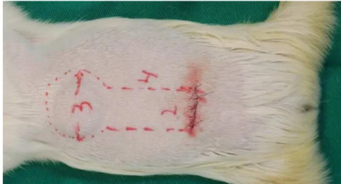
All surgical procedures were performed under general anesthesia by intraperitoneal injection of 100 mg/kg of ketamine hydrochloride (Vetanarcol™, 50 mg/ml, König S/A, Sao Paulo, Brazil) associated to 10 mg/kg of xylazine (Kensol™, 20 mg/ml, König S/A, Sao Paulo, Brazil). Under anesthesia, each animal was placed in the prone position. After epilation of the dorsum from tail up to cervical region, the skin was prepared and draped in a sterile fashion. A surgical transverse incision of 2 cm, symmetrically crossing the midline above lumbar region was done 3-cm above the hind limbs. Following, a tunnel was built above the panniculus carnosus, up to the shoulder blades. The implant (Figure 1) was inserted through the incision and placed on a pocket between scapulas, approximately 3 cm apart from the incision (Figure 2). Incision was closed using 4-0 polyamide interrupted sutures.
FIGURE 2 – Prosthesis implanted in the animal dorsum.
Follow-up and macroscopic assessment
After surgical procedures, animals were placed on a warm environment at 25ºC until complete recovering from anesthesia, and afterwards re-allocated to their respective boxes. Animals were daily observed for their level of activity, respiratory distress, diarrhea, starvation or weight gain, and refusal of food of food or water (indirect signals of infection or sepsis). The macroscopic aspects of wound and pocket implant were carefully inspected for dislocation or extrusion of the prostheses, as well for abscess, hematoma, seroma or wound dehiscence.If any sign of suffering was seen, the veterinarian was authorized to interrupt the research and to euthanize the animal.
Microscopic assessment
The involving skin, subjacent tissues, capsules and implants were removed en bloc and preserved for later
qualitative-quantitative analysis of histologic samples sectioned 5 to 7µm and stained with hematoxylin-eosin (H&E) and histochemical Sirius Red dye (SR) saturated with picric acid as described elsewhere27-30. SR-stained slides were used to analysis of collagen morphology and deposition under polarization.
Qualitative histologic analysis
H&E stained slides were used to establish the type and intensity of inlammation by scoring and to measure capsule thickness. Grading was done by an independent pathologist. Briely, acute inlammation was characterized by the indings of neutrophyls, polymorphonuclear cells, vascular congestion and diapedesis; as to the chronic inlammatory iniltrate, the presence of mononuclear cells (lymphocytes, plasmocytes and monocytes). As described elsewhere31 the two kinds of inlammation were
classiied by scores (0=absent; 1=discrete; 2=moderate, and 3=intense), three slides / animal. Collected data were used to calculate the average score from each group by timepoint.
Quantitative analysis of capsule thickness
H&E-stained sections were examined by light microscopy. Microscopic digital images were obtained as
previously described28. The points of greatest thickness were
located by scanning the sections at a magniication of x100, with the selection of three regions of interest (ROIs): a middle point correspondent to maximal thickness and two lateral adjacent points 50 micra apart from the middle point. Each digitized image (640 x 480 pixels) corresponded to a microscope ield
of 0.29 mm2 (one pixel =0, 53 micrometer). Digital images
were processed and analyzed for capsule thickness using the calibrated free hands measurement tools of the public domain NIH ImageJ program (developed at the U.S. National Institute of Health, Baltimore-MD, and available at htttp://rsb.info.nih.gov/ nih-image/). Three slides were examined per animal and three ROIs in each section were measured by digital planimetry (total = 54 observations by subgroup). The averages mean thickness/ capsule/group/ that were measured at that ROIs of capsules were expressed as mean ± standard deviation (SD).
Quantitative collagen analysis
New histological sections of 7 micrometers thickness were cut from the parafin blocks that were utilized for the histopathological study. These sections were taken from the point of greatest capsular thickness from inner layer to outer lamina of tissue. Three slides of three SR-stained samples of the capsular layer of each animal were used to assess morphometry and density of collagen type III – Col 3 (ibrilar, non-polymerized) and type I – Col 1(thick, polymerized) under an optic polarized-light microscope.
Polarized images (100X, each field area = 0.29 mm2)
were captured with a high-resolution digital camera (Sony™ DSC H-10016.1 MP, Japan) coupled to a polarized microscope. Digitized images were transferred to a personal computer and saved to disc as bmp files (640 x 480 pixels) based on RGB (red, green, blue) color mode, forming an image bank. The computer was equipped with the SAMM, an image analysis software developed and validated by the Federal University of Ceará, Brazil32-33. Due to their anisotropic properties, under
polarization, collagen type I (thicker and bundled mature collagen fibers) turns red (reddish birrefringence) and collagen type III (fibrilar, thinner and dispersed immature collagen) turns green. Based on the color spectrum, the “separate colors” tool was used to split images into color channels (red, green,
blue)28,32. in order to analyze by differential colorimetric
method the deposition of the collagen fibers. The results were expressed as percentages of areas of collagen type I (Col 1) or collagen type III (Col 3) positive pixels per recorded field (the total area of the histologic cut). Subsequently, Col 1/Col 3 ratios were calculated for each group. The average mean percentage for each capsule thin section was determined by calculating the average of means obtained from reading the three ROIs. In our pilot-study, these assessments were performed in two occasions separated by a lapse time of three months by two observers without knowledge of the clinic pathological stage and the results from the histological study using H.E. staining. No significant inter-observer and intra-observer variability has been noticed.
Statistical analysis
In the descriptive analysis, data were summarized as
means, medians, standard deviations, standard errors, 1stand
3rd quartiles, minimum and maximum values. Scores from the
histological assessment of inlammation were analyzed with Kruskal-Wallis test with Dunn’s multiple comparison tests when necessary. Capsule thickness data were analyzed by one factor analysis of variance (ANOVA). Collagen type I and type III deposition percentage were analyzed with Kruskal-Wallis test with Dunn’s multiple comparison tests when necessary. Col 1/Col 3 ratios were analyzed with Sudent’s t test. The used statistical programs were: Statistical Package for Social Sciences (SPSS) version 18.0 and GraphPad Prism 5.0. For evaluating normality conditions of the variables the Shapiro-Wilks test was used. Statistical signiicance was set for a conidence interval of 95% (p<0.05).
Results
Macroscopic analysis
Microscopic analysis
Capsule morphology and inlammation scores
A tri-laminar structure of the capsule was found in all specimens both in H&E and picrosirius stained samples. There was an inner cellular layer adjacent to the implant, an intervening lamina comprising loose connective tissue including an internal vascular supply and a rich cellular presence, and an outer layer collagenous layer of densely packed collagen ibres aligned parallel
to the prosthese surface. The inner layer exhibited an epithelial-like single layer composed from ibrocytes and histiocytes, the so called synovial-type metaplasia (STM). The STM was less
pronounced in propranolol group at 21st post-operative day,
although without statistical signiicance. Also, in some capsules villous hyperplasia was found (Figure 3). Except for the apparent lesser vascularization (not quantiied) in the middle layer from propranolol-treated group as compared to controls we did not observed gross differences between capsules.
FIGURE 3 – Microscopic images of capsules. (A) Tri-layered structure of capsule. (B) Negative image showing the three layers with bright intervening lamina. (C) Zooming of tri-layered capsule. (D) Synovial-type metaplasia with villous hyperplasia of inner capsular layer (x100).
All capsules tended to become less cellular with time. Propranolol-treated capsules demonstrated less inlammatory acute response moderate/intense in about 70% of samples at 7thpost-operative (PO) day; absente/discrete in about 84%
at 14th PO day, but persistent at 21st day (33%), and no giant
cells anytime (Table 1). Also, chronic inlammation was absent
Group Acute
inlammation Post-operative day (%)7th 14th 21st
Control Absent/Discrete 33.3 66.7 83.3
Moderate/Intense 66.7 33.3 16.7
Propranolol Absent/Discrete 33.3 83.3 66.7
Moderate/Intense 66.7 16.7 33.3
TABLE 1 – Intensity of acute inlammatory reaction.
Group Chronic
inlammation Post-operative day (%)7th 14th 21st
Control Absent/Discrete 66.7 83.3 0.0
Moderate/Intense 33.3 16.7 100
Propranolol Absent/Discrete 83.3 66.7 83.3
Moderate/Intense 16.7 33.3 16.7
TABLE 2 – Intensity of chronic inlammatory reaction.
in 100% of propranolol-treated capsules at 21st PO day. Acute
inlammation was practically resolved by 21st PO day in the
control group. However, 100% of tissue samples from this group
harvested at 21st day exhibited a moderate to intense chronic
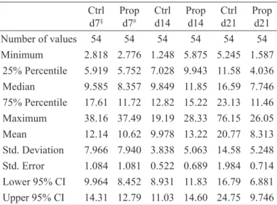
Capsule thickness
Propranolol was highly effective in reducing capsule thickness at 7th and 21stdays periods when compared to control
group (Figure 4 and Table 3).
There was a trend to increasing collagen type I/type III ratios along the time in control group while in propranolol-treated group these ratios exhibited lower and almost constant values along the time. was higher in control group. Proportionally, with the evolution of the post-operatory period, type I collagen increased, and was present in both groups (Figure 7). The highest proportion of type III collagen in propranolol-treated group was of statistical signiicance at all time-points when both groups were compared; also it was signiicant into each group comparison.
FIGURE 4 – Thickness measures of the capsular reaction Bars represent control and propranalol groups at 7th, 14th and
21st timepoints. Signiicant differences were encountered on 7th
(p<0.001) and 21st timepoints (p<0.05), when comparing control
and propranolol groups. ANOVA test. ns = non signiicant.
Ctrl d7§
Prop d7#
Ctrl d14
Prop d14
Ctrl d21
Prop d21
Number of values 54 54 54 54 54 54
Minimum 2.818 2.776 1.248 5.875 5.245 1.587
25% Percentile 5.919 5.752 7.028 9.943 11.58 4.036
Median 9.585 8.357 9.849 11.85 16.59 7.746
75% Percentile 17.61 11.72 12.82 15.22 23.13 11.46
Maximum 38.16 37.49 19.19 28.33 76.15 26.05
Mean 12.14 10.62 9.978 13.22 20.77 8.313
Std. Deviation 7.966 7.940 3.838 5.063 14.58 5.248
Std. Error 1.084 1.081 0.522 0.689 1.984 0.714
Lower 95% CI 9.964 8.452 8.931 11.83 16.79 6.881
Upper 95% CI 14.31 12.79 11.03 14.60 24.75 9.746
TABLE 3 – Descriptive statistics of collagen type I deposition.
§ Ctrl: control group; #: Prop: propranolol group.
Collagen density
Propranolol was effective in impairing deposition of collagen type I ibers into capsule tissue surrounding silicone implants (p-value < 0.0001, one-way ANOVA, Kruskal-Wallis test) when compared to control group (Figure 5).In the initial phase of the capsular formation (7th day) the presence of collagen
type I (reddish) was almost the same for all groups. Density of capsular collagen type III is illustrated in Figure 6.
FIGURE 5 – Density of capsular collagen type I Boxes represent control and propranalol groups at 7th, 14th and 21st timepoints.
Signiicant differences were encountered on 14th (p<0.001) and 21st
timepoints (p<0.0001), when comparing control and propranalol groups.ANOVA test.
FIGURE 6 – Density of capsular collagen type III Boxes represent control and propranalol groups at 7th, 14th and 21st timepoints. No
signiicant differences were encountered when comparing timepoints.
Figure 8 depicts signiicant picrosirius red-stained images with and without polarization at 7th, 14th and 21st timepoints.
FIGURE 8 – Photomicrography of capsules obtained from propranolol-treated animals (right) or untreated (left) and stained with Sirius Red under polarization. Collagen type I ibers stain reddish (orange or red). Collagen type III stain greenish (x100).
Discussion
Adverse capsular contracture (ACC) should be regarded as an exaggeration of normal wound healing6. In patient
populations, this condition is accompanied by overt symptoms
and loss of esthetic results of breast augmentation13 Since the
irst reports on capsular contracture in the literature, a number of preclinical studies have been conducted in an attempt to prevent its development, using different drugs (such as leukotriene receptor antagonists, antibiotics, anti-neoplastic and antiibroticagents,
steroids and non-steroidal anti-inlammatory drugs) administered locally or systemically, but with disappointing results3-9,13-16. Thus,
there is currently no proven pharmacological therapy for ACC13.
Special attention should be paid to the possibility of identifying an oral drug for the treatment of a complication that has serious physical, psychological and inancial consequences and for which the only effective treatment is surgery. Propranolol
is commonly used in the treatment of cardiovascular diseases19.
competitive antagonist of beta-1 and beta-2 adrenergic receptors19-21.
Epinephrine or norepinephrine adrenoceptors, stress-released catecholamines,have been implicated in the pathophysiology of delayed wound healing of cutaneous lesions, acting by delaying the migration and activation of macrophages and neutrophils, inhibiting keratinocyte migration and re-epithelialization in vitro
and in vivo wound models22-23. Besides alteration of cellular
activity related to inlammation, catecholamines stimulate vasoconstriction that causes hypoxia at the wound area and the increase of endothelial cell proliferation, vascular endothelial growth factor (VEGF) expression and angiogenesis. Also, catecholamines promote inhibition of eosinophil inlux23-24.
High concentrations of catecholamines may delay ibroblasts proliferation, myoibroblastic differentiation and collagen deposition24-26. Strack et al.25 demonstrated that propranolol was
effective in reducing inlammation and liver ibrosis progression in a model of nonsinusoidal ibrosis. The transcript level of proibrogenic cytokines TNF-alfa, TGF-beta and CTGF was reduced >50% in ibrotic portal areas. Also, propranolol treatment results in levels of proinlammatory mediators (angiotensinogen and endothelin-1) of vasoconstriction under sympathetic nervous system control.
In this study, the option for silicone instead of saline prostheses was due to the greater incidence of capsular contracture found in the use of the former27. PubMed database was extensively searched for reviewing models of capsular contracture. Capsular contracture is a refractory human-speciic condition, such as keloids are. The proposed models found have provided valuable information about pathogenesis and treatment possibilities of ACC. The length of the observation time (three weeks) used in this study was based on other studies dealing with wound healing. As for the method of measuring the thickness of the capsule and the collagen type I and type III deposition, the picrosirius polarization microscopy method plus digital measurement, previously described in another context was utilized28-31. The use
of conventional histology method to assess inlammation was necessary because it remains the most utilized method to assess capsular inlammation32-37. Baker’s classiication and applanation
tonometry did not seem very appropriate to investigate contractures or ibrosis in rodent models16-17. Nowadays, the use of implants
with smooth surfaces is no longer acceptable due to the fact that they behave differently regarding the occurrence of capsular contracture11 Therefore, it was decided to use one type of textured
prostheses so that responses to the use of propranolol could be veriied eficiently without excessive number of variables. In this study, all samples from both groups exhibited a tri-layered
capsule as described elsewhere by several scholars in human capsules and animal capsules surrounding textured implants11,33-37. Various authors mention that synovial-like metaplasia present in the interface capsule-textured implant is due to mechanic friction between implanted materiel and the peri-prosthetic tissue37-39. The tissue invasion of STM directed to the coating texture (aka, villous hyperplasia) was found in all capsules examined, although without signiicant difference between groups. Despite these fact, propranolol was capable of reduce the thickness of capsular reaction tissue at all timepoints, suggesting an anti-proliferative or cytotoxic effect of the drug.
Lyras40, analyzing the tissue reaction to rough and
smooth silicone implants in a rodent model detected intense acute inlammatory reaction to textured implants. Here, in non-propranolol-treated group, this kind of reaction was absent or discrete in 1/3 of capsules at 7th post-operative (PO) day, 2/3
at 14th PO day and 5/6 at 21st PO day, conirming the indings
already described for textured surface41-43. In our study we was
found that in propranolol-treated capsules the acute inlammatory reaction was absent or discrete in 1/3, 5/6 and 2/3 at 7th, 14th and
21st post-operative day, whereas in control group this reaction was
moderate or intense in 2/3, 1/3 and 1/6 at the same periods. As to chronic inlammatory reaction, in control group this reaction was moderate or intense in 1/3 at 7th PO day, 1/6 at 14th PO day
and 100% at 21st PO day, while in propranolol group an absent or
discrete chronic inlammatory reaction was found in 5/6, 2/3 and 5/6 of the capsules at 7th, 14th and 21st PO day respectively. Clearly,
propranolol exerted an anti-inlammatory effect that persisted through all the experiment.
The effect of propranolol in reducing capsular inlammation and thickness, and in modulating collagen deposition on textured implants observed in this experiment is consistent with indings from previous studies of others ibrosis
conditions19, which showed that the application of nonselective
beta-adrenoceptors antagonists inhibits maturation of collagen, reduces the number of vessels, collagen density, number of mast cells, eosinophils and myoibroblasts compared with control22,23,25.
These concerted actions of propranolol provide a new opportunity to support pharmacotherapeutic approaches to human adverse capsular contracture.
Conclusion
References
1. International Society for Aesthetic Plastic Surgery. Available from http://www.surgery.org.
2. Brazilian Society of Plastic Surgery Available from http://www. cirurgiaplastica.org.br.
3. Tang L, Eaton JW. Natural responses to unnatural materials: A molecular mechanism for foreign body reactions. Mol Med. 1999
Jun;5(6):351-8. PMID: 10415159.
4. Thevenot PT, Baker DW, Weng H, Sun MW, Tang L. The pivotal
role of ibrocytes and mast cells in mediating ibrotic reactions to biomaterials. Biomaterials. 2011 Nov;32(33):8394-403. doi: 10.1016/j.biomaterials.2011.07.084.
5. Jones KS. Assays on the inluence of biomaterials on allogeneic
rejection in tissue engineering.Tissue Eng Part B Rev. 2008
Dec;14(4):407-17. doi: 10.1089/ten.teb.2008.0264.
6. Soder BL, Propst JT, Brooks TM, Goodwin RL, Friedman HI, Yost MJ, Gourdie RG. The connexin43 carboxyl-terminal peptide ACT1 modulates the biological response to silicone implants.
Plast Reconstr Surg. 2009 May;123(5):1440-51. doi: 10.1097/ PRS.0b013e3181a0741d.
7. Adams WP Jr. Capsular contracture: what is it? What causes it? How can it be prevented and managed? Clin Plast Surg. 2009;36(1):119-26. doi: 10.1016/j.cps.2008.08.007.
8. Berry MG, Cucchiara V, Davies DM. Breast augmentation: part II – adverse capsular contracture. J Plast Reconstr Aesth Surg. 2010
Dec;63:2098-107. doi: 10.1016/j.bjps.2010.04.011.b 2010.
9. McCurdy JA, Jr. Capsular contracture following augmentation mammoplasty: etiology and pathogenesis. In: Shifman M (editor).
Breast augmentation. Berlin-Heidelberg: Springer; 2009. p.525-40.
10. Barnsley GP, Sigurdson LJ, Barnsley SE. Textured surface breast implants in the prevention of capsular contracture among breast augmentation patients: a meta-analysis of randomized controlled
trials. Plast Reconstr Surg. 2006 Jun;117(7):2182-90. PMID: 16772915.
11. Araco A, Caruso R, Araco F, Overton J, Gravante G. Capsular contractures: a systematic review. Plast Reconstr Surg. 2009
Dec;124(6):1808-19. doi: 10.1097/PRS.0b013e3181bf7f26.
12. Embrey M, Adams EE, Cunningham B, Peters W, Young VL, Carlo GL. A review of the literature on the etiology of capsular contracture and a pilot study to determine the outcome of capsular contracture
interventions. Aesthetic Plast Surg. 1999 May-Jun;23(3):197-206.
PMID: 10384019.
13. Hidalgo DA, Spector JA. Breast augmentation. Plast Reconstr Surg.
2014 Apr;133(4):567e-83e. doi: 10.1097/PRS.0000000000000033.
14. Fernandes JR, Salinas HM, Broelsch GF, McCormack MC, Meppelink AM, Randolph MA, Redmond RW, Austen WG Jr.Prevention of capsular contracture with photochemical tissue
passivation. Plast Reconstr Surg. 2014 Mar;133(3):571-7. doi: 10.1097/01.prs.0000438063.31043.79.
15. McCoy BJ, Person P, Cohen IK. Collagen production and types in
ibrous capsules around breast implants.Plast Reconstr Surg. 1984 Jun;73(6):924-7. PMID: 6374707.
16. Moreira M, Fagundes DJ, de Jesus Simões M, de Oliveira MC, Dos
Santos Previdelli IT, Moreira AC. Zairlukast pocket delivery impairs
the capsule healing around textured implants in rats. Aesthetic Plast
Surg. 2009 Jan;33(1):90-7. doi: 10.1007/s00266-008-9245-4. 17. Bastos EM, Neto MS, Alves MT, Garcia EB, Santos RA, Heink T,
Pereira JB, Ferreira LM. Histologic analysis of zairlukast’s effect
on capsule formation around silicone implants. Aesthetic Plast Surg.
2007 Sep-Oct;31(5):559-65. PMID: 17576504.
18. Scuderi N, Mazzocchi M, Fioramonti P, Palumbo F, Rizzo MI, Monarca C, Onesti MG. Treatment of the capsular contracture
around mammary implants: our experience. G Chir. 2008
Aug-Sep;29(8-9):369-72. PMID: 18834572.
19. de Mesquita CJ. About strawberry, crab claws, and the Sir
James Black’s invention. Hypothesis: can we battle keloids
with propranolol? Med Hypotheses. 2010 Feb;74(2):353-9. doi:
10.1016/j.mehy.2009.08.035.
20. Szychta P, Stewart K, Anderson W. Treatment of infantile hemangiomas with propranolol: clinical guidelines. Plast
Reconstr Surg. 2014 Apr;133(4):852-62. doi: 10.1097/ PRS.0000000000000007.
21. Amos T, Stein DJ, Ipser JC. Pharmacological interventions for preventing post-traumatic stress disorder (PTSD). Cochrane
Database Syst Rev. 2014 Jul 8;7:CD006239. doi: 10.1002/14651858.
CD006239.pub2.
22. Souza BR, Cardoso JF, Amadeu TP, Desmoulière A, Costa AM. Sympathetic denervation accelerates wound contraction but delays reepithelialization in rats. Wound Repair Regen. 2005
Sep-Oct;13(5):498-505. PMID: 16176458.
23. Bible LE, Pasupuleti LV, Alzate WD, Gore AV, Song KJ, Sifri ZC, Livingston DH, Mohr AM. Early propranolol administration to severely injured patients can improve bone marrow dysfunction.
J Trauma Acute Care Surg. 2014 Jul;77(1):54-60. doi: 10.1097/
TA.0000000000000264.
24. Ellsworth DL, Coady SA, Chen W, Srinivasan SR, Boerwinkle E, Berenson GS. Interactive effects between polymorphisms in the beta-adrenergic receptors and longitudinal changes in obesity. Obes
Res. 2005 Mar;13(3):519-26. PMID: 15833937.
25. Strack I, Schulte S, Varnholt H, Schievenbusch S, Töx U, Wendland K, Steffen HM, Drebber U, Dienes HP, Odenthal M.
β-Adrenoceptorblockade in sclerosing cholangitis of Mdr2 knockout mice: antiibroticeffects in a model of nonsinusoidal ibrosis. Lab Invest. 2011 Feb;91(2):252-61. doi: 10.1038/labinvest.2010.162. 26. Kisseleva T, Brenner DA. Mechanisms of ibrogenesis. ExpBiol Med
(Maywood). 2008 Feb;233(2):109-22. doi: 10.3181/0707-MR-190. 27. Schaub TA, Ahmad J, Rohrich RJ.Capsular contracture with breast
implants in the cosmetic patient: saline versus silicone--a systematic
review of the literature. Plast Reconstr Surg. 2010 Dec;126(6):2140-9. doi: 10.1097/PRS.0b013e3181f2b5a2.
28. Mesquita CJG,Leite JAD, Fechine FV, Rocha JLC, Leite JGS, Leite Filho JAD, Barbosa Filho RA. Effect of imiquimod on partial-thickness burns. Burns. 2010;36:97-108. doi: 10.1016/j.
burns.2009.04.022.
29. Junqueira LC, Cossermelli W, Brentani R. Differential staining of
collagens type I, II and III by Sirius Red and polarization microscopy.
Arch Histol Jpn. 1978;41:267–74. PMID: 82432.
30. Junqueira LC, Bignolas G, Brentani RR. Picrosirius staining plus polarization microscopy, a speciic method for collagen detection in tissue sections. Histochem J. 1979 Jul;11:447–55. PMID: 91593. 31. Montes GS, Junqueira LC. The use of the picrosirius polarization
method for the study of the biopathology of collagen. Mem Inst
Oswaldo Cruz. 1991;86(Suppl. 3):1–11. PMID: 1726969.
32. Jamacaru FVF. Quantiicação de angiogênese corneana in vivo
através de processamento de imagens digitais [Tese]. Universidade
Federal do Ceará; 2006.
33. Balderrama CM, Ribas-Filho JM, Malafaia O, Czeczko NG, Dietz UA, Sakamoto DG, Bittencourt LP. Healing reaction to mammary prostheses covered by textured silicone and silicone foam in rats.
Acta Cir Bras. 2009 Sep-Oct;24(5):367-76.
doi.org/10.1590/S0102-86502009000500006.
34. Kamel M, Protzner K, Fornasier V, Peters W, Smith D, Ibanez D. The peri-implant breast capsule: An immunophenotypic study of capsules taken at explantation surgery. J Biomed Mater Res.
35. Bergmann PA, Tamouridis G, Lohmeyer JA, Mauss KL, Becker B, Knobloch J, Mailänder P, Siemers F. The effect of a bacterial contamination on the formation of capsular contracture with polyurethane breast implants in comparison with textured silicone implants: an animal study. J Plast Reconstr Aesthet Surg. 2014
Oct;67(10):1364-70. doi: 10.1016/j.bjps.2014.05.040.
36. Smahel J. Histology of the capsules causing constrictive ibrosis around breast implants. Br J Plast Surg. 1977 Oct;30(4):324-9.
PMID: 145256.
37. Ko CY, Ahn CY, Ko J, Chopra W, Shaw WW. Capsular synovial
metaplasia as a common response to both textured and smooth
implants. Plast Reconstr Surg. 1996 Jun;97(7):1427-35. PMID: 8643727.
38. Haddad Filho D, Zveibel DK, Alonso N, Gemperli R. Comparison between textured silicone implants and those bonded with
expanded polytetraluoroethylene in rats. Acta Cir Bras. 2007 May-Jun;22(3):187-94. doi.org/10.1590/S0102-86502007000300006.
39. Brohim RM, Foresman PA, Hildebrandt PK, Rodeheaver GT. Early tissue reaction to textured breast implant surfaces. Ann Plast Surg.
1992 Apr;28(4):354-62. PMID: 1596069.
40. Lyras I. Tissue reaction to rough and smooth silicone implants. A comparative and analytical experimental study in rats. Rev Soc Bras
Cir Plast. 1993;8(1):131-41.
41. Clugston PA, Perry LC, Hammond DC, Maxwell GP. A rat model for capsular contracture: the effects of surface texturing. Ann Plast
Surg. 1994 Dec;33(6):595-9. PMID: 7880048.
42. Lesesne CB. Textured surface silicone breast implants: histology in
the human. Aesthetic Plast Surg. 1997 Mar-Apr;21(2):93-6. PMID:
9143423.
43. Batra M, Bernard S, Picha G. Histologic comparison of breast implant shells with smooth, foam, and pillar microstructuring in a rat model from 1 day to 6 months. Plast Reconstr Surg. 1995
Feb;95(2):354-63. PMID: 7824615.
Acknowledgements
SILIMED® for donating the mini-implants used in this
experiment and Prof. Dr. Marto Leal for his expertise in statistical analysis.
Correspondence:
Sérgio Botelho Guimarães Rua Barão de Aratanha, 1465
60050-071 Fortaleza – CE Brasil
Tel.: (55 85)3226-2400 [email protected]
Received: Sep 15, 2014
Review: Nov 17, 2014
Accepted: Dec 18, 2014
Conlict of interest: none
Financial source: none
1Research performed at Experimental Surgery Lab (LABEX), Medical