Intensive care bedside echocardiography: true or a
distant dream?
Ecocardiograia à beira do leito em terapia intensiva: uma
realidade ou um sonho distante?
INTRODUCTION
In the last 10 years the systematic introduction of semi-continued echocardiography in intensive care units and in some services has made this method a valuable tool for severely ill patients management. Con-ditions where early diagnosis is sometimes decisive for a patient man-agement, e.g. in cardiac tamponade and/or aortal dissecation, reiterate its implementation relevance and need.(1-2) Currently available equip-ments portability, and patient-focused training (point of care echocar-diography) justify its use by intensivist physicians. Some recommenda-tions suggested for its use are in chart 1.
Appropriate perfusion and optimized oxygen supply are the major intensive care objectives in circulatory dysfunction patients. Cardiac output (CO) is a variable closely related to venous return, which is defined as the difference between right atrium pressure and mean sys-temic filling pressure (MSFP). Consequently, the CO should be pro-portional to the blood flow arriving the heart, and, depending on the Frank-Starling curve patient’s situation,(3) preload increase by a volume test may either increase or not the cardiac output. In hypovolemic conditions, probably the increased venous return will increase right ventricle preload, and consequently the left ventricle’s, thus optmizing the systolic volume. This situation is said preload dependent, and the patient will be considered as a volume test responder. A frequent
in-Uri Adrian Prync Flato1, André
Luis de Campos2, Matheus Ribas
Trindade2, Hélio Penna Guimarães3,
Marcelo Luiz Campos Vieira4,
Fernando Brunori5
1. Physician – Intensive Care Unit – Instituto Dante Pazzanese de Cardiologia - São Paulo (SP), Brazil. 2. Physician - Intensive Care Unit - Hospital Universitário da Universidade de Marília – UNIMAR – Marília (SP), Brazil.
3. Physician – Internal Medicine Intensive Care Unit - Universidade Federal de São Paulo – UNIFESP - São Paulo (SP), Brazil.
4. Physician - Echocardiography Service - Instituto do Coração (InCor) da Faculdade de Medicina da Universidade de São Paulo – USP - São Paulo (SP), Brazil.
5. Physician – Intensive Care Unit - Hospital Leforte - São Paulo (SP), Brazil.
ABSTRACT
During the last few years, tech-nological development and acquired experience advanced and the echocar-diogram has become an important and useful tool in intensive care unit en-vironment. Data obtained from semi quantitative Doppler echocardiogra-phy (transthoracic and transesopha-geal) evaluation has contributed to an appropriate patient monitoring and management. Echocardiography as a
diagnostic, prognostic and monitoring method for luid responsiveness assess-ment has become available nowadays since hand-carried ultrasound devices are portable and cheaper. Adequate training and development of appropri-ateness criteria for use of echocardiog-raphy in intensive care unit may lead to a standard use as a bedside tool.
Keywords: Echocardiography; In-tensive care; Heart arrest; Inservice training; Point-of-care systems
Received from the Intensive Care Unit - Instituto Dante Pazzanese de Cardiologia - São Paulo (SP), Brazil.
Submitted on July 18, 2009 Accepted on January 4th, 2010
Author for correspondence:
Uri Adrian Prync Flato Instituto Dante Pazzanese de Cardiologia
Av. Dr. Dante Pazzanese, 500 – 3º andar CEP: 04012-180- São Paulo (SP), Brazil.
tensive care question is knowing the patients’ “vol-ume status”, and how to evaluate if tissue perfusion targeted therapeutic interventions are towards the right direction, i.e. if they are beneficial to the pa-tient. With such questions, we perform CO moni-toring as a valuable tool for critically ill patients evaluation. A fundamental aspect in these patients is the CO determination, which can’t be reliably de-termined by physical examination. The Swan Ganz catheter remains as the current gold standard, how-ever its use has been reduced based on the last two years published scientific evidence.(4) Thus, new non-invasive, safe, reliable and reproducible CO monitoring technologies, among them echocardiog-raphy, may mean considerable advantage on these patients management in comparison with the Swan Ganz catheter use.(5) Other non-invasive CO moni-toring methods are listed on chart 2.
In addition to static measures evaluation, its implementation has allowed analyzing the systolic volume variation (dynamic respiratorycycle-related measurements), which is proportional to pulse-pres-sure (PP) and delta PP.(6-8) Another echocardiogra-phy tool is the possibility of estimating left ventric-ular filling pressure (LVFP), usually correlated with pulmonary wedge capilary pressure (PWCP), in the absence of significant valve changes, such as moder-ate to severe mitral valve incompetence. For this, the relationship between the trans-mitral (pulsed doppler) flow measure, positioned above the mi-tral valve closure (ventricular cavity), wave (E), i.e. rapid filling phase, and the tissue doppler (E’) mea-sure, positioned on the septal or lateral mitral ring is used. The E/E’ <8 ratio predicts a LVFP < 12 mmHg; E/E’ > 15 predicts LVFP > 18 mmHg, and intermediate values are in a grey zone.(9) Left atrium
Chart 1 – Intensive care echocardiogram uses
Indications Echocardiography evaluation
Hemodynamic instability Fluid-responsiveness evaluation
Ventricular functioning Systolic and Diastolic ventricular function evaluation
Systolic volume and cardiac output calculation Segmentar LV and RV analysis
Hypovolemia Inferior vena cava collapsing ratio
Right ventricular illing DVP-PSAP evaluation
Cardiac tamponade RV diastolic collapse, and RA systolic
Paradoxical ventricular septum movement
Inspiratory right and left illing and aortal and pulmonary TVI variation
Pulmonary thromboembolism RV dilation and dysfunction
Pulmonary artery hypertension
Direct RV and/or pulmonary trunk thrombus view
Severe valve dysfunction Mitral incompetence (low area, vena contracta, PISA)
Mitral stenosis (PHT, transvalvar gradient, valve area)
Aortal incompetence (vena contracta, PHT)Aortal stenosis (transvalvar gradient, valve area, continuity)Tricuspid incompetence (low area)
Intracardiac shunts Interatrial and interventricular defects
Persistent ductus arteriosus Echo with microbubbles
Infective endocarditis Vegetations, valve dysfunctions
Cardiorespiratory arrest Pseudo PEA and PEA diferentiation
Diferential CRA diagnosis CRA prognosis
Pacemaker positioning Pacemaker location and implantation
pressure (LAP) may be estimated calculating the mitral regurgitation velocity integral in non-mitral valve disease patients. Practically, the non-invasive and validated gradient between left ventricle and left atrium is measured (4x peak mitral regurgita-tion velocity) in heart failure patients.(10)
Other echocardiographic measurements may help evaluating fluid-responsiveness, such as infe-rior cava vein distensibililty rate (ICVD), supeinfe-rior cava vein collapsing rate (SCVC), right ventricu-lar systolic pressure (RVSP), right atrium pressure (RAP), and diastolic and end-systolic left ventricle
area after volume infusion (Chart 3).
The echocardiogram dynamic both pressure and/or volume measurements advantages as compared to the static ones reside on that systolic and diastolic func-tion, and valve changes, do not significantly interact with data interpretation, and thus with therapeutic choice. By inferior vena cava collapsing rate (IVCC), the inferior vena cava (IVC) diameter by subcostal echocardiographic section, aligning the cursor on mode M (motion) 2 cm from RA, we can estimate the right atrium pressure (Chart 4 and Figure 1).(9) Obvi-ously there are situations where we can’t evaluete RAP
Chart 2 – Non-invasive cardiac output monitoring methods
Methods Advantages Disadvantages
Esophageal doppler (CardioQ) Tolerated with awaken patient Continued monitoring
Operator-dependent
Probe can displace, changing accuracy
NICO (partial CO2 re-inhala-tion and measuring CO by indi-rect FICK technique
Minimally invasive Good CO accuracy
Needs OTI and MV MV changes may change CO Hemodynamic stability needed Transesophageal echocardiogram Good OC correlation with
termo-dilution
Fluid-responsiveness evaluation
Not tolerated by awaken patients Operator-dependent
May cause esophageal injury
horacic electric bioimpedance No vascular access needed Electrode positioning subject to error Pulse contour evaluation (Lidco,
PiCCO, FloTrac)
Minimally invasive CO Arterial curve artifacts may interphere on measures eicacy
Anaysis prejudiced with arrhythmia
CO – cardiac output; OTI – orotracheal intubation; MV – mechanic ventilation; PiCCO - pulse contour cardiac output’; Lidco - lithium dilution cardiac output’; NICO – non invasive cardiac output.
Chart 3 – Static and dynamic echocardiogram hemodynamic variables
Echocardiography variable Formula Static/Dynamic
parameter
∆IVC (%) IVC (Dmax – Dmin) / Dmin x 100 Dynamic
∆SVC (%) VCS (Dmax - Dmin) / Dmax x 100 Dynamic
Systolic volume (TEE) LV exit way section area x VTIAo Dynamic
∆Systolic volume (TTE or TEE) +
Passive leg raising
LV exit way section area x VTIAo, following lower limbs elevation maneuver
Dynamic
Left ventricle end-diastolic area LE diastole planimetric area (transversal section) Static
RVSP 4 x (Vmax IT)² + RAP Static
Cardiac Output (LV exit way section area x VTIAo) x heart rate Static
LAP LAP = 1.24 (E/E’) + 1.9
4x mitral regurgitation peak velocity
Static
LVFP=PWCP E/E’ < 8 - LVFP < 12 mmHg
E/E’ >15 - LVFP >18 mmHg
Static
by IVC respiratory phasic range, such as in case of right ventricle (RV) dysfunction and cardiac tampon-ade. Inferior vena cava is a predominantly extra-tho-racic vessel (intra-abdominal) related with right heart chambers suffering amplitude variation according to the respiratory cycle and systolic volume. During spontaneous inspiration there is a reduced intra-tho-racic pressure and increased venous return. Its ampli-tude variation is related with RAP measurement, how-ever there are not yet literature evidences for its use as fluid-responsiveness index in spontaneous breathing or mechanicaly assisted ventilation patients.
Chart 4 – Inferior vena cava evaluation as hemodynamic in-dex for spontaneously breathing patients
IVC size (cm)
Collapsing rate (%) RAP (right atrium
pressure)
<1.5 cm 100% collapsed 0-5 mmHg
1.5-2.5cm >50% collapsed 5-10 mmHg
1.5-2.5cm <50% collapsed 10-15 mmHg
>2.5cm <50% collapsed 15-20 mmHg
>2.5cm No change >20 mmHg
IVC – inferior vena cava; RAP – right atrium pressure.
Source: Otto CM. Echocardiographic evaluation of left and right ven-tricular systolic function. In: Otto CM, editor. Textbook of clinical echocardiography. 2nd ed. Philadelphia: WB Saunders; 2000.
In mechanic ventilation patients, the IVC diam-eter variation is reversed, i.e., during the inspira-tory phase there is increased intra-thoracic pressure and transference of pressures from the right atrium and, consequently, to the communicating vessels. In this case the vena cava has its diameter increased due to two main mechanisms:
1- RV preload reduction
2- RV increased post-load (positive pressure) associated with increased LV preload secondary to blood emptying from pulmonary bed and conse-quent systolic volume increase
This heart-lung coupling causes extra-thoracic vessels changes (both arterial and venous), which may represent volume replacement needs. In this situation, under mechanic ventilation, a percentage of inferior vena cava variation (%∆IVC) and or su-perior (%∆SVC) is used as parameter for volume infusion responsiveness and to identify them as ei-ther responders or non-responders.(11,12)
In practice we use a %∆IVC cutoff value above 12% according to Feissel et al.(13) or above 18% according to Barbier et al.(14) Regarding the %∆SVC,(15) we use a higher cutoff value equal to 36% in controlled mechanic ventilation patients for
ICV – inferior vena cava.
Figure 1 – A) Inferior vena cava measuring, mode M, IVC 30% collapsed. B) IVC ingurgitated secondary to cardiac tamponade. C) Formula used for prediction of right atrial pressure in spontaneous breathing patients.
A) B)
C)
Inferior vena cava collapsing index (IVCCI%) =
estimate RAP
fluid-responsiveness determination.
Information from echocardiographic analysis should be always considered before the patient’s clinical picture.
Recently, De Backer et al. Studied the respira-tory rate (RR >30) interference on systolic volume evaluation in relation to dynamic indices. Their data showed that %∆SVC had no respiratory rate interference, and is perhaps a more suitable param-eter in this settings, such e.g., in patients with acute respiratory distress syndrome (ARDS) and/or rel-evant metabolic acidosis patients.(16)
The CO measurement may be performed using transthoracic echocardiography (TTE) by mea-suring the left ventricle exit way times the Dop-pler measured aortal valve time-velocity integral (VTIAo). The systolic volume found with this method is then multiplied times the heart rate, thus finding the CO.(17-18)
A growing set of evidence-based interventions is in place to guide intensive care clinical practice. As previously described, several and variable hemo-dynamic monitoring methods are available from echocardiogram. However, we show in the chart 5 the most consistent fluid-responsiveness related evidences, and their cutoff values.
In this context, several articles search for answers to help and study the fluid treatment effects in
se-verely ill patients, with emphasis on echocardiog-raphy use. Despite Swan Ganz catheter use contro-versies, echocardiogram is a non-invasive test, easy to perform, with low morbidity and additional to other monitoring tools.(19) A point to be highlighted is that the previously described studies had retro-spectively evaluated the predictive value of hemo-dynamic indices after volume administration, and not necessarily the clinical outcome, which should be evaluated in prospective, randomized and larger samples trials. Second, we should bear in mind that not necessarily a patient responsive to a volume test (%IVC 25%) actually needs fluid. A practical exam-ple would be an anesthetized patient with preserved microcirculation parameters and rated as volume-responder in who excessive hydration can entail ve-nous-capillary congestion and increased morbidity. (20) This deserves reflection as the patient’s global
clinical status and use of bedside echocardiographic data. Hemodynamic echocardiographic informa-tion use confirms the hospital’s efforts on safety monitoring and measuring of ICU options, which can be focused on the structure, process and health care results.(21)
Another much useful echocardiographic fash-ion in the intensive care setting is transesophageal echocardiography. Its indications and diagnostic ac-curacy are reported on chart 6.(22-24) In conditions,
Chart 5 – Dynamic echocardiographic luid-responsiveness indices
Echo Hemodynamic index
Objective Formula Cutof value Author/Year Reference
TEE Aortal peak veloci-ty variation (delta Vpeak)
Aortal peak velocity du-ring respiratory cycle
V p m a x A o - V p m i n A o / 0 , 5 (VpmaxAo+VpminAo)
12% Feissel M
/Mon-net X -2001
39
TEE Superior vena cava collapsing rate
SVC variation during MV respiratory cycle
(maximal diameter – minimal diameter/maximal Demeter) x 100
36% Vieillard- Baron
A - 2004
15
TTE Inferior vena cava distensibility rate
SVC variation during MV respiratory cycle
(maximal diameter – minimal diameter/ minimal diameter) x 100
12%/18% ( m e d i a n 15%)
Feissel M 2002 Barbier C 2004
13-14
TEE PLR aortal blood low variation
Diference between
aortal low velocity af-ter PLR maneuver and baseline
Aortal low (PLR) – baseline aortal low
10% Monnet X -2006 40
TTE PLR systolic volu-me variation
Variation > 15% sys-tolic volume after PLR maneuver and baseline
Ao LVEW section area (longi-tudinal axis) x VTIAo (apical)
12.5% Lamia -2007 41
e.g. during post heart surgery period, TEE is some-times imperative due to difficult transthoracic im-ages acquisition. The initial choice for transthoracic or transesophageal echocardiogram depends on the heart structure to be evaluated, as well as the clini-cal setting involved (surgiclini-cal center, intensive care unit, pre-hospital).(25)
Chart 6 - Transthoracic and transesophageal echocardio-gram characteristics and accuracy
Indication TTE TEE
Aortal dissection + +++
Infective endocarditis evaluation + +++
Intracardiac thrombus + ++
Prosthetic valve + ++
Atrial apendix + +++
Obese patient + ++
Emphysema patient + ++
Hight PEEP MV + ++
Surgery tubes or incisions +- +++
Post cardiac surgery period + +++
+ - less indicated; +++ - more indicated; +- - eventually indicated. TTE – transthoracic echocardiography; TEE – transesophageal echo-cardiography; MV - mechanic ventilation; PEEP – positive end-expi-ratory pressure.
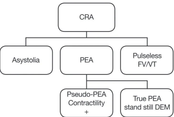
In the intensive care unit, an echocardiogram may be used as an ancillary method for cardiorespi-ratory arrest (CRA) differential diagnosis, specifi-cally in pulseless electric activity (PEA) and asysto-lia, where spontaneous circulation return depends on reversion of the primary cause (hypovolemia, hypoxia, hiperkalemia, cardiac tamponade, pulmo-nary thromboembolism).
Particularly on PEA, with echocardiographic support, two forms were described: true PEA (no heart contractility associated to no pulse) and pseu-do-PEA (myocardial contractility present, with no pulse) (Figure 2). This differentiation is important due to prognostic implications. The implemen-tation of this new format during a CRA is being developed by well designed protocols, appropriate training and mainly, with no thoracic compres-sions interruptions.(26-28) Perhaps, the introduction of new technologies improve the extra-hospital and hospital survival, unchanged for three decades. (29,31) Blaivas et al.(32) evaluated 169 non-arrhythmic
CRA patients (PEA asystolia) using echocardiogram during resuscitation maneuvers and demonstrated 100% mortality in patients with cardiac stand still.
Other authors confirmed these findings, and sug-gested that echocardiogram cardiac stand still in non-arrhythmic CRA is, perhaps, enough to stop cardiopulmonary resuscitation efforts.(33,34) With an agile and more accurate clinical status benefit-ing from a particular intervention identification, maybe resuscitation odds may be significantly im-proved. So far we only have some echocardiogram use in CRA cases and series reports. Nevertheless this is a field to be explored with a perspective of future guidelines implementation. Another impor-tant echocardiogram monitoring and diagnosis re-lated point is identifying its effectiveness and clini-cal feasibility with specific situations protocols, such as shock, CRA and sepsis. Based on these con-cepts, some studies mentioned in the text may be summarized on chart 7, evidencing its feasibility. Among the studies we highlight FEER and BEAT as promising and reproducible.
Training and education in intensive care echo-cardiography
Echocardiography systematization and train-ing within the intensive care unit depends on solid guidelines and continued medical education imple-mentation. The support of national and internation-al associations is fundamentinternation-al for these principles certification conduction. Currently, in France 90% of intensive care units have echocardiography train-ing in a 2 years-long program. Another interesttrain-ing fact is that in England, 90% of intraoperative TEE are conducted by anesthesiologists. This absolutely
CRA
Asystolia PEA
Pseudo-PEA Contractility
+
Pulseless FV/VT
True PEA stand still DEM
CRA – cardiorespiratory arrest; PEA – pulseless electric activity; VF – ventricular ibrilation; VT – ventricular tachycardia; EMD – Eletric-mechanical dissociation.
does not mean that the intensivist physician will replace echocardiographists in the ICU, but that will use this tool to respond specific and contex-tualized questions. Were recently published by the World Interactive Network Focused on Critical Ul-trasound (WINFOCUS) recommendations on the use of echocardiography in the intensive care set-tings, and also by the British Echocardiography So-ciety and American College of Chest Physicians and French Speaking Reanimation Society.(35-38) These guidelines recommend rational degrees of compe-tency and training and how the echocardiogram should be performed. They propose three distinct levels of education, relating the echocardiography use in emergency conditions, as during CRA, and the need of requesting an expert evaluation when indicated and needed. The time to complete each phase depends on each institution’s training, i.e., if there is a formal ICU echocardiography training during intensive medicine specialization, presence of preceptor echocardiography-expert cardiologists, and the ways of measuring students’ performance based on different international associations’ rules. WINFOCUS suggested a 2-year supervised train-ing period, with at least 50 cases recorded yearly to achieve Level 2 competence (Chart 8). It is im-portant highlighting the activity limits for different medical professionals, and the different horizons of
Chart 7- Protocols using intensive care echocardiogram as diagnostic and therapeutic tools
Bedside echocardiogram protocols
Protocol Scenario Objective Variables Results Reference Author/
Year BEAT – Bedside
echocar-diographic assessment in trauma/critical care
Hemody-namic
TTE hemody-namic variables measuring
B= pump (cardiac index), E= pericardial efusion A= area or heart function T= tank, volume status (IVCI)
Good PAC and CVP correlation
18 Gunst M -
2008
Sepsis-Echo – Focused trai-ning for goal-oriented han-dheld
echocardiography perfor-med by noncardiologist re-sidents in the ICU
Sepsis Complement
early therapy with echocar-diogram
Measuring heart contrac-tility and inferior vena cava variation in sepsis
Feasible and pro-mising in sepsis set-tings
25 Vignon P -
2007
FEER – Focused echocar-diographic evaluation in resuscitation
management
CRA Evaluation of
non-arrhyth-mic CRA cau-ses
Portable echocardiogram use for ruling out
tam-ponade, hypervolemia,
pneumothorax, pulmona-ry thromboembolism.
Reduce CRA time with appropriate treatment and ma-nagement standardi-zation during CRA.
26 Breitkreutz
R - 2007
ICU – intensive care unit; IVCI – inferior vena cava index; TTE – transthoracic echocardiogram; TEE – transthoracic echocardiogram; PAC – pul-monary artery catheter; CVP – central venous pressure.
Chart 8 – WINFOCUS36 guidelines-based proposed
intensi-ve care echocardiography competence leintensi-vels
Level 3 Echocardiography expert (invasive procedures, echocardiography investigator)
Level 2 Performs TEE and TTE, reference for Level 1, cardiovascular abnormalities diagnosis, possible investigation training
Level 1 Standard thoracic images acquisition (TEE, TTE), normal and abnormal recognition, need of an expert evaluation recognition, compare with other monitoring techniques
Emergency ECHO
TTE standard images acquisition, relating du-ring CRA according to the ACLS algorithm, re-cognition for requesting expert evaluation.
CRA – cardiorespiratory arrest; TTE – transthoracic echocardiogram; TEE – transesophageal echocardiogram; ACLS – advanced cardio life support.
Source: Price S, Via G, Sloth E, Guarracino F, Breitkreutz R, Catena E, Talmor D; World Interactive Network Focused On Critical Ultra-Sound ECHO-ICU Group. Echocardiography practice, training and accreditation in the intensive care: document for the World Interactive Network Focused on Critical Ultrasound (WINFOCUS). Cardiovasc Ultrasound. 2008;6:49.
FINAL COMMENTS
The use of echocardiography in the inten-sive care and emergency settings is nowadays real in some European countries and North American centers. Possibly in a near future we can have the same rational in Latin American intensive care and emergency centers, such as predicting fluid respon-siveness in critical ill patient.(39-41) The both theo-retical and practical qualifications and appropriate training are fundamental stones for this tool imple-mentation. By scientific research and cooperation between both national and international medical associations, we can improve our daily practice, of-fering our patients better treatment and hoping our dream comes true.
RESUMO
Nos últimos anos, com o avanço tecnológico e a experiência ad-quirida, o ecocardiograma tem se tornado uma ferramenta impor-tante e cada vez mais utilizada no ambiente de terapia intensiva. As informações obtidas, através da ecocardiograia transtorácica e da ecocardiograia transesofágica corroboraram com o monitoramen-to e o cuidado centrado no paciente. Seu papel como ferramenta de diagnóstico, prognóstico e monitoramento da resposta à infusão de luidos (luído-responsividade) tornaram-se disponíveis nos dias de hoje, em razão da portabilidade e diminuição dos custos na aqui-sição dos equipamentos. O treinamento adequado, assim como o desenvolvimento de diretrizes relacionadas à utilização do ecocar-diograma na unidade de terapia intensiva, possibilitarão a padroni-zação deste método assim como sua implementação à beira do leito.
Descritores: Ecocardiograia; Cuidados intensivos; Parada car-díaca; Capacitação em serviço; Sistemas automatizados de assistên-cia junto ao leito
REFERENCES
1. Stamos TD, Soble JS. he use of echocardiography in the critical care setting. Crit Care Clin. 2001;17(2):253-70, v. Review.
2. Heidenreich PA. Transesophageal echocardiography (TEE) in the critical care patient. Cardiol Clin. 2000;18(4):789-805, ix.
3. Guyton AC, Lindsey AW, Abernathy B, Richardson T. Ve-nous return at various right atrial pressures and the normal venous return curve. Am J Physiol; 1957;189(3):609-15. 4. Wiener RS, Welch HG. Trends in the use of the
pulmona-ry artepulmona-ry catheter in the United States, 1993-2004. JAMA. 2007;298(4):423-9.
5. Hett DA, Jonas MM. Non-invasive cardiac output moni-toring. Curr Anaesth Crit Care. 2003;14(4):187-91. 6. Jardin F, Valtier B, Beauchet A, Dubourg O, Bourdarias
JP. Invasive monitoring combined with two-dimensional echocardiographic study in septic shock. Intensive Care Med. 1994;20(8):550-4.
7. Jardin F, Fourme T, Page B, Loubières Y, Vieillard-Baron A, Beauchet A, Bourdarias JP. Persistent preload defect in severe sepsis despite luid loading: A longitudinal echo-cardiographic study in patients with septic shock. Chest. 1999;116(5):1354-9.
8. Michard F, Boussat S, Chemla D, Anguel N, Mercat A, Lecarpentier Y, et al. Relation between respiratory changes in arterial pulse pressure and luid responsiveness in septic patients with acute circulatory failure. Am J Respir Crit Care Med. 2000;162(1):134-8.
9. Garcia MJ, Ares MA, Asher C, Rodriguez L, Vandervo-ort P, homas JD. An index of early left ventricular illing that combined with pulsed Doppler peak E velocity may estimate capillary wedge pressure. J Am Coll Cardiol.
1997;29(2):448-54.
10. Nagueh SF, Middleton KJ, Kopelen HA, Zoghbi WA, Quiñones MA. Doppler tissue imaging: a noninvasi-ve technique for evaluation of left noninvasi-ventricular relaxation and estimation of illing pressures. J Am Coll Cardiol. 1997;30(6):1527-33.
11. Otto CM. Echocardiographic evaluation of left and right ventricular systolic function. In: Otto CM, editor. Text-book of clinical echocardiography. 2nd ed. Philadelphia: WB Saunders; 2000. p. 120–1.
12. Mitaka C, Nagura T, Sakanishi N, Tsunoda Y, Amaha K. Two-dimensional echocardiographic evaluation of inferior vena cava, right ventricle and left ventricle during positive-pressure ventilation with varying levels of positive end-expiratory pressure. Crit Care Med. 1989;17(3):205-10. 13. Feissel M, Michard F, Mangin I. Respiratory change in
inferior vena cava diameter predict luid responsiveness in septic shock (abstract). Am J Respir Crit Care Med. 2002;165(Suppl):A712.
14. Barbier C, Loubières Y, Schmit C, Hayon J, Ricôme JL, Jardin F, Vieillard-Baron A. Respiratory changes in inferior vena cava diameter are helpful in predicting luid respon-siveness in ventilated septic patients. Intensive Care Med. 2004;30(9):1740-6.
15. Vieillard-Baron A, Chergui K, Rabiller A, Peyrouset O, Page B, Beauchet A, Jardin F. Superior vena cava collap-sibility as a gauge of volume status in ventilated septic pa-tients. Intensive Care Med. 2004;30(9):1734-9.
16. De Backer D, Taccone FS, Holsten R, Ibrahimi F, Vincent JL. Inluence of respiratory rate on stroke volume varia-tion in mechanically ventilated patients. Anesthesiology. 2009;110(5):1092-7.
determina-tion of cardiac output in man. Clinical validadetermina-tion. Circula-tion. 1983;67(3):593-602.
18. Gunst M, Ghaemmaghami V, Sperry J, Robinson M, O’Keefe T, Friese R, Frankel H. Accuracy of cardiac func-tion and volume status estimates using the bedside echo-cardiographic assessment in trauma/critical care. J Trauma. 2008;65(3):509-16.
19. Teboul JL, Monnet X. Prediction of volume responsiveness in critically ill patients with spontaneous breathing activi-ty. Curr Opin Crit Care. 2008,14(3):334-9.
20. Michard F, Teboul JL. Predicting luid responsiveness in ICU patients: a critical analysis of the evidence. Chest. 2002;121(6):2000-8.
21. Berenholtz SM, Pustavoitau A, Schwartz SJ, Prono-vost PJ. How safe my intensive care unit? Methods for monitoring and measurement. Curr Opin Crit Care. 2007;13(6):703-8.
22. Moore AG, Eagle KA, Bruckman D, Moon BS, Malouf JF, Fattori R, et al. Choice of computed tomography, transe-et al. Choice of computed tomography, transe-Choice of computed tomography, transe-sophageal echocardiography, magnetic resonance imaging, and aortography in acute aortic dissection: International Registry of Acute Aortic Dissection (IRAD). Am J Car-diol. 2002;89(10):1235-8.
23. Poelaert J, Goarin JP. Indications de l’échocardiographie Doppler chez les patients en état critique. In: Vignon P, Goarin JP, editors. Echocardiographie Doppler en réani-mation, anesthesia et médecine d’urgence. Paris: Elsevier; 2002. p. 17-30.
24. Voga G, Bennett D, Matamis D, Rhodes A, for the Section of Cardiovascular Hemodynamics, ESICM. he use of echocardiography in European intensive care units [Abs-tract] Intensive Care Med. 2002; 28(Suppl 1):S18. 25. Vignon P, Dugard A, Abraham J, Belcour D, Gondran G,
Pepino F, et al. Focused training for goal-oriented hand-held echocardiography performed by noncardiologist residents in the intensive care unit. Intensive Care Med. 2007;33(10):1795-9.
26. Breitkreutz R, Walcher F, Seeger FH. Focused echocardio-Focused echocardio-graphic evaluation in resuscitation management: concept of an advanced life support-conformed algorithm. Crit Care Med. 2007;35(5 Suppl):S150-61.
27. Hernandez C, Shuler K, Hannan H, Sonyika C, Likou-rezos A, Marshall J. C.A.U.S.E.: Cardiac arrest ultra-sound exam--a better approach to managing patients in primary non-arrhythmogenic cardiac arrest. Resuscitation. 2008;76(2):198-206.
28. Sloth E, Jakobsen CJ, Melsen NC, Ravn HB. he resus-citation guidelines in force-time for improvement towards causal therapy? Resuscitation. 2007;74(1):198-9.
29. International Liaison Committee on Resuscitation. 2005 International Consensus on Cardiopulmonary Resuscita-tion (CPR) and Emergency Cardiovascular Care (ECC) Science With Treatment Recommendations. Circulation. 2005;112(22 Suppl):III-1–III-136.
30. American Heart Association in collaboration with Inter-national Liaison Committee on Resuscitation. Guidelines 2000 for Cardiopulmonary Resuscitation and Emergency Cardiovascular Care. Circulation. 2000;102(8 Suppl):I1– I384.
31. Standards for cardiopulmonary resuscitation (CPR) and emergency cardiac care (ECC). 3. Advanced life support. JAMA. 1974;227(7) Suppl:852-60.
32. Blaivas M, Fox JC. Outcome in cardiac arrest patients found to have cardiac standstill on the bedside emer-gency department echocardiogram. Acad Emerg Med. 2001;8(6):616-21.
33. Salen P, Melniker L, Chooljian C, Rose JS, Alteveer J, Reed J, Heller M. Does the presence or absence of sono-graphically identiied cardiac activity predict resuscitation outcomes of cardiac arrest patients? Am J Emerg Med. 2005;23(4):459-62.
34. Salen P, O’Connor R, Sierzenski P, Passarello B, Pancu D, Melanson S, et al. Can cardiac sonography and capnogra-phy be used independently and in combination to predict resuscitation outcomes? Acad Emerg Med. 2001;8(6):610-5.
35. Fox K. A position statement: echocardiography in the cri-tically ill. On behalf of a Collaborative Working Group of the British Society of Echocardiography (BSE). J Intensive Care Soc. 2008;9(2):197-8.
36. Price S, Via G, Sloth E, Guarracino F, Breitkreutz R, Ca-tena E, Talmor D; World Interactive Network Focused On Critical UltraSound ECHO-ICU Group. Echocardiogra-phy practice, training and accreditation in the intensive care: document for the World Interactive Network Focu-sed on Critical Ultrasound (WINFOCUS). Cardiovasc Ultrasound. 2008;6:49.
37. Boyd JH, Walley KR. he role of echocardiography in hemodynamic monitoring. Curr Opin Crit Care. 2009;15(3):239-43. Review.
38. Mayo PH, Beaulieu Y, Doelken P, Feller-Kopman D, Har-rod C, Kaplan A, et al. American College of Chest Phy-sicians/La Société de Réanimation de Langue Française statement on competence in critical care ultrasonography. Chest. 2009;135(4):1050-60.
39. Feissel M, Michard F, Mangin I, Ruyer O, Faller JP, Teboul JL. Respiratory changes in aortic blood velocity as an in-dicator of luid responsiveness in ventilated patients with septic shock. Chest. 2001;119(3):867-73.
40. Monnet X, Rienzo M, Osman D, Anguel N, Richard C, Pinsky MR, Teboul JL. Passive leg raising predicts luid responsiveness in the critically ill. Crit Care Med. 2006;34(5):1402-7.