Article/Artigo
1. Department of Pathology Science, Biological Science Center, State University of Londrina, Londrina, PR, Brazil. 2. Health Science Research Center, University North of Paraná, Londrina, PR, Brazil. 3. Medical Mycology Research Center, Chiba University, Chiba, Japan.
Address to: Dra. Eiko Nakagawa Itano. Depto Ciências Patológicas/CCB/UEL. Campus Universitário, 86051-970 Londrina, PR, Brasil.
Phone: 55 43 3371-4469; Fax: 55 43 3371-4207 e-mail: [email protected]
Received in 21/01/2010 Accepted in 27/07/2010
INTRODUCTION
High molecular mass fraction in clinical isolates of
Paracoccidioides
brasiliensis
Fração de alta massa molecular em isolados clínicos de
Paracoccidioides brasiliensis
Andréa Longoni Fredrich
1, Luciene Airy Nagashima
1, Wander Rogério Pavanelli
1, Audrey de Souza Marquez
2,
Mari Sumigawa Kaminami
1, Nilson de Jesus Carlos
1, Ayako Sano
3, Mario Augusto Ono
1and Eiko Nakagawa Itano
1ABSTACT
Introduction: Diferent serum levels of the IgG/IgE for Paracoccidioides brasiliensis high mass
molecular (hMM) fraction (~366kDa) in the acute and chronic forms of the disease have been reported. Considering the nonexistence of hMM fraction investigation involving clinical isolates of
P. brasiliensis, the present study aimed to investigate the presence of the hMM fraction (~366kDa)
in cell free antigens (CFA) from P. brasiliensis clinical isolates. Methods: CFA from 10 clinical
isolates and a reference strain (Pb18) were submited to SDS-polyacrylamide gel electrophoresis (SDS-PAGE) followed by gel image capturing and densitometer analysis. Additionally, CFA from 20 isolates and Pb18 were analyzed by capture ELISA (cELISA) using polyclonal (polAb) or monoclonal (mAb) antibodies to the hMM fraction. Results: he presence of the hMM component was observed in CFA of all samples analyzed by SDS-PAGE/densitometry and by cELISA. In addition, Pearson’s correlation test demonstrated stronger coeicients between hMM fraction levels using pAb and mAb (R = 0.853) in cELISA. Conclusions: he soluble hMM fraction was present in all the P. brasiliensis clinical isolates analyzed and the reference strain Pb18, which could
be used as a source of this antigen. he work also introduces for irst time, the cELISA method for
P. brasiliensis hMM fraction detection. Analysis also suggests that detection is viable using polAb
or mAb and this methodology may be useful for future investigation of the soluble hMM fraction (~366kDa) in sera from PCM patients.
Key-words: Paracoccidioidomycosis. Soluble antigen. Capture ELISA. Monoclonal antibodies. IgG.
RESUMO
Introdução: Diferentes níveis sorológicos de IgG/IgE contra a fração de alta massa molecular (hMM) (~366kDa) de Paracoccidioides brasiliensis têm sido encontrados na PCM aguda e
crônica. Considerando a inexistência de investigação sobre esta fração em isolados clínicos de P. brasiliensis, o objetivo deste estudo foi investigar a presença da fração hMM (~366kDa)
no preparado livre de células (CFA) de P. brasiliensis obtidos de isolados clínicos. Métodos:
CFA de 10 isolados e de cepa de referência (Pb18) foram submetidas à eletroforese em gel de SDS-poliacrilamida (SDS-PAGE) seguida de captura de imagem e análise por densitometria. Adicionalmente, CFA de 20 isolados e de Pb18 foram analisados por ELISA captura (cELISA) utilizando anticorpos policlonal (polAb) ou monoclonal (mAb) para fração hMM. Resultados:
A presença do componente de hMM foi observada em todas as amostras analisadas por SDS-PAGE/densitometria e por cELISA. Adicionalmente, o teste de correlação de Pearson
demonstrou forte relação entre os níveis de fração hMM usando pAb e mAb (R = 0.853) no cELISA. Conclusões: Conclui-se que a fração hMM está presente em todos os isolados clínicos de P. brasiliensis analisados e no isolado referencial, sugerindo a possibilidade dos mesmos
serem utilizados como fonte desta fração antigênica. Este trabalho também introduz pela primeira vez o método de cELISA para detecção da fração hMM de P. brasiliensis, sugerindo
que detecção utilizando anticorpos polAb ou mAb é viável e essa metodologia poderá ser útil para investigação futura desta fração solúvel (~366kDa) em soros de pacientes com PCM.
Palavras-chaves: Paracoccidioidomicose. Antígeno solúvel. ELISA de captura. Anticorpos monoclonais. IgG.
Paracoccidioidomycosis (PCM), a deep mycosis endemic in Latin America, is caused by the thermally dimorphic fungus Paracoccidioides brasiliensis,
which develops as yeast at body temperature and as mycelium at room temperature. P. brasiliensis
causes natural infections by inhalation of conidia or mycelial elements1. Most exposed subjects
develop an asymptomatic infection, although some individuals present clinical manifestations that can vary from benign and localized to severe and disseminated forms2. Two forms of the disease are
distinguished: the acute or subacute and chronic form. he acute form is more severe and rare, while the chronic form occurs more frequently and mostly afects adult males3,4.
Usually, PCM diagnosis is inferred from indirect evidence obtained via serological tests and clinicallyrelevant antigens have been identiied and adapted for use inimmunoassays for the detection of specific antibodies5. For this purpose, several
fungal components have already been identiied. he antigens most frequently identiied in PCM patient sera are the glycoproteins of 43kDa (97-100%)6-8, the
main PCM diagnostic antigen6,7,9,10, 160kDa (78%)
and 70kDa (60%)8.
he other alternative laboratory approach for diagnosis of PCM is the detection of circulating
P. brasiliensis antigens. The inhibition ELISA
methodology (inh-ELISA) is ableto detect gp43 in 96.3% of PCM patients, mainly in those withthe acute form of the disease (100%)11. Gp70 has also
been detected in the urine or in cerebrospinal luid (CSF) of PCM patients12,13. Gómez et al14,15 reported
the use of inh-ELISA and detected gp87 circulating antigen in sera from patients withactive disease.
Puccia et al9 demonstrated polydispersed
high-molecular mass glycoprotein, with heterogeneous electrophoresis migration. From this heterogeneous electrophoresis migration Marquez et al16 isolated
Fredrich AL et al - High molecular mass fraction and Paracoccidioides brasiliensis
METHODS
RESULTS or cell free antigens (CFA) preparations. Moreover, diferent serum
levels of the IgG/IgE to the hMM fraction was veriied in sera of acute and chronic PCM patients and the authors suggested the analysis as a new characteristic to diferentiate between these two clinical forms of the disease16.
Taking into account the lack of data regarding the soluble hMM fraction (~366kDa) in P. brasiliensis clinical isolates and the
immunological methodology for identifying it, in the present study, the hMMAg antigen was investigated in diferent clinical isolates and capture ELISA (cELISA) was introduced. In principle, observation veriied that all the P. brasiliensis samples analyzed produced the
hMM fraction (~366kDa). In addition, this hMM fraction was detected by cELISA using monoclonal or polyclonal antibodies to the hMM fraction.
Fungal isolates
Clinical isolates of P. brasiliensis of the chronic form of PCM
disease were obtained: 17 isolates (LDR1 to LDR17) from Londrina State University (MOOI/CCS, HC, HC, Londrina State University, Londrina, Paraná) patients (2000 to 2006); two isolates (RC-Wang and RC-Hori) from Chiba University, Chiba, Japan; one isolate (EPM-01) and reference strain (Pb18) from UNIFESP, São Paulo, Brazil. P. brasiliensis strains were maintained on potato dextrose agar
(Difco Laboratories, MI, USA) slants at room temperature. Prior to experiments, samples of the isolates were inoculated onto a slant of brain heart infusion agar (BHI, Difco Laboratories) supplemented with 1% dextrose and cultured at 35ºC to produce the yeast form and maintained by subculturing at 35ºC at 5-day intervals on Sabouraud agar (Micromed, Rio de Janeiro, RJ, Brazil).
Cell free antigens preparation
Yeast cells were collected and the CFA samples were obtained according to Camargo et al17, modiied by the addition of PMSF
protease inhibitor at 2.5mM to the supernatant. The protein concentration was determined by the Lowry method18, adjusted to
3mg/mL and stored in -80ºC freezer until ready to use.
Cell free antigens analysis by SDS-PAGE
Cell free antigens (3mg/mL) samples, obtained as described above, were mixed with the reducing sample bufer (62.5mM Tris-HCl, pH 6.8, 2% sodium dodecyl sulfate (SDS), 10% glycerol, 10%
β-mercaptoethanol and 0.05% bromophenol blue) and boiled for 3 min. he antigens were separated by SDS-polyacrylamide gel electrophoresis (7.5%), in tris-glycine bufer, pH 8.2, at 125v. Protein standards with the following molecular masses were used: myosin (201.1kDa); β-galactosidase (115.7kDa); bovine serum albumin (93.6kDa); ovalbumin (50.3kDa); carbonic anhydrase (37.3kDa). Using specific densitometer software, Glob-Al Scan (Cellogel Electrophoresis Co, Milan, Italy), silver stained dehydrated SDS-electrophoresis gel image was captured, the speciic cutof point of that deines the area of the hMM fraction band (~366kDa) was indicated and the result automatically calculated and expressed in percentages.
Capture ELISA for IgG-hMM fraction
ELISA immunoplates sensitized with rabbit IgG anti-hMM fraction (25µg/ml) were incubated with CFA samples at 30μg/ml at 37ºC for 1h and then with polyclonal mouse IgG anti-hMM
(IgG pAb) (30μg/ml), anti-mouse IgG peroxidase conjugate and OPD (100μL well). he absorbance was read at 492nm. Additionally, cELISA for hMM fraction level determination was performed as described, substituting polyclonal mouse IgG anti-hMM fraction for monoclonal IgG anti-hMM fraction (IgG mAb) as secondary antibodies. Polyclonal rabbit and mouse antibodies were produced by using the hMM fractions obtained, according to Pavanelli et al19 for immunizations. A monoclonal
antibody was obtained by spleen cells (from immunized BALB/c mice with the hMM fraction) fused with P3U1 cell line by using PEG. Hybridomas were screened by ELISA with hMM fraction (~366kDa) and cloned by limiting dilutions. Pristane-primed BALB/c mice were injected i.p. with hybridoma and IgG puriied (Sepharose-G protein column) from ascite luid. he cutof was determined as the mean plus 2 standard deviations of the absorbance obtained with control: primary rabbit IgG anti-hMM, secondary mouse pAb or mAb IgG and peroxidase conjugate without the CFA sample.
Statistical analysis
Statistical comparisons were performed by analysis of variance (ANOVA) and by the Tukey test. All values are reported as the mean ± SD of the mean, with signiicance assumed in the range of p<0.05. Pearson’s correlation was applied between hMM fraction levels using IgG pAb and mAb anti-hMM fraction and a signiicant correlation was considered when r ≥ 0.50.
Ethical
his study was approved by the Internal Scientiic Commission and the Bioethics in Research Commitee of the State University of Londrina (Londrina, PR, Brazil).
CFA analysis by SDS-PAGE
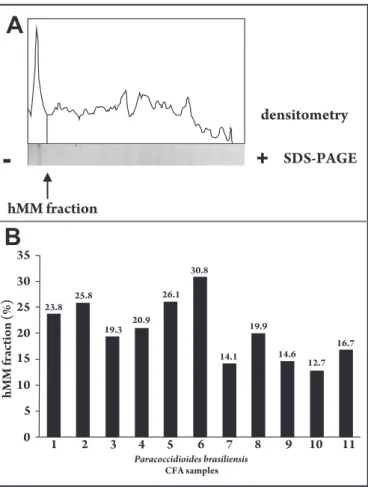
he presence of the hMM fraction was observed in CFA from all the clinical isolates and reference Pb18 by PAGE. he SDS-PAGE was submited to densitometry in an atempt to quantify the hMM fraction. he results show that 20.5 ± 5.7% (clinical isolates) and 16.7% (Pb18) from CFA correspond to the hMM fraction
(Figure 1). his study indicates that this antigen is present in diferent
clinical isolates from chronic patients and could be used as source of the hMM fraction.
Capture ELISA for hMM fraction levels in CFA samples
Various researchers have atempted to detect circulating antigensin PCM patients using polyvalent antigens or antibodies in diferentassays. For this purpose, the presence of the hMM fraction in CFA samples was determined using cELISA expressed as optical density units (OD). Analysis of the result veriied the presence of the hMM fraction in Pb18, a strain characterised as highly virulent, and in all the clinical isolates of P. brasiliensis, with the use of polAb or mAb to the hMM fraction
(Table 1).All values greater than the values obtained in the control systems of polyclonal/polyclonal or monoclonal/monoclonal with free samples of CFA plus 2 x standard (0.118 ± 0.008 and 0.086 ± 0.005, respectively) were considered as positive.
Correlation analysis
TABLE 1 - High molecular mass fraction (~ 366 KDa) levels in CFA from clinical isolates Paracoccidioides brasiliensis (Pb) by capture ELISA, expressed in optical density units.
Reference strain Pb pAb-hMM mAb-hMM
Pb18 0.600 0.596
LDR1 0.659 0.655
LDR2 0.617 0.664
LDR3 0.587 0.574
LDR4 0.531 0.458
LDR5 0.434 0.437
LDR6 0.574 0.583
LDR7 0.548 0.582
LDR8 0.591 0.679
Clinical isolates LDR9 0.570 0.610
RC-WANG 0.547 0.542
LDR10 0.513 0.545
EPM-01 0.533 0.490
LDR11 0.454 0.532
RC-HORI 0.430 0.431
LDR12 0.506 0.478
LDR13 0.533 0.560
LDR14 0.487 0.490
LDR15 0.430 0.437
LDR16 0.523 0.599
LDR17 0.602 0.591
Mean±SEM 0.537±0.064 0.549±0.076
ELISA immunoplates sensitized with rabbit IgG anti-hMM were incubated with CFA samples and then with polyclonal mouse IgG anti-hMM (pAb-hMM) or with monoclonal mouse IgG anti-hMM (mAb-hMM). Ater incubation with anti-mouse IgG peroxidase conjugate, absorbance was read at 492nm. All values higher than the controls: polyclonal/polyclonal (0.118 ± 0.008) or polyclonal/monoclonal (0.086 ± 0.005), with free CFA sample, plus 2 x standard were considered positive.
Paracoccidioides brasiliensis
FIGURE 1 - he hMM fraction in CFA from P. brasiliensis by SDS-PAGE and densitometry. A) P. brasiliensis CFA sample was separated using 7.5% SDS-polyacrylamide gel electrophoresis (SDS-PAGE), silver stained and dehydrated. The gel image was captured and the specific cutoff point of hMM fraction (~366kDa) was marked and equivalent area was automatically calculated and expressed in percentages of hMM fraction. B) Percentage of hMM fraction in relation to all other fractions in CFA from clinical isolates of P. brasiliensis: (1) LDR13, (2) LDR11, (3) EPM-01, (4) LDR16, (5) LDR17, (6) Hori, (7) RC-Wang, (8) LDR12, (9) LDR14, (10) LDR10 and reference strain (11) Pb18.
FIGURE 2 - Pearson’s correlation tests between hMM fraction levels by capture ELISA (cELISA) using monoclonal (mAb) or polyclonal (polAb). he cELISA results showed a stronger correlation between hMM fraction levels with IgG pAb or mAb anti-hMM as a secondary antibody (r= 0.85).
DISCUSSION
In this work, the hMM fraction was detected in all the
P. brasiliensis clinical isolates and the reference strain Pb18 by
SDS-PAGE, suggesting that all these P. brasiliensis could be used as a source
of this fraction.
For diagnostic purposes, the presence of the target antigen in all isolates of the P. brasiliensis is important. he present study indicates that
this antigen is present in diferent clinical isolates from chronic patients as the common P. brasiliensis antigen. However, further studies involving
larger sample of isolates are required to conirm these indings. Panunto-Castelo et al8 identiied hMM antigens with 172 or
160kDa in exoAg from three P. brasiliensis isolates (DGO, C-9 and
BAT). In this study, the considered hMM band identiied in CFA from the P. brasiliensis isolates presented ~366kDa and is, therefore,
a diferent hMM antigen.
The detection of circulating antigens is a useful approach for serodiagnosis for monitoring PCM treatment. he gp43 glycoprotein, one of the most important immunodominant antigens of P. brasiliensis,
has been extensively investigated, particularly in relation to diagnosis20-22.
Gp43 has been detected at higher levels in PCM patient sera as circulating antigens22 and in CSF and bronchoalveolar lavage (BAL) luid samples
from PCM patients11. However, recently the existence of a P. brasiliensis
isolate presenting diferences in this major antigen coding gene gp4323,24
has been demonstrated and speculation of the possibility of a new species in the genus Paracoccidioides has been noted. Considering genetic diferences in gp43 according to P. brasiliensis isolate, it has become
important to investigate other P. brasiliensis antigens for diagnosis.
Besides gp4311, the 87-kDa molecule15, gp7013 and the high molecular
mass antigen with 160kDa25 were also introduced as potential candidates
he authors declare that there is no conlict of interest. CONFLICT OF INTEREST
FINANCIAL SUPPORT
REFERENCES Panuto-Castelo et al8 demonstrated that the hMM antigens
(172 or 160kDa) are highly reactive with serum IgG of patients with acute or chronic PCM, indicating their potential application in the diagnosis and follow-up of the disease. In addition, Coltri et al25 characterized this antigen as a protein of 160kDa, designated
paracoccin, with selective binding to immobilized GlcNAc and able to interact with laminin.
In this study, the association between SDS-PAGE and densitometry analysis shows that approximately 16-20% of P. brasiliensis CFA antigens
correspond to the hMM fraction. Considering the proportion and as a soluble antigen present in CFA, we speculate that its presence may also be observed as a soluble antigen during infection, similar to other antigens, such as gp43 or gp70, present in CFA and in the serum11,13,22,26, with the
potential for diagnosis and follow-up of the PCM patients. hus, the hMM fraction is also important when considering the distinct isotypic humoral immune response to hMM antigens with ~336kDa observed in the acute and chronic forms of the PCM disease, which suggest that it has potential as a new biomarker for diferentiating these two clinical forms16.
In this study cELISA was introduced to detect the hMM fraction in clinical isolates and the reference Pb18 strain was introduced and the presence of the hMM fraction was detected in all the samples analyzed, in agreement with SDS-PAGE. his study is qualitative and the results are expressed as optical density. cELISA has the advantage of being able to process large numbers of samples at the same time and presents high sensitivity and a high speciicity. he analysis was performed using polAb or mAb to the hMM fraction as a secondary antibody and the results showed a stronger correlation between hMM fraction levels obtained using pAb or mAb anti-hMM. cELISA with speciic polAb and mAb was used in previous studies by our group to determine circulating soluble gp43 levels in PCM22 and determining
plasmatic hMM fraction levels in PCM patients by cELISA will be the object of future investigations.
Unexpectedly, the results of ELISA showed homogeneity. We believe that regardless of diferences in isolates or strains, these components are beter preserved and produced more homogeneously, as observed with other antigens reported in the literature8. he same isolates showed
less homogeneity in relation to gp43 determined by capture ELISA (data not shown) and the heterogeneity observed in the percentage of the hMM fraction in the electrophoresis was due to variations in the other components present in CFA (data not shown).
In conclusion, the soluble hMM fraction was present in all the P. brasiliensis clinical isolates analyzed and the reference strain
Pb18, which could be used as source of this fraction. he work also introduced for irst time the capture ELISA method for P. brasiliensis
hMM fraction (~366kDa) detection.
his work was supported inancial by the Financiadora de Estudos e Projetos (FINEP), the Secretaria de Estado da Ciência, Tecnologia e Ensino Superior (SETI/PR), the Fundação Araucaria/PR, Pró-reitoria de Extensão (PROEX/UEL), the Pró-Pró-reitoria de Pesquisa (PROPPG/UEL) and the Coordenação de Aperfeiçoamento de Pessoal de Nível Superior (CAPES).
1. Restrepo A, Tobón AM. Paracoccidioides brasiliensis. In: Mandell GL, Bennet JE, Dollin R, editors. Principles and Practice of Infectious Diseases. 6th ed. Philadelphia: Elsevier; 2005. p. 3062-3068.
2. Borges-Walmsley MI, Chen D, Shu X, Walmsley AR. The pathobiology of
Paracoccidioides brasiliensis. Trends Microbiol 2002;10:80-87.
3. Shikanai-Yasuda MA, Telles Filho FQ, Mendes RP, Colombo AL, Moreti ML, Grupo de Consultores do Consenso em Paracoccidioidomicose. Consenso em Paracoccidioidomicose. Rev Bras Med Tropical 2006; 39:297-310.
4. Franco M, Montenegro MR, Mendes RP, Marques SA, Dillon NL, Mota NGS. Paracoccidioiodomycosis: a recently proposed classiication of its clinical forms. Rev Soc Brasil Med Trop1987; 20:129-132.
5. Ferreira-da-Cruz MF, Galvão-Castro B, Daniel-Ribeiro CT. Isolation and antigenicity of a 45-kilodalton Paracoccidioides brasiliensis immunodominant antigen. Infect Immun 1992; 60:2667-2671.
6. De-Camargo Z, Unterkircher C, Campoy SP, Travassos LR. Production of
Paracoccidioides brasiliensis exoantigens for immunodiffusion tests. J Clin
Microbiol 1988; 26:2147-2151.
7. Taborda CP, Camargo ZP. Diagnosis of paracoccidioidomycosis by passive haemagglutination assay of antibody using a puriied and speciic antigen-gp43. J Med Vet Mycol 1993; 31:155-160.
8. Panunto-Castelo A, Freitas-da-Silva G, Bragheto IC, Martinez R, Roque-Barreira MC. Paracoccidioides brasiliensis exoantigens: recognition by IgG from patients with diferent clinical forms of paracoccidioidomycosis. Microbes Infect 2003; 5:1205-1211.
9. Puccia R, Schenkman S, Gorin PA, Travassos LR. Exocellular components of
Paracoccidioides brasiliensis: identiication of a speciic antigen. Infect Immun
1986; 53:199-206.
10. Puccia R, Travassos LR. 43-kilodalton glycoprotein from Paracoccidioides brasiliensis: immunochemical reactions with sera from patients with paracoccidioidomycosis, histoplasmosis, or Jorge Lobo’s disease. J Clin Microbiol 1991; 29:1610-1615.
11. Marques-da-Silva SH, Colombo AL, Blota MHSL, Lopes LD, Queiroz-Telles F, Camargo ZP. Detection of circulating gp43 antigen in serum, cerebrospinal luid and bronchoalveolar lavage of patients with paracoccidioidomycosis. J Clin Microbiol 2003; 41:3675-3680.
12. Salina MA, Shikanai-Yasuda MA, Mendes RP, Barravieira B, Mendes-Giannini MJS. Detection of circulating Paracoccidioides brasiliensis antigen in urine of
paracoccidioidomycosis patients before and during treatment. J Clin Microbiol 1998; 36:1723-1728.
13. Marques-da-Silva SH, Matos Grosso D, Lopes JD, Colombo AL, Blota MHSL, Queiroz-Telles F, et al. Detection of Paracoccidioides brasiliensis gp70 circulating
antigen and follow-up of patients undergoing antimycotic therapy. J Clin Microbiol 2004; 42:4480-4486.
14. Gómez BL, Figueroa JL, Hamilton AJ, Ortiz B, Robledo MA, Hay RJ, et al. Use of monoclonal antibodies in diagnosis of paracoccidioidomycosis: new strategies for detection of circulating antigens. J Clin Microbiol 1997; 35:3278-3283. 15. Gómez BL, Figueroa JI, Hamilton AJ, Diez S, Rojas M, Tobón AM, et al. Restrepo
A. Antigenemia in patients with paracoccidioidomycosis: detection of the 87-kilodalton determinant during and ater antifungal therapy. J Clin Microbiol 1998; 36:3309-3316.
16. Márquez AS, Vicentini P, Ono MA, Watanabe MAE, Camargo ZP, Itano EN. Reactivity of antibodies from patients with acute and chronic paracoccidioidomycosis to a high-mass molecular antigen from Paracoccidioides brasiliensis. J Clin Labor Anal 2005; 19:199-204.
17. Camargo ZP, Taborda CP, Rodrigues EG, Travassos LR. he use of cell-free antigens of Paracoccidioides brasiliensis in serological tests. J Med Vet Mycol
1991; 29:31-38.
18. Lowry OH, Rosebrough NJ, Farr AL, Randall RJ. Colorimetric assays: Lowry´s method for protein determination. J Biol Chem 1951; 193:265-275. 19. Pavanelli WR, Kaminami MS, Gerez JR, Sano A, Ono MA, Camargo ICC, et al.
Protection induced in BALB/c mice by the high-molecular-mass (hMM) fraction of Paracoccidioides brasiliensis. Mycopathologia 2007; 163:117-128.
20. Camargo ZP, Unterkircher C, Travassos L. Identiication of antigenic polypeptides of Paracoccidioides brasiliensis by immunoblotting. J Med Vet Mycol 1989; 27:407-412.
21. Brummer E, Castañeda E, Restrepo A. Paracoccidioidomycosis: an update. Clin Microbiol Rev 1993; 6:89-117.
22. Miura CSN, Estevão D, Lopes JD, Itano EN. Levels of speciic antigen (gp43), speciic antibodies, and antigen-antibody complexes in saliva and serum of paracoccidioidomycosis patients. Med Mycol 2001; 37:423-428.
23. Carrerro L, Nino-Vega G, Teixeira MM, Carvalho MJA, Soares CM, Pereira M, et al. New Paracoccidioides brasiliensis isolate reveals unexpected genomic variability in this human pathogen. Fungal Genet Biol 2008; 45:605-612. 24. Takayama A, Itano EN, Sano A, Ono MA, Kamei K. An atypical Paracoccidioides
brasiliensis clinical isolate. Med Mycol 2009; 47:01-09.
25. Coltri KC, Casabona-Fortunato AS, Gennari-Cardoso ML, Pinzan CF, Ruas LP, Mariano VS, et al. Paracoccin, a GlcNAc-binding lectin from Paracoccidioides brasiliensis, binds to laminin and induces TNF-alpha production by macrophages. Microbes Infect 2006; 8:704-713.
26. Stambuk BU, Puccia R, De-Almeida ML, Travassos LR, Schenkman S. Secretion of the 43kDa glycoprotein antigen by Paracoccidioides brasiliensis. J Med Vet Mycol